Oral Session
Parameter Mapping & Relaxometry
Joint Annual Meeting ISMRM-ESMRMB & ISMRT 31st Annual Meeting • 07-12 May 2022 • London, UK

17:00 |
0178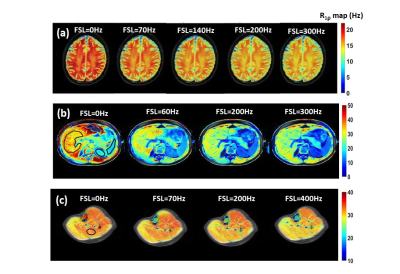 |
R1ρ Dispersion Imaging of Normal Tissues in Human Subjects at 3T Video Not Available
Fatemeh Adelnia1,2, Zhongliang Zu1,2, Feng Wang1,2, Saikat Sengupta1,2, Kevin D Harkins1,2, and John C Gore1,2,3
1Vanderbilt University Institute of Imaging Science, Nashville, TN, United States, 2Department of Radiology and Radiological Sciences, Vanderbilt University Medical Center, Nashville, TN, United States, 3Department of Biomedical Engineering, Vanderbilt University, Nashville, TN, United States
R1ρ dispersion over a range of weak locking fields has the potential to reveal information on microvascular geometry and density based on diffusion effects. Using an efficient protocol for data acquisition and theoretical fits to a simple model we present estimates of novel parameters derived from R1ρ dispersion measurements that are in the range expected for microvasculature structure. Our in vivo results support the application of R1ρ dispersion at low locking fields in human studies. However, further validation such as ex vivo studies need to be performed.
|
|
| 17:12 | 0179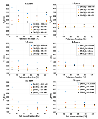 |
Further insight on T1 relaxation in fat-water mixtures
Véronique Fortier1,2 and Ives R. Levesque2,3
1Medical Imaging, McGill University Health Center, Montreal, QC, Canada, 2Medical Physics Unit, McGill University, Montreal, QC, Canada, 3Research Institute of McGill University Health Center, Montreal, QC, Canada
The impact of the fat fraction in fat-water mixtures on the fat and water T1s is an open question, with variable observations reported in phantoms, in vivo in bone marrow, and in liver. This work investigated variations of fat and water T1 as a function of fat content in phantoms in the presence of a water relaxation enhancing agent (MnCl2). Fat fraction and MnCl2 impacted both the water and fat T1s, in different ways. Our observations may shed light on variable trends of fat and water T1 as a function of fat content reported in the literature.
|
|
| 17:24 | 0180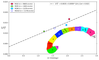 |
Is the intra-axonal T2 relaxation time related to the axonal calibre? A diffusion-relaxation and histological data study
Muhamed Barakovic1,2,3,4, Marco Pizzolato4,5, Cristina Granziera1,2, Jean-Philippe Thiran4,6,7, Derek K. Jones3,8, and Erick Jorge Canales-Rodríguez4
1Translational Imaging in Neurology (ThINk) Basel, Department of Biomedical Engineering, University Hospital Basel and University of Basel, Basel, Switzerland, 2MS Center and Research Center for Clinical Neuroimmunology and Neuroscience Basel, University Hospital Basel and University of Basel, Basel, Switzerland, 3Cardiff University Brain Research Imaging Centre, Cardiff University, Cardiff, Wales, United Kingdom, 4Signal Processing Laboratory 5 (LTS5), École Polytechnique Fédérale de Lausanne, Lausanne, Switzerland, 5Department of Applied Mathematics and Computer Science, Technical University of Denmark, Kgs. Lyngby, Denmark, 6Radiology Department, Centre Hospitalier Universitaire Vaudois and University of Lausanne, Lausanne, Switzerland, 7Centre d’Imagerie Biomédicale (CIBM), EPFL, Lausanne, Switzerland, 8Mary MacKillop Institute for Health Research, Faculty of Health Sciences, Australian Catholic University, Melbourne, Australia
The main limitation of current axon diameter mapping techniques is that the diffusion MRI (dMRI) signals from axons smaller than 2.0 μm are practically undistinguished from each other, even for the most advanced human scanners. Consequently, there is a resolution limit for the in vivo estimation of axon diameters from dMRI data. Therefore, it would be desirable to find another source of MRI contrast sensitive to the axonal calibre. This proof-of-concept study used a surface-based relaxation model to investigate whether the intra-axonal T2 estimated in a human brain is related to the inner axon radius measured from histological data.
|
|
| 17:36 | 0181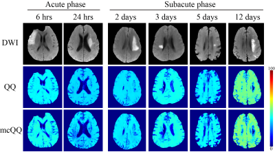 |
Multi-echo Complex QSM+qBOLD (mcQQ) for Oxygen Extraction Fraction (OEF) mapping
Junghun Cho1, Jinwei Zhang2, Pascal Spincemaille1, Hang Zhang2, Thanh D. Nguyen1, Ajay Gupta1, and Yi Wang1,2
1Radiology, Weill Cornell Medicine, New York, NY, United States, 2Biomedical Engineering, Cornell University, Ithaca, NY, United States
Quantitative mapping of oxygen extraction fraction (OEF) is critical to evaluate brain tissue viability and function in neurologic disorders. An integrated model of QSM and qBOLD (QSM+qBOLD or QQ) has been developed to map OEF from a routine gradient echo MRI without the need for vascular challenges, and QQ inversion can be obtained using deep learning. This study proposes a multi-echo complex QQ (mcQQ) that improves fidelity to data noise characteristics using complex domain. The proposed mcQQ provided more accurate OEF in simulation and an improved sensitivity to OEF abnormalities in ischemic stroke patients, compared to the current QQ method.
|
|
| 17:48 | 0182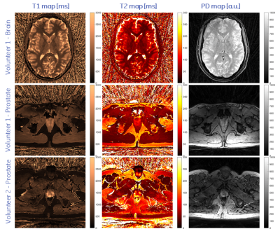 |
Quantitative Parameter Mapping of Prostate using Stack-of-Stars and QTI Encoding Video Not Available
Rolf F Schulte1, Carolin M Pirkl1, Pablo Garcia-Polo2, Matteo Cencini3,4, Michela Tosetti3,4, Luis Marti-Bonmati5, and Marion I Menzel1,6
1GE Healthcare, Munich, Germany, 2GE Healthcare, Madrid, Spain, 3IRCCS Stella Maris, Pisa, Italy, 4IMAGO7 Foundation, Pisa, Italy, 5Hospital Universitario y Politécnico La Fe, Valencia, Spain, 6Technische Hochschule Ingolstadt, Ingolstadt, Germany
Quantitative MRI offers diagnostic insights into tissue and enables characterisation of diseases, while reducing variability between operators, sites and vendors. A stack-of-stars sequence using Quantitative Transient State Imaging (QTI) was optimised and implemented to map T1, T2 and Proton Density in human prostates. Resulting in vivo multi-parametric maps were of high quality, while validation in the NIST phantom show good agreement with T1 and T2 reference values.
|
|
| 18:00 | 0183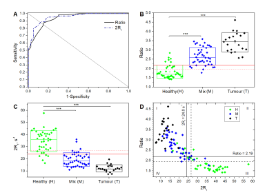 |
Low field NMR Relaxometry Studies of Tumours with applications in Intraoperative Tumour Margin Assessment in Breast-Conserving Surgery
Simona Baroni1, Valeria Bitonto1, Maria Rosaria Ruggiero1, Alessandra Pittaro2, isabella Castellano2, Riccardo Bussone3, Lionel M. Broche4, David J. Lurie4, Silvio Aime1,5, and Simonetta Geninatti Crich1
1Department of Molecular Biotechnology and Health Sciences - Molecular Imaging Center, University of Turin, Torino, Italy, 2Department of Medical Sciences, University of Turin, Torino, Italy, 3Breast Unit, Ospedale Cottolengo, Torino, Italy, 4University of Aberdeen, Aberdeen, United Kingdom, 5IRCCS SDN, Napoli, Italy
A new methodology, based on low field NMR relaxometry, has been employed to characterize breast tumour tissue and to tackle the important clinical need for the assessment of tumour margins during breast-conserving surgery. The method relies on the acquisition of proton longitudinal relaxation rate at low magnetic field strengths which are affected by the changes in the composition of the mammary gland tissue occurring during the development of neoplasia. A sensitivity, specificity, and accuracy of 92%, 85%, and 89%, respectively, were achieved. The information obtained has the potential to support the surgeon in real-time margin assessment.
|
|
| 18:12 | 0184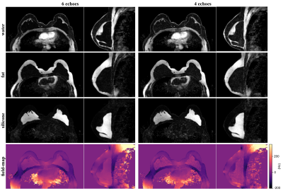 |
Hierarchical multi-resolution graph-cuts for water–fat–silicone separation in breast MRI
Jonathan K. Stelter1,2, Christof Boehm1, Stefan Ruschke1, Kilian Weiss3, Maximilian N. Diefenbach1, Mingming Wu1, Tabea Borde1, Marcus R. Makowski1, Eva M. Fallenberg1, and Dimitrios C. Karampinos1
1Department of Diagnostic and Interventional Radiology, Technical University of Munich, Munich, Germany, 2Department of Physics, Technical University of Munich, Garching, Germany, 3Philips Healthcare, Hamburg, Germany
A water- and fat-suppressed sequence is typically used to verify the integrity of silicone breast implants. The feasibility of using chemical shift encoding-based MRI for in vivo water–fat–silicone separation was recently shown. In this work, a hierarchical multi-resolution graph-cut framework is developed and proposed for water–fat–silicone separation in breast MRI. Robustness and competitive processing times are achieved by performing graph-cuts using varying spatial resolution. Results show successful water–fat–silicone separation for 4 echoes (the minimum of required echoes) without water–fat–silicone swaps and an improved silicone contrast compared to the conventional sequence.
|
 Back to Meeting Home
 Back to the Program-at-a-Glance
The International Society for Magnetic Resonance in Medicine is accredited by the Accreditation Council for Continuing Medical Education to provide continuing medical education for physicians.
 Back to Meeting Home
 Back to the Program-at-a-Glance
 View the Presentation