Oral Session
Thermometry & Electrical Property Mapping
Joint Annual Meeting ISMRM-ESMRMB & ISMRT 31st Annual Meeting • 07-12 May 2022 • London, UK

Oral Session
Thermometry & Electrical Property Mapping
| 14:45 | 0700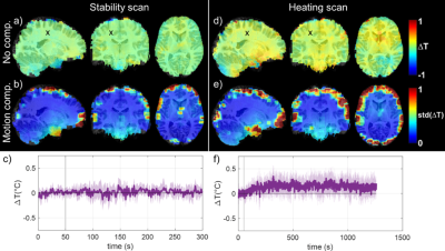 |
Measuring RF field-induced temperature variations in brain MRI exams with motion compensated MRT and field monitoring.
Caroline Le Ster1, Franck Mauconduit1, Christian Mirkes2, Alexandre Vignaud1, and Nicoal Boulant1
1Université Paris-Saclay, CNRS, BAOBAB, Neurospin, CEA, Gif-sur-Yvette, France, 2Skope MRT, Zurich, Switzerland
Sub-degree brain temperature rise occurring through RF power deposition during the MR exam was measured with MR thermometry at 7T using a multi-slice EPI sequence and concurrent field monitoring. This sequence was first tested in vitro with field perturbations, an optical probe being used for ground truth measurements, and the accuracy of the method reached 0.02°C. In vivo, this methodology was complemented by a motion compensation step and precision reached 0.05°C. After 20-min of scanning at maximal SAR, temperature rise induced by RF power deposition throughout the inner brain reached 0-0.2°C, well below the values predicted by thermal models.
|
|
| 14:57 | 0701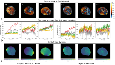 |
High-precision MR thermometry of RF heating in the upper thigh at 7T using a multi-echo water-fat separation model
Mathijs W. I. Kikken1, Bart R. Steensma2, Cornelis A. T. van den Berg2, and Alexander J. E. Raaijmakers1
1Biomedical Engineering - Medical Imaging Analysis, Eindhoven University of Technology, Eindhoven, Netherlands, 2Center for Image Sciences - Computational Imaging Group, University Medical Center Utrecht, Utrecht, Netherlands A multi-echo MRT approach is presented for application in RF safety assessment and validation of thermal simulations. By water-fat separation, more accurate determination of the drift field is possible. The method was tested in the thigh at 7T, using multi-transmit coils. Precision and accuracy were improved considerably compared to a previous single-echo fat-referenced method (precision: 0.09 vs 0.19 °C). Comparison of measured temperature distributions to simulated counterparts show good relative agreement in three subjects for multiple RF shim settings. Strikingly, simulated heating magnitudes mostly underestimated the observed heating with varying extent, suggesting a role for subject-specific parameters, such as perfusion. |
|
| 15:09 | 0702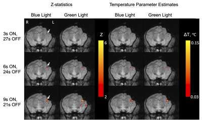 |
Detection of Laser-Associated Heating in the Brain During Simultaneous BOLD-fMRI and Optogenetic Stimulation
Huiwen Luo1,2, Zhangyan Yang1,2, Pai-feng Yang2,3, Feng Wang2,3, Jamie L Reed2,3, Li Min Chen2,3, John C Gore1,2,3, and William A Grissom 1,2,3
1Biomedical Engineering, Vanderbilt University, Nashville, TN, United States, 2Vanderbilt University Institute of Imaging Science, Vanderbilt University, Nashville, TN, United States, 3Radiology and Radiological Sciences, Vanderbilt University, Nashville, TN, United States
Optogenetic stimulation with BOLD-fMRI has been applied in studies of brain circuits, by selectively exciting optical fluorophores with wavelength-specific light. However, the light used for stimulation may induce non-negligible heating effects. Because BOLD fMRI is mainly based on relatively long TE gradient-recalled echo acquisitions that are also suitable for measuring temperature changes with the proton resonance frequency (PRF) shift, we can monitor simultaneously the neural effects and laser-associated heating. We proposed a processing pipeline to calculate temperatures from BOLD images based on PRF shift thermometry and detect small temperature rises induced by laser-associated heating with general linear modeling.
|
|
| 15:21 | 0703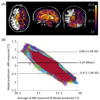 |
Comparison of whole brain biophysical model predictions and MR thermometry measurements in healthy humans
Dongsuk Sung1, Benjamin B. Risk2, Peter A. Kottke3, Jason W. Allen1,4,5, Fadi Nahab5, Andrei G. Fedorov3,6, and Candace C. Fleischer1,4,6
1Department of Biomedical Engineering, Georgia Institute of Technology and Emory University, Atlanta, GA, United States, 2Department of Biostatistics and Bioinformatics, Emory University, Atlanta, GA, United States, 3Woodruff School of Mechanical Engineering, Georgia Institute of Technology, Atlanta, GA, United States, 4Department of Radiology and Imaging Sciences, Emory University School of Medicine, Atlanta, GA, United States, 5Department of Neurology, Emory University School of Medicine, Atlanta, GA, United States, 6Petit Institute for Bioengineering and Bioscience, Georgia Institute of Technology, Atlanta, GA, United States
To advance our understanding of thermal dynamics in the human brain, a thermal modeling framework was previously developed to facilitate temperature predictions in the absence of clinical thermometry. Here, predicted brain temperatures using our fully conserved model were compared with MR thermometry in 21 healthy human subjects. Bland-Altman plots demonstrated agreement between predictions and MR-measurements for average temperature values, but some differences were observed at the lowest and highest temperatures. Regional variations were similar between predicted and measured temperatures. We anticipate our modeling framework will form the necessary baseline for predicting injury-induced brain temperature changes in patients.
|
|
| 15:33 | 0704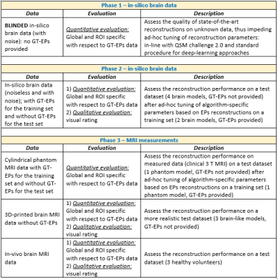 |
The first MR Electrical Properties Tomography (MR-EPT) reconstruction challenge
Stefano Mandija1,2 and Cornelis A.T. van den Berg1,2
1Department of Radiotherapy, UMC Utrecht, Utrecht, Netherlands, 2Computational Imaging Group for MR Diagnostics and Therapy, UMC Utrecht, Utrecht, Netherlands
This work announces the initial design of the first Electrical Properties (EPs) reconstruction challenge and opens up the call for ideas to finalize its implementation and to define the organizing committee members. Several EPs reconstruction methods have been presented in the last decade. However, as emerged from the last EPs workshop (IMEP-Utrecht’19), testing on common data has not been performed yet, nor data are publicly available. The scope of this challenge is therefore to benchmark current reconstructions on the same data. These data will also be made available to favor benchmarking of new methods in the future.
|
|
15:45 |
0705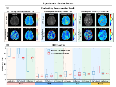 |
Feasibility study for conductivity reconstructions from spin-echo images using artificial neural network with simulation data in 3T MR system
Kyu-Jin Jung1, Stefano Mandija2,3, Jun-Hyung Kim1, Chuanjiang Cui1, Sanghyeok Choi1, Jaeuk Yi1, Mina Park4, Cornelis A.T. van den Berg2,3, and Dong-Hyun Kim1
1Department of Electrical and Electronic Engineering, Yonsei University, Seoul, Korea, Republic of, 2Department of Radiotherapy, UMC Utrecht, Utrecht, Netherlands, 3Computational Imaging Group for MR Diagnostics and Therapy, UMC Utrecht, Utrecht, Netherlands, 4Department of Radiology, Gangnam Severance Hospital, Yonsei University College of Medicine, Seoul, Korea, Republic of
Phase-based Electrical properties tomography is a non-invasive imaging technique that uses MRI systems to measure the tissue conductivity. However, the conductivity reconstruction process causes problems such as noise amplification and boundary artifact. To address such limitations, several DL-based reconstruction methods were proposed. Building upon these works, we propose an ANN-based conductivity reconstruction method trained only on simulation dataset. The proposed method was studied with the aim of: (a) approaching ground-truth conductivity values, (b) noise-robustness, (c) higher image resolution, (d) generalization to clinical data. The feasibility was investigated on simulations and TSE in-vivo data (one healthy volunteer, two meningioma cases).
|
 Back to Meeting Home
 Back to the Program-at-a-Glance
The International Society for Magnetic Resonance in Medicine is accredited by the Accreditation Council for Continuing Medical Education to provide continuing medical education for physicians.
 Back to Meeting Home
 Back to the Program-at-a-Glance
 View the Presentation