Oral Session
Non-Proton
Joint Annual Meeting ISMRM-ESMRMB & ISMRT 31st Annual Meeting • 07-12 May 2022 • London, UK

09:15 |
0628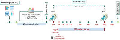 |
Real-time observation of postprandial hepatic glucose metabolism with interleaved 2H Metabolic Imaging and 13C-MRS at 7 T
Simone Poli1,2, Ahmed Fahiem Emara3, Edona Ballabani3, Angeline Buser3, Michele Schiavon4, David Herzig3, Chiara Dalla Man4, Luc Tappy3, Roland Kreis1,2, and Lia Bally3
1Magnetic Resonance Methodology, Institute of Diagnostic and Interventional Neuroradiology, University of Bern, Bern, Switzerland, 2Translational Imaging Center, sitem-insel, Bern, Switzerland, 3Department of Diabetes, Endocrinology, Nutritional Medicine and Metabolism UDEM, Insel Hospital, University Hospital Bern, Bern, Switzerland, 4Department of Information Engineering (DEI), University of Padova, Padova, Italy
The liver takes a central role in the regulation of glucose homeostasis by storing glucose in the fed state and releasing it during food abstinence. As a consequence, the liver is critically involved in glucose homeostasis disorders. We propose a novel non-invasive approach combining Deuterium Metabolic Imaging (DMI) with 13C-MRS at 7 T to dynamically map hepatic glucose metabolism. Preliminary results support technical feasibility and provide first insights into distinct hepatic glucose profiles in patients with dysregulated glucose homeostasis. The proposed approach may open new avenues for a better understanding of pathophysiology of glucose dysregulation and development of targeted treatments.
|
|
| 09:27 | 0629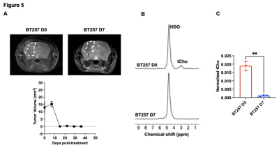 |
Deuterated choline tracks choline kinase α activity and enables non-invasive assessment of response to therapy in gliomas in vivo. Video Permission Withheld
Celine Taglang1, Georgios Batsios1, Meryssa Tran1, Anne-Marie Gillespie1, Sabrina Ronen1, and Pavithra Viswanath1
1Radiology, University of California San Francisco, San Francisco, CA, United States
Elevated choline phospholipid metabolism is a hallmark of cancer. Here, we show that silencing choline kinase a (CKα), the rate-limiting enzyme in choline phospholipid biosynthesis, abrogates total choline (tCho) production from [2H9]-choline in patient-derived glioma models, indicating that tCho production from [2H9]-choline predominantly reflects CKα activity. Importantly, we show that [2H9]-choline metabolism to tCho serves to delineate tumor from normal brain in mice bearing orthotopic patient-derived low-grade gliomas. Furthermore, [2H9]-choline informs on response to therapy, at early timepoints when anatomical alterations cannot be detected, pointing to the ability of [2H9]-choline to assess pseudoprogression, which is a challenge in glioma imaging.
|
|
| 09:39 | 0630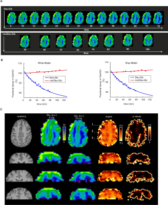 |
Deuterium labeling enables non-invasive 3D proton MR imaging of glucose and neurotransmitter metabolism in the human brain Video Not Available
Petr Bednarik1, Dario Goranovic1, Alena Svatkova2, Fabian Niess1, Lukas Hingerl1, Bernhard Strasser1, Dinesh Deelchand3, Benjamin Spurny-Dworak4, Siegfried Trattnig1, Gilbert Hangel1,5, Thomas Scherer2, Rupert Lanzenberger4, and Wolfgang Bogner1
1High Field MR Center, Medical University of Vienna, Vienna, Austria, 2Department of Medicine III, Medical University of Vienna, Vienna, Austria, 3Center for Magnetic Resonance Research, University of Minnesota, Minneapolis, MN, United States, 4Department of Psychiatry ans Psychotherapy, Medical University of Vienna, Vienna, Austria, 5Department of Neurosurgery, Medical University of Vienna, Vienna, Austria
Recent studies have proposed deuterium (2H)-Magnetic Resonance Spectroscopic Imaging (MRSI) as a reliable, non-invasive, and safe method to quantify the human metabolism of 2H-labeled substrates such as glucose and their downstream metabolism and address the major drawbacks of positron emission tomography or carbon (13C)-MRS. Here, we pioneered a dynamic proton 3D (1H)-MRSI for indirect 2H-measurements in humans. In contrast to 2H-MRS(I), the method provides higher sensitivity and chemical specificity to differentiate glutamate, glutamine, and gamma-aminobutyric acid deuterated at specific molecular positions while simultaneously mapping both labeled and unlabeled metabolites without specialized hardware after peroral ingestion of 2H-labeled glucose.
|
|
| 09:51 |
0631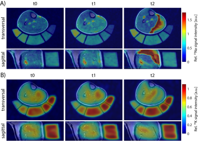 |
39K/23Na MRI detects changes in muscular ion concentrations after exercise and in delayed-onset muscle soreness
Lena V. Gast1, Laura-Marie Baier1, Oliver Chaudry2, Christian R. Meixner1, Max Müller1, Klaus Engelke2,3, Michael Uder1, Armin M. Nagel1,4, and Rafael Heiß1
1Institute of Radiology, University Hospital Erlangen, Friedrich-Alexander-Universität Erlangen-Nürnberg (FAU), Erlangen, Germany, 2Department of Medicine 3, University Hospital Erlangen, Friedrich-Alexander-Universität Erlangen-Nürnberg (FAU), Erlangen, Germany, 3Institute of Medical Physics, Friedrich-Alexander-Universität Erlangen-Nürnberg (FAU), Erlangen, Germany, 4Division of Medical Physics in Radiology, German Cancer Research Center (DKFZ), Heidelberg, Germany In this work, we investigated changes in the tissue potassium and sodium concentrations (TPC/TSC) of calf muscle tissue after exercise and in delayed-onset muscle soreness (DOMS) using combined 39K/23Na MRI. 14 healthy subjects were examined at baseline, directly after performing eccentric exercises and again 48h after exercise. Sodium and potassium concentrations showed inverse evolution, with changes in TSC being approximately twice as high as in TPC. Moreover, TPC was elevated in sore muscles without obvious edema, while TSC had returned to baseline or below. Thus, combined 39K/23Na MRI might help to elucidate physiological processes associated with DOMS and muscle fatigue. |
|
| 10:03 |
0632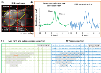 |
3D Deuterium Metabolic Imaging of the Human Liver at 7T using Low-rank and Subspace Modeling
Kyung Min Nam1, Ayhan Gursan1, Nam G. Lee2, Jannie Wijnen1, Dennis Klomp1, Jeanine Prompers1, Alex Bhogal1, and Arjan Hendriks1
1Radiology, University of Medical Center Utrecht, Utrecht, Netherlands, 2Biomedical Engineering, University of Southern California, Los Angeles, CA, United States
Deuterium metabolic imaging (DMI) is an emerging technique to spatially map metabolism in vivo through the intake of deuterium (i.e., 2H or D) labeled substrates such as [6,6′-2H2]-glucose. Although DMI has the potential to become a powerful tool to assess liver metabolism, it has limitations due to its long scan time, and low signal-to-noise ratio (SNR) for high spatial resolution in the human body. In this work, we demonstrated the feasibility of low-rank and subspace modeling (LRSM) reconstruction to increase SNR by reducing spectral noise, allowing high spatiotemporal resolution for 3D DMI of the human liver at 7T.
|
|
| 10:15 | 0633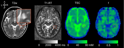 |
Heterogeneity of multiple sclerosis lesions evidenced by 7T multiparametric sodium MRI
Mohamed Mounir EL MENDILI1,2, Bertrand AUDOIN1,2,3, Ben RIDLEY 1,2, Armin NAGEL4, Soraya GHERIB1,2, Lauriane PINI1,2, Patrick VIOUT1,2, Maxime GUYE1,2, Audrey RICO3, Clemence BOUTIERE3, Jean-Philippe RANJEVA 1,2, Jean PELLETIER1,2,3, Adil MAAROUF1,2,3, and Wafaa ZAARAOUI1,2
1Aix Marseille Univ, CNRS, CRMBM, Marseille, France, 2APHM, Hôpital de la Timone, CEMEREM, Marseille, France, 3APHM, Hôpital de la Timone, Pôle de Neurosciences Cliniques, Service de Neurologie, Marseille, France, 4University Hospital Erlangen, Institute of Radiology, Erlangen, Germany Inflammatory demyelinated lesions are a hallmark of multiple sclerosis (MS) and are the pathological substratum of clinical relapses. After their occurrence, neuropathological changes within lesions and lesion repair are variable and unpredictable. Chiefly, recovery from the first relapses is a key element of the long-term prognosis. Thus, in vivo exploration of lesion repair is of paramount importance. The multi-TE sodium 7T MRI approach revealed that maintained sodium homeostasis within lesion is associated with a better recovery from relapses in MS. Sodium MRI appears to be a promising tool to assess in vivo preservation of neuronal function in MS. |
|
| 10:27 | 0634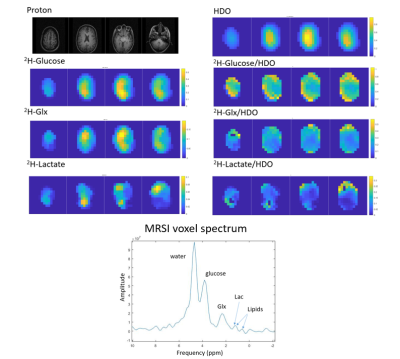 |
Comparison of clinical deuterium metabolic imaging (DMI) and hyperpolarized carbon-13 imaging at 3 T in the normal brain
Alixander S Khan1,2, Joshua D Kaggie1,2, Mary A McLean1,2, Rolf F Schulte3, Matthew J Locke1, Amy Frary1, and Ferdia A Gallagher1,2
1Department of Radiology, University of Cambridge, Cambridge, United Kingdom, 2Cancer Research UK Cambridge Centre, University of Cambridge, Cambridge, United Kingdom, 3GE Healthcare, Munich, Germany Deuterium metabolic imaging (DMI) and hyperpolarized 13C-pyruvate MRI (HP-13C-MRI) are two promising approaches for non-invasive and non-ionizing imaging of tissue metabolism. Here we directly compare these two techniques at 3 T for the first time in humans. DMI using [6,6’-2H2]glucose, and 13C-MRI using hyperpolarized [1-13C]pyruvate, were undertaken in five healthy volunteers. The ratio of 13C-lactate/13C-bicarbonate (mean ±S.D. = 5.05 ±0.89) with HP-13C-MRI was higher than the equivalent 2H-lactate/2H-Glx ratio (0.43 ±0.19) with DMI, which can be explained by differences in tracer administration, subsequent timing of acquisition, and tissue physiology. The results demonstrate the two methods provide different yet complementary data. |
|
| 10:39 | 0635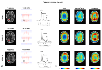 |
Deuterium Metabolic Imaging of the Human Brain in vivo at 7T
Eulalia Seres Roig1, Henk M. De Feyter1, Terence Nixon1, Loreen Ruhm2,3,4, Nikolai Avdievich2, Anke Henning2,4, and Robin A. de Graaf1
1Department of Radiology and Biomedical Imaging, Yale University, New Haven, CT, United States, 2High-Field MR Center, Max Planck Institute for Biological Cybernetics, Tuebingen, Germany, 3IMPRS for Cognitive and Systems Neuroscience, Eberhard-Karls University of Tuebingen, Tuebingen, Germany, 4Advanced Imaging Research Center, UT Southwestern Medical Center, Dallas, TX, United States
Brain energy metabolism, for which glucose is the main fuel, is essential not only for normal brain function but also for its active role in the mechanisms that underly brain diseases. Deuterium Metabolic Imaging (DMI) is a promising MR modality for investigating whole brain energy metabolism in humans at ultra-high field. In this study, we explore the potential of DMI in the human brain in vivo at 7T. We present whole brain 3D DMI maps of well-resolved deuterated (2H) metabolite resonances of water, glucose and glutamate/glutamine (Glx) following oral administration of [6,6’-2H2]-labelled glucose.
|
 Back to Meeting Home
 Back to the Program-at-a-Glance
The International Society for Magnetic Resonance in Medicine is accredited by the Accreditation Council for Continuing Medical Education to provide continuing medical education for physicians.
 Back to Meeting Home
 Back to the Program-at-a-Glance
 View the Presentation