Oral Session
High-Field MR
Joint Annual Meeting ISMRM-ESMRMB & ISMRT 31st Annual Meeting • 07-12 May 2022 • London, UK

| 16:45 | 0381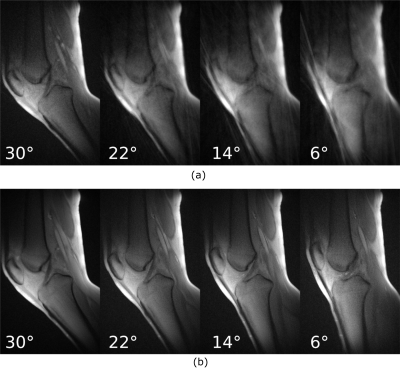 |
High resolution CINE imaging of guided knee motion using golden-angle radial MRI and a novel knee loading device
Martin A Aleksiev1, Martin Krämer2, Nicholas M Brisson3, Marta B Maggioni1, Georg N Duda3, and Jürgen R Reichenbach1
1Medical Physics Group, Institute of Diagnostic and Interventional Radiology, Jena University Hospital - Friedrich Schiller University Jena, Jena, Germany, 2Institute of Diagnostic and Interventional Radiology, Jena University Hospital - Friedrich Schiller University Jena, Jena, Germany, 3Julius Wolff Institute, Berlin Institute of Health at Charité - Universitätsmedizin Berlin, Berlin, Germany
An MRI-safe device guiding knee motion/loading with a rotary encoder was used during MRI measurements of multiple knee flexion-extension cycles using radial gradient echo imaging with the golden-angle as a radial increment. Reproducibility of knee motion was investigated and anatomical CINE images were reconstructed for different knee angles by synchronizing the encoder data with the MRI data and selective gating, enabling evaluation of musculoskeletal conditions.
|
|
| 16:57 | 0382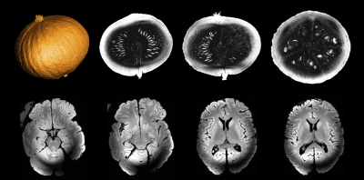 |
Progress on the commissioning of the Iseult 11.7T whole-body MRI: first images
Jerome Allard1, Alexis Amadon2, Guy Aubert1, Jean Belorgey1, Christophe Berriaud1, Cédric Bonnelye2, Romain Boudoin1, Nicolas Boulant2, Thierry Boussuge1, Fawzi Boumezbeur2, Philippe Brédy1, Peter Dietz3, Guillaume Dilasser1, Yannick Drouen1, Olivier Dubois1, Sandrine Faict-Bastin1, Ignacio Gonzalez Insua3, Vincent Gras2, Quentin Guihard1, Patrice Guiho1,
Jean-Christophe Guillard1, Vincent Jannot1, François-Paul Juster1, Felix Koeber3, Brice Koestel4, Denis Le Bihan2, René Leboeuf1, Cécile Lerman2, Frédéric Leprêtre2, Ange Lotodé1, François Nunio1, Lionel Quettier1, Hermann Landes5, Hervé Lannou1, Aurélien Massire4, Franck Mauconduit2, Frédéric Molinié1, Hubert Neyrial1, Cédric Péron1, Luc Renou4,
Arnaud Roger1, Philippe Rouffiat4, Wolfram Ruth3, Thierry Schild1, Patrick Sieber3, Mathieu Santin6,7, Martin Schroeder3, Loris Scola1, Armand Sinanna1, Vadim Stepanov1, Mathieu Szmigiel1, Olivier Tellier1, Robert Touzery1, Pierre Védrine1, Alexandre Vignaud2, Christian Walter1, and Karsten Wiclow3
1University of Paris-Saclay, CEA, Irfu, Gif sur Yvette, France, 2University of Paris-Saclay, CEA, CNRS, BAOBAB, NeuroSpin, Gif sur Yvette, France, 3Siemens Healthcare GmbH, Erlangen, Germany, 4Siemens Healthcare SAS, Saint-Denis, France, 5Ensimtech, Buckenhof, Germany, 6Sorbonne University, Institut du Cerveau - Paris Brain Institute - ICM, INSERM, CNRS, Paris, France, 7ICM, Centre de NeuroImagerie de Recherche – CENIR, Paris, France
The Iseult project started in 2001 and is a collaborative effort involving CEA and the University of Freiburg in academics, Guerbet, Siemens Healthineers and Bruker Biospin as industrial partners. One central aspect of the project has been the design by CEA of a whole-body magnet of 11.7T field strength with a 90-cm wide bore. After nearly 20 years of research and development, prototyping, integration and tests, first images have been acquired with this unique MRI scanner. Some key validation results are presented here.
|
|
17:09 |
0383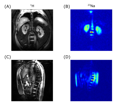 |
First in-vivo 23Na human imaging at 10.5T using a combined 23Na-loop 1H-dipole transceiver array
Simon Schmidt1, Arcan Erturk1, Russell L Lagore1, Andrea Grant1, Armin Nagel2, Stefan Zbyn1, and Gregory J Metzger1
1Center for Magnetic Resonance Research, University of Minnesota, Minneapolis, MN, United States, 2Friedrich-Alexander-Universität Erlangen-Nürnberg, Erlangen, Germany Performing sodium and proton imaging in the same study can provide a wealth of unique multinuclear insights for the study of pathologies and their treatment. However, the coils to perform such studies in the human body are limited and none were validated for human use at 10.5T until now. While both nuclei can benefit from increased SNR at 10.5T sodium suffers from low SNR due to the lower gyromagnetic ratio and natural abundance in the human body compared to 1H. First in-vivo results are presented for a 10.5T sodium-proton torso array following a safety validation study determining safe operating limits. |
|
17:21 |
0384 |
Combined 23Na and pTx-1H Imaging for Renal MRI at 7 Tesla Video Not Available
Christian R. Meixner1, Nico Egger1, Lena V. Gast1, Andreas K. Bitz2, Titus Lanz3, Ralph Kimmlingen4, Laurent Ruck1, Jürgen Herrler5, Michael Uder1, and Armin M. Nagel1,6
1Institute of Radiology, University Hospital Erlangen, Friedrich-Alexander- Universität Erlangen-Nürnberg (FAU), Erlangen, Germany, 2Faculty of Electrical Engineering and Information Technology, University of Applied Sciences, Aachen, Germany, 3Rapid Biomedical GmbH, Rimpar, Germany, 4Siemens Healthcare GmbH, Erlangen, Germany, 5Department of Neuroradiology, University Hospital Erlangen, Friedrich-Alexander- Universität Erlangen-Nürnberg (FAU), Erlangen, Germany, 6Division of Medical Physics in Radiology, German Cancer Research Center (DKFZ), Heidelberg, Germany In the human kidneys, the cortico-medullary sodium gradient plays an important role as it is essential for the concentration of urine and changes may indicate renal malfunction. However, quantitative 23Na MRI requires high-quality 1H MRI, since this facilitates co-registration and correction of partial volume effects, which is needed for body imaging in particular. We therefore show the feasibility of high-quality renal proton and sodium imaging using an 8Tx/16Rx 1H transceiver array that can be combined with a 23Na four-rung birdcage coil in a single setup. |
|
| 17:33 |
0385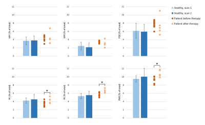 |
In-vivo 31P MRSI in healthy and malignant human pancreas at 7 Tesla
Leonard W.F. Seelen1,2, Lieke van den Wildenberg1, Ayhan Gursan1, Tijl A. van der Velden1, Mark Gosselink1, Martijn Froeling1, Wybe J.M. van der Kemp1, Firdaus A.A. Mohamed Hoesein3, Nadia Haj Mohammad4, I. Quintus Molenaar2, Hjalmar C. van Santvoort2, Dennis W.J. Klomp1, and Jeanine J. Prompers1
1Dept. of Radiology, University Medical Center Utrecht, Utrecht, Netherlands, 2Dept. of Surgery, UMC Utrecht Cancer Center and St Antonius Hospital Nieuwegein: Regional Academic Cancer Center Utrecht, Utrecht University, Utrecht, Netherlands, 3Dept. of Radiology, University Medical Center Utrecht, Utrecht University, Utrecht, Netherlands, 4Dept. of Medical Oncology, UMC Utrecht Cancer Center, Regional Academic Cancer Center Utrecht, Utrecht University, Utrecht, Netherlands Early response assessment for patients with pancreatic cancer receiving chemotherapy is limited. Detection of alterations in 31P metabolite levels during treatment could change this perspective. However, to date 31P MRS has not yet been demonstrated in the human pancreas in vivo. Here we show in-vivo 31P MRSI data in the pancreas of healthy subjects, using a 31P whole-body transmit coil in combination with a 16-channel receive array at 7T, and show moderate to good test-retest reliability. In addition, we demonstrate the feasibility of performing 31P MRSI in a patient with pancreatic cancer before and after chemotherapy. |
|
| 17:45 |
0386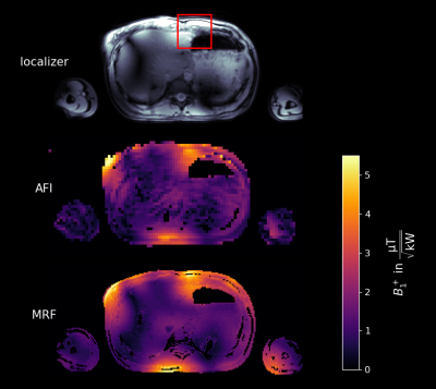 |
Low power free-breathing absolute B1+ mapping in the human body at 7T using magnetic resonance fingerprinting
Max Lutz1, Christoph Stefan Aigner1, Sebastian Dietrich1, Sebastian Flassbeck2,3, Constance G. F. Gatefait1, Christoph Kolbitsch1, and Sebastian Schmitter1,4,5
1Physikalisch-Technische Bundesanstalt, Braunschweig and Berlin, Germany, 2Dept. of Radiology, Center for Biomedical Imaging, New York, NY, United States, 3Center for Advanced Imaging Innovation and Research, New York, NY, United States, 4Medical Physics in Radiology, German Cancer Research Center (DKFZ), Heidelberg, Germany, 5Center for Magnetic Resonance Research, University of Minnesota, Minneapolis, MN, United States
At 7T, power restrictions are a major limitation to accurately map the absolute transmit magnetic field (B1+) in the body. To overcome this, we investigate an absolute B1+ mapping method with low RF power using magnetic resonance fingerprinting (MRF). Measurements are done in a phantom at 3T and in-vivo in the liver at 7T. Resulting maps are compared to the actual flip angle (AFI) method. The obtained results show good agreement between the two methods, while the MRF approach seems to perform better in regions of low B1+ amplitude. Motion robustness introduced by a radial acquisition scheme enables free-breathing measurements.
|
|
| 17:57 | 0387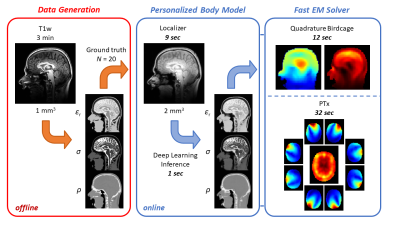 |
Fast Subject-Specific Local SAR and B1+ Prediction for PTx at 7T using only an Initial Localizer Scan
Wyger Brink1, Marius Staring2, Rob Remis3, and Andrew Webb1
1C.J. Gorter Center for High Field MRI, dept. of Radiology, Leiden University Medical Center, Leiden, Netherlands, 2Division of Image Processing, dept. of Radiology, Leiden University Medical Center, Leiden, Netherlands, 3Circuits and Systems group, dept. of Microelectronics, Delft University of Technology, Delft, Netherlands
This work presents a method for local SAR and B1+ prediction using only a 9 seconds long localizer as input. The total procedure can be executed in less than 30 seconds for a birdcage setup and less than 45 seconds for an eight-channel PTx configuration, enabling seamless integration into the MR workflow.
|
|
| 18:09 |
0388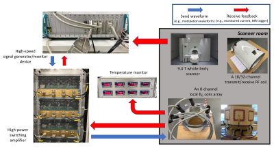 |
Image acceleration with an 8-channel local B0 coil array compatible with parallel imaging in a 9.4T human MR scanner
Rui Tian1, Alexander Loktyushin1, Kai Buckenmaier1, Theodor Steffen1, and Klaus Scheffler1,2
1High-Field MR center, Max Planck Institute for Biological Cybernetics, Tübingen, Germany, 2Department for Biomedical Magnetic Resonance, University of Tübingen, Tübingen, Germany A recent idea of spread spectrum MRI utilizes rapid modulations of localized magnetic fields to speed up image acquisitions. To push this concept further, we experimentally demonstrated 2D FLASH scans at 9.4T accelerated by 3-fold with modulation of a newly built 8-channel local B0 coil array, and 6-fold together with parallel imaging. Moreover, for FLASH and single shot spiral acquisitions, we designed and simulated another two modulation schemes for the B0 coil array to further investigate the advantages of independent control over each local coil for imaging acceleration. |
 Back to Meeting Home
 Back to the Program-at-a-Glance
The International Society for Magnetic Resonance in Medicine is accredited by the Accreditation Council for Continuing Medical Education to provide continuing medical education for physicians.
 Back to Meeting Home
 Back to the Program-at-a-Glance
 View the Presentation