Oral Session
Breast
Joint Annual Meeting ISMRM-ESMRMB & ISMRT 31st Annual Meeting • 07-12 May 2022 • London, UK

| 17:00 | 0145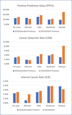 |
High Spatiotemporal Resolution Breast MR Increases Cancer Detection and Reduces Unnecessary Biopsies in Patients with Marked BPE
Sarah Eskreis-Winkler1, Linden Dixon1, Ragni Jindal1, Janice Sung1, Kimberly Feigin1, Elizabeth J. Sutton1, Danny Martinez1, and Katja Pinker1
1Memorial Sloan Kettering Cancer Center, New York, NY, United States
Contrast-enhanced breast MRI is a powerful tool for breast cancer detection, but can be limited in the setting of marked background parenchymal enhancement (BPE), which can mask underlying lesions. We found that among patients with marked BPE, positive predictive value (PPV2) and cancer detection rate (CDR) were statistically significantly increased when the high spatial/high temporal resolution protocol, rather than the standard protocol, was used. The interval cancer rate was not significantly different between these groups.
|
|
| 17:12 | 0146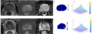 |
Detection of Prostate Cancer using Diffusion-Relaxation Correlation Spectrum Imaging with Support Vector Machine Model – A Feasibility Study Video Permission Withheld
Xiaobin Wei1, Guangyu Wu1, and Ke Xue2
1Renji Hospital, School of Medicine, Shanghai Jiao Tong University, Shanghai, China, 2MR Collaboration, Central Research Institute, United Imaging Healthcare, Shanghai, China
For clinical prostate examination, magnetic resonance imaging (MRI) is a significant imaging modality. In this study, the feasibility of diffusion-relaxation correlation spectrum imaging (DR-CSI) combined with a support vector machine (SVM) model for detecting PCa in vivo was initially explored, and its diagnostic performance was evaluated and compared with the Prostate Imaging-Reporting and Data System (PI-RADS) score based on multiparametric MRI (MP-MRI). The DR-CSI combined with SVM model may suggest additional clinical value and potential to improve the detection of PCa.
|
|
| 17:24 | 0147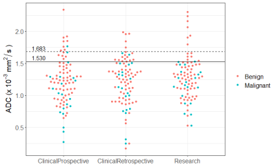 |
Clinical validation of ADC thresholds established by the ECOG-ACRIN A6702 trial to reduce unnecessary biopsies in breast MR screening exams
Debosmita Biswas1, Andrea Winters1, Anum S Kazerouni1, Inyoung Youn1,2, Janie M Lee1, Sara H Javid1, Habib Rahbar1, and Savannah C Partridge1
1Radiology, University of Washington, Seattle, WA, United States, 2Radiology, Kangbuk Samsung Hospital, Seoul, Korea, Republic of
Numerous single-site studies have established that diffusion-weighted MRI (DWI) can improve breast MRI performance, but clinical implementation has been limited by lack of standardization. The ECOG-ACRIN multicenter A6702 trial determined two pre-specified ADC thresholds from standardized acquisition that could reduce unnecessary biopsies by up to 36%. Since lesion ADCs were computed using centralized offline analysis, whether similar performance could be achieved in clinical settings is not yet established. We demonstrate that implementing the ADC threshold specified by the ECOG-ACRIN trial in a clinical breast imaging setting could provide high sensitivity and potentially reduce unnecessary biopsies for screening breast MRI.
|
|
| 17:36 | 0148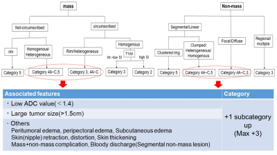 |
The diagnostic performance of modified BI-RADS using high-resolution diffusion weighted images (HR-DWI), T1WI and T2WI
Rie Ota1, Masako Kataoka1, Mami Iima1, Ayami Ohno Kishimoto2, Maya Honda3, Kanae Kawai Miyake1, Yosuke Yamada4, Yasuhide Takeuchi4, Masakazu Toi5, and Yuji Nakamoto1
1Department of Diagnostic Imaging and Nuclear Medicine, Kyoto University graduate school of medicine, Kyoto, Japan, 2Department of Diagnostic Radiology, Kyoto Katsura Hospital, Kyoto, Japan, 3Department of Diagnostic Radiology, Kansai Electric Power Hospital, Osaka, Japan, 4Department of Diagnostic Pathology, Kyoto University Hospital, Kyoto, Japan, 5Department of Breast Surgery, Kyoto University Hospital, Kyoto, Japan
This study aimed to evaluate the diagnostic performance of non-contrast protocol combining high-resolution diffusion weighted images (HR-DWI) with rs-EPI, T1WI and T2WI, using modified BI-RADS. In total, 98 breast lesions (60 malignant and 38 benign lesions) were categorized and compared to the pathological diagnosis. Area under the ROC curve (AUC)s of the non-contrast protocol were 0.90 (95% confidence interval (95%CI) : 0.82-0.95) for both readers. When excluding cases in which ADC value could not be measured, the AUC values were improved. Non-contrast protocol with modified BI-RADS can be used to evaluate breast lesions.
|
|
| 17:48 | 0149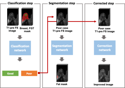 |
Assessment and improvement of the quality of fat saturation in breast MRI using deep-learning with synthetic data
Junghwa Kang1, Ga Eun Park2, Sung Hun Kim2, and Yoonho Nam1
1Division of biomedical engineering, Hankuk university of foreign studies, gyunggi-do,yongin-si, Korea, Republic of, 2Department of Radiology, Seoul St. Mary's Hospital, Seoul, Korea, Republic of
We propose a fully automatic method to assess and improve the quality of the fat saturation in breast MR images. For this purpose, three deep neural networks were trained using both actual and synthetic breast MR data. Firstly, the poorly fat saturated cases were classified using a binary classification network. Then, the poorly fat saturated regions were localized using a segmentation network. Lastly, for the poor cases, the remaining fat signals were retrospectively suppressed using a correction network. The results showed that our networks successfully identified the poor cases and suppressed the remaining fat signals.
|
|
| 18:00 | 0150 |
Deep Learning for Triage of Normal Breast MRI Exams to an Abbreviated Interpretation Worklist
Arka Bhowmik1, Natasha Monga1, Kristin Belen1, Danny Martinez1, Elizabeth J. Sutton1, Katja Pinker-Domenig1, and Sarah Eskreis-Winkler1
1Department of Radiology, Memorial Sloan Kettering Cancer Center, New York, NY, United States
In this abstract, we develop and evaluate a deep learning model to identify completely normal screening breast MRIs for triage to an abbreviated interpretation worklist, a workflow that misses no cancers and markedly reduces radiologist interpretation times. In our held out test set, the algorithm triaged 20% of all screening exams to the abbreviated worklist and 80% to full interpretation worklist for radiologist without missing any cancer exams (100% sensitivity), which reduced the total projected reading time for exams from 148 hours to 119 hours.
|
|
| 18:12 | 0151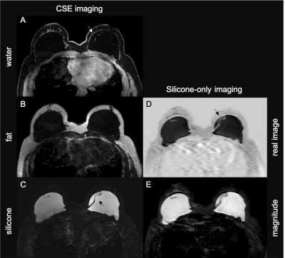 |
Silicone Implant and Fibrous Capsule Assessment Based on Water-Fat-Silicone Images from a Chemical Shift Encoding-Based Species Separation
Tabea Borde1, Antonia Wiedemann1, Jonathan Stelter1, Christof Böhm1, Stefan Ruschke1, Kilian Weiss1, Mingming Wu1, Marcus R. Makowski1, Dimitrios C. Karampinos1, and Eva M. Fallenberg1
1Technical University of Munich, Munich, Germany
With continuously rising breast augmentation procedures worldwide, there is an increasing clinical need for an early and accurate detection of implant complications. In clinical practice, silicone implants are mainly visualized by silicone-only acquisitions which can be limited by a low signal-to-noise ratio and poor resolution. The present work proposes chemical shift encoding-based multi-echo gradient-echo imaging in combination with a robust graph-cuts-based water-fat-silicone separation. The resulting multi-contrast imaging at high isotropic resolution enables a more precise delineation of the implant capsule and a better evaluation of the implant’s integrity compared to conventional silicone-only acquisition.
|
|
| 18:24 | 0152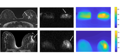 |
Field-Cycling imaging identifies breast cancer at low magnetic field strengths
Vasiliki Mallikourti1, James Ross1, Oliver Maier2, Gareth Davies1, Gerald Lip3, Markus Bödenler4, Rudolf Stollberger5, Yazan Masannat3, and Lionel Broche1
1Aberdeen Biomedical Imaging Centre, University of Aberdeen, Aberdeen, United Kingdom, 2Institute of Medical Engineering, Graz University of Technology, Graz, Austria, 3Breast unit, Aberdeen Royal Infirmary, Aberdeen, United Kingdom, 4Institute of eHealth, University of Applied Sciences FH JOANNEUM, Graz, Austria, 5BioTechMed-Graz, Graz, Austria
Field-Cycling imaging (FCI) can image over a range of low magnetic field strengths (0.2 T to 0.2 mT) through rapid switching between magnetic field levels. This allows measuring the field-depended changes of the longitudinal T1 relaxation time, known as T1 dispersion, exploring new approaches for breast tumour contrast. FCI images were acquired from patients with breast tumours to generate multi field-T1 maps and T1 dispersions. The T1 maps exhibited visible contrast of the tumour region. T1 dispersion profiles from tumours differed from those in healthy breast tissues, showing that FCI can detect breast tumours at low field strengths.
|
 Back to Meeting Home
 Back to the Program-at-a-Glance
The International Society for Magnetic Resonance in Medicine is accredited by the Accreditation Council for Continuing Medical Education to provide continuing medical education for physicians.
 Back to Meeting Home
 Back to the Program-at-a-Glance
 View the Presentation