Oral Session
Female Pelvis
Joint Annual Meeting ISMRM-ESMRMB & ISMRT 31st Annual Meeting • 07-12 May 2022 • London, UK

17:00 |
0723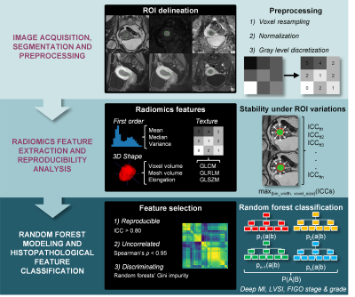 |
Development of MRI-based 3D Radiomics Signatures for Preoperative Risk Stratification of Patients with Histology-proven Endometrial Cancer
Thierry L. Lefebvre1,2, Yoshiko Ueno3,4, Anthony Dohan5,6, Avishek Chatterjee1,7, Martin Vallières1,8, Eric Winter-Reinhold9, Sameh Saif3, Ives R. Levesque1,10, Xing Ziggy Zeng11, Reza Forghani3,9, Jan Seuntjens1, Philippe Soyer5,6, Peter Savadjiev3,12, and Caroline Reinhold3,9
1Medical Physics Unit, McGill University, Montreal, QC, Canada, 2Cancer Research UK Cambridge Institute, University of Cambridge, Cambridge, United Kingdom, 3Department of Diagnostic Radiology, McGill University, Montreal, QC, Canada, 4Department of Radiology, Kobe University Graduate School of Medicine, Kobe, Japan, 5Department of Radiology, Cochin Hospital, AP-HP.Centre, Paris, France, 6Faculté de Médecine, Université de Paris, Paris, France, 7Department of Precision Medicine, GROW - School for Oncology and Developmental Biology, Maastricht University, Maastricht, Netherlands, 8Department of Computer Science, Université de Sherbrooke, Sherbrooke, QC, Canada, 9Augmented Intelligence & Precision Health Laboratory, Research Institute of McGill University Health Centre, Montreal, QC, Canada, 10Research Institute of McGill University Health Centre, Montreal, QC, Canada, 11Department of Obstetrics and Gynecology, McGill University Health Centre, Montreal, QC, Canada, 12School of Computer Science, McGill University, Montreal, QC, Canada
Radiomics analysis on standard MRI prior to surgery holds potential to help identifying high-risk histopathological features of endometrial carcinoma, including FIGO stage, deep myometrial invasion, lymphovascular space invasion and tumor grade, thus supporting preoperative risk stratification for optimal patient management. This dual-center retrospective study evaluated the role of radiomics to assess high-risk phenotypes of endometrial cancer in women who underwent 1.5-T MRI before hysterectomy. Radiomics-based machine learning models provided consistent clinically acceptable performance for differentiating early from advanced FIGO stage endometrial carcinoma and for differentiating low- from high-risk histopathological markers in two independent datasets from different institutions on preoperative MRI.
|
|
| 17:12 | 0724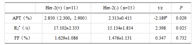 |
Evaluation of Her-2 gene expression in endometrial carcinoma by APTw and mDIXON-Quant Video Not Available
Changjun Ma1, Shifeng Tian2, Lihua Chen2, Nan Wang2, Qingwei Song2, Xiaoxiao Zhang3, and Ailian Liu2
1Department of Radiology, the First Affiliated Hospital of Dalian Medical University, Dalian,China, China, 2Department of Radiology, the First Affiliated Hospital of Dalian Medical University, Dalian, China, 3Philips Healthcare, Beijing, China, Beijing, China
Overexpression in malignant tumors such as ovarian cancer, endometrial carcinoma (EC), etc., can cause abnormal cell proliferation and lead to malignant transformation and is related to tumor angiogenesis and tumor metastasis. This study revealed that amide proton transfer weighted (APTw) and mDIXON-Quant would be useful to evaluate the expression of Her-2 gene in EC.
|
|
| 17:24 | 0725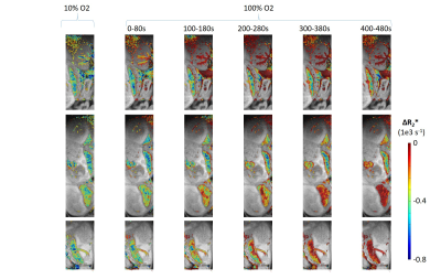 |
Visualizing oxygen transport in real time in the murine placenta at 15.2T
Talia Harris1, Michal Neeman2, and Lucio Frydman3
1Department of Chemical Research Support, Weizmann Institute of Science, Rehovot, Israel, 23Department of Biological Regulation, Weizmann Institute of Science, Rehovot, Israel, 3Department of Chemical and Biological Physics, Weizmann Institute of Science, Rehovot, Israel
We demonstrate that with the high signal to noise and sensitivity to oxygenation at 15.2 T we can visualize the transport of oxygen across the murine placenta with temporal resolution of 20s. Changes in the R2* of the placenta and embryo heart, observed upon switching from 100% O2 to 10% and switching back to 100% O2, exhibit different dynamic behavior than the maternal blood vessels, specifically a delayed response compared to the maternal blood vessels and a much slower response when switching from 10% O2 to 100% O2.
|
|
17:36 |
0726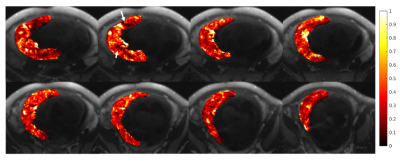 |
Maternal Blood Volume Measurements of Human Placenta with Fetal Growth Restriction using Ferumoxytol-Enhanced MRI
Ruiming Chen1, Sean B. Fain1,2, Ronald R. Magness3, Kathleen M. Antony4,5,6, J. Igor Iruretagoyena4, Ian M. Bird4, Dinesh M. Shah4, Oliver Wieben1,2,7, and Kevin M. Johnson1,7
1Medical Physics, University of Wisconsin - Madison, Madison, WI, United States, 2Biomedical Engineering, University of Wisconsin - Madison, Madison, WI, United States, 3Obstetrics & Gynecology, University of South Florida, Tampa, FL, United States, 4Obstetrics & Gynecology, University of Wisconsin - Madison, Madison, WI, United States, 5Comparative Biosciences, University of Wisconsin - Madison, Madison, WI, United States, 6Wisconsin National Primate Research Center, University of Wisconsin - Madison, Madison, WI, United States, 7Radiology, University of Wisconsin - Madison, Madison, WI, United States
Fetal growth restriction (FGR) has been associated with insufficient development of utero-placental vasculature, which could be identified using maternal placental blood volume measurements. This preliminary study uses ferumoxytol-enhanced MRI to measure placental blood volume of three FGR subjects using variable flip angle T1-mapping. Mean values of maternal fractional, placental, and total blood volumes are reported, as well as changes in placental R1 values between two time points after contrast injection. We observe local placental heterogeneities, including regions with low factional blood volume in the subjects.
|
|
| 17:48 | 0727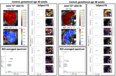 |
Quantifying Placental Structure and Function over Gestation with Combined T2*-diffusion MRI
Paddy J. Slator1, Jana Hutter2,3, Laurence H. Jackson2,3, Alison Ho4, Lisa Story4, Lucy C. Chappell4, Mary Rutherford2, Joseph V. Hajnal2,3, and Daniel C. Alexander1
1Centre for Medical Image Computing, Department of Computer Science, University College London, London, United Kingdom, 2Centre for the Developing Brain, School of Biomedical Engineering and Imaging Sciences, King's College London, London, United Kingdom, 3Biomedical Engineering Department, School of Biomedical Engineering and Imaging Sciences, King's College London, London, United Kingdom, 4Department of Women and Children's Health, School of Life Course Sciences, King's College London, London, United Kingdom
We quantify placental structure and function over gestation with combined diffusion-relaxometry MRI. We apply a simultaneous T2*-diffusion acquisition protocol to a cohort of 65 pregnant participants, and hence estimate T2* and ADC maps, T2*-ADC spectra, and map T2*-ADC spectrum components with a data-driven technique, InSpect. We show InSpect is more sensitive to placental maturation than separate T2* and ADC mapping alone.
|
|
| 18:00 | 0728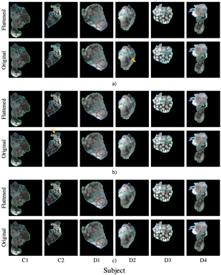 |
Automatic Segmentation of Twin Regions in Mo-Di Placentae Based on Geometric Analysis of Spatiotemporal BOLD MRI Signals
Tal Shnitzer1, S. Mazdak Abulnaga1, Carolina Bibbo2, P. Ellen Grant3, Polina Golland1, Justin Solomon1, and Esra Abaci Turk3
1Computer Science and Artificial Intelligence Laboratory, Electrical Engineering and Computer Science, Massachusetts Institute of Technology, Cambridge, MA, United States, 2Maternal and Fetal Medicine, Brigham and Women’s Hospital, Harvard Medical School, Boston, MA, United States, 3Fetal-Neonatal Neuroiaging and Developmental Science Center, Boston Children's Hospital, Harvard Medical School, Boston, MA, United States
We propose an automatic segmentation method for delineating functional regions of the placenta responsible for each twin in Mo-Di placentae. The study of differences in MRI biomarkers between identical twins promises to elucidate placental function and fetal development. We combine temporal information from BOLD MRI time series and spatial information from the umbilical cord insertion in a flattened placenta representation. We demonstrate alignment of the automatic segmentation results with expert manual delineations and subsequent agreement of dynamic MRI signals in the identified regions with those derived from expert segmentations. Our method enables automatic localized analysis of the placenta.
|
|
| 18:12 |
0729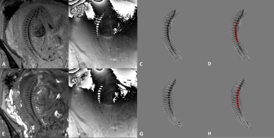 |
Quantifying Fetal Vertebral Body Calcium Changes Using Quantitative Susceptibility Mapping
Kiarash Ghassaban1,2, Cong Sun3, Guangbin Wang3,4, and Ewart Mark Haacke1,2
1Department of Radiology, Wayne State University, Detroit, MI, United States, 2SpinTech Inc., Bingham Farms, MI, United States, 3Department of Radiology, Shandong Provincial Hospital, Shandong University, Jinan, China, 4Department of Radiology, Shandong Provincial Hospital Affiliated to Shandong First Medical University, Jinan, China
In vivo fetal vertebral calcium content was quantified using local quantitative susceptibility mapping (QSM) collected as part of a STAGE double flip angle (FA) protocol. A strong decreasing average vertebral susceptibility as a function of gestational age was seen in 54 normal fetuses including 37 cases with QSM data available at both FA’s and 17 cases with QSM at one FA totaling 91 independent datasets. An almost one-to-one linear correlation was seen between the QSM values from both FA’s. These results provide strong evidence of an in vivo MR-based quantitative technique to measure calcium development pre-term.
|
 Back to Meeting Home
 Back to the Program-at-a-Glance
The International Society for Magnetic Resonance in Medicine is accredited by the Accreditation Council for Continuing Medical Education to provide continuing medical education for physicians.
 Back to Meeting Home
 Back to the Program-at-a-Glance
 View the Presentation