Oral Session
Advances in Prostate Imaging
Joint Annual Meeting ISMRM-ESMRMB & ISMRT 31st Annual Meeting • 07-12 May 2022 • London, UK

| 09:15 | 0221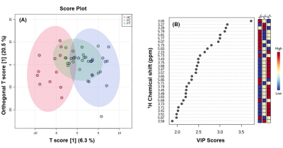 |
Mapping of Metabolite Profile of Urine of Patients with Chronic Prostatitis, Prostate Cancer and Healthy Controls: A Pilot NMR Metabolomic Study
Naranamangalam R Jagannathan1,2,3, R Ravikanth Reddy1, Arun Kumar Shirshetty4, Samsudeen Marwan5, Deepak Masanam4, and Ananathakrishanan Sivaraman5
1Dept. of Radiology, Chettinad Hospital & Research Institute, Kelambakkam, India, 2Dept. of Electrical Engineering, Indian Institute of Technology, Chennai, India, 3Radiology, Sri Ramachandra Inst. of Higher Education and Research, Chennai, India, 4Dept. of Urology, Chettinad Hospital & Research Institute, Kelambakkam, India, 5Chennai Urology and Robotics Institute Hospital, Chennai, India
The present study aims to evaluate a comprehensive metabolic profiling of patients with chronic prostatitis (PT) by investigating their urine samples and compare it with the prostate cancer (PCa) patients and healthy controls (HC) using 1H nuclear magnetic resonance (NMR) metabolomics. The metabolic profile of chronic prostatitis patients falls between that seen for PCa and HC. This pilot study showed that NMR metabolomics may be useful to differentiate patients with PT from PCa and HC.
|
|
| 09:27 | 0222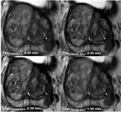 |
Highly Accelerated T2 and Diffusion Weighted Imaging of the Prostate by Combining Compressed SENSE, Sparse Sampling and Deep Learning
Felix N. Harder1, Kilian Weiss2, Thomas Amiel3, Johannes M. Peeters4, Robert Tauber3, Marcus R. Makowski1, Andreas P. Sauter1, Jürgen E. Gschwend3, Dimitrios C. Karampinos1, and Rickmer F. Braren1
1Institute of Diagnostic and Interventional Radiology, Technical University of Munich, School of Medicine, Munich, Germany, 2Philips GmbH Market DACH, Hamburg, Germany, 3Department of Urology, Technical University of Munich, School of Medicine, Munich, Germany, 4Philips Healthcare, Best, Netherlands
Since prostate MRI is increasingly applied and yet limited by long acquisition times, we prospectively investigated the performance of a novel reconstruction algorithm, combining compressed sensing, parallel imaging and deep learning (C-SENSE AI) in patients with histologically proven prostate cancer. Highly accelerated T2w and DWI sequences were compared to clinical standard of reference T2w and DWI. C-SENSE AI enabled 58% acceleration in T2w imaging and 47% acceleration in DWI of the prostate while obtaining significantly better image quality and tumor detection. C-SENSE AI seems particularly interesting in view of the need for accelerated prostate MRI with preserved high image quality.
|
|
| 09:39 | 0223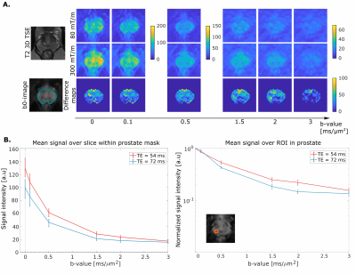 |
300 mT/m diffusion MRI 'below the neck': First results in healthy prostate
Malwina Molendowska1, Marco Palombo1, Fabrizio Fasano2,3, Derek K. Jones1, Daniel C. Alexander4, Eleftheria Panagiotaki4, and Chantal Tax1,5
1Cardiff University Brain Research Imaging Centre (CUBRIC), Cardiff University, Cardiff, United Kingdom, 2Siemens Healthcare Ltd, Camberly, United Kingdom, Camberly, United Kingdom, 3Siemens Healthcare GmbH, Erlangen, Germany, Erlangen, Germany, 4Centre for Medical Image Computing, University College London, London, United Kingdom, 5Image Sciences Institute, University Medical Center Utrecht, Utrecht, Netherlands
This work showcases the first ever use of ultra-strong gradient in ’below the neck’ human MRI. It assesses the benefits, compared to more commonly-available gradient strengths, of 300mT/m gradients for comprehensive characterization of the prostate gland. Leveraging such gradient amplitudes enables unique tissue contrasts, with a potential to improve specificity and/or sensitivity of current MRI methods for prostate cancer characterisation and detection.
|
|
| 09:51 | 0224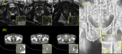 |
A multimodality, MRI positive-contrast fiducial marker for prostate cancer treatment: first-in-patient experience with proton therapy Video Permission Withheld
Jeremiah Sanders1, Li Wang2, Falk Poenisch3, Narayan Sahoo3, Xiaorong Zhu3, Jingfei Ma1, Seungtaek Choi4, Quynh Nguyen4, Henry Mok4, Chad Tang4, Katina Crabtree4, Sean McGuire4, Karen Hoffman4, Shalin Shah4, and Steven Frank4
1Imaging Physics, UT MD Anderson Cancer Center, Houston, TX, United States, 2Experimental Radiation Oncology, UT MD Anderson Cancer Center, Houston, TX, United States, 3Radiation Physics, UT MD Anderson Cancer Center, Houston, TX, United States, 4Radiation Oncology, UT MD Anderson Cancer Center, Houston, TX, United States
Carbon and gold fiducial markers (FMs) are the most commonly used FMs for prostate radiotherapy. However, these markers do not produce positive contrast on MRI, and can cause large magnetic susceptibility artifacts on MRI. These properties of carbon and gold FMs make their identification on MRI challenging and complicate treatment planning processes that use co-image registration of MRI with CT for radiotherapy treatments. A novel multimodality FM has been developed that produces positive contrast on four imaging modalities, including MRI. This work presents the first-in-patient experience using the multimodality FM for proton therapy of prostate cancer.
|
 Back to Meeting Home
 Back to the Program-at-a-Glance
The International Society for Magnetic Resonance in Medicine is accredited by the Accreditation Council for Continuing Medical Education to provide continuing medical education for physicians.
 Back to Meeting Home
 Back to the Program-at-a-Glance
 View the Presentation