Oral Session
Perfusion: From Head to Toe
Joint Annual Meeting ISMRM-ESMRMB & ISMRT 31st Annual Meeting • 07-12 May 2022 • London, UK

| 14:45 | 0070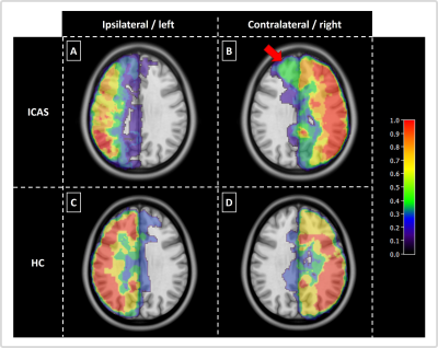 |
Perfusion territory shifts in asymptomatic carotid artery stenosis measured by super-selective arterial spin labelling
Gabriel Hoffmann1,2, Miriam Reichert1, Jens Göttler1, Michael Helle3, Kim van de Ven4, Claus Zimmer1,2, Moritz Hernandez Petzsche1,2, Hans Liebl1,5, Nico Sollmann1,6, Christine Preibisch1,2, and Stephan Kaczmarz1,2,7
1Department of Diagnostic and Interventional Neuroradiology, School of Medicine, Klinikum rechts der Isar, Technical University of Munich, Munich, Germany, 2TUM-Neuroimaging Center, Klinikum rechts der Isar, Technical University of Munich, Munich, Germany, 3Philips Research, Hamburg, Germany, 4Philips Healthcare, Best, Netherlands, 5Department of Radiology, Neuroradiology and Minimal-Invasive Therapy, Klinikum Bogenhausen, Munich, Germany, 6Department of Diagnostic and Interventional Radiology, University Hospital Ulm, Ulm, Germany, 7Philips Healthcare, Hamburg, Germany
Vessel-selective imaging is promising to examine collateral blood supply in asymptomatic internal carotid artery stenosis (ICAS). Established modalities like digital subtraction angiography are invasive, not quantitative and associated with potential complication risks. A viable non-invasive alternative is super-selective arterial spin labelling, providing perfusion territories of individual arteries. We present data from seven asymptomatic ICAS patients and four age-matched healthy controls. We compared individual perfusion territory maps to an atlas of vascular territories and evaluated intra-hemispheric differences, allowing for quantitative assessment of stenotic mal-perfusion as well as compensatory collateral blood supply from the contralateral ICA.
|
|
| 14:57 | 0071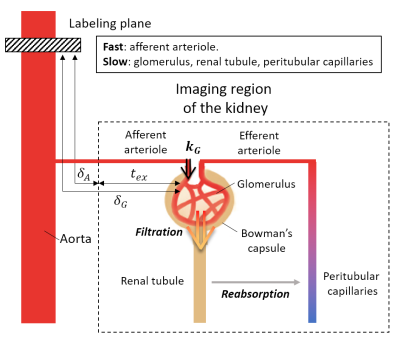 |
Measuring glomerular blood transfer rate in kidney using diffusion-weighted arterial spin labeling
Hyun-Seo Ahn1 and Sung-Hong Park1
1Department of Bio and Brain Engineering, Korea Advanced Institute of Science and Technology, Daejeon, Korea, Republic of Imaging methods for the non-invasive measurement of renal function, such as glomerular filtration rate (GFR), are promising. In this study, we propose a renal perfusion kinetic model to estimate the blood transfer rate constant ($$$k_G$$$) in glomerulus with the multi-slice multi-delay diffusion-weighted arterial spin labeling sequence. Mean cortical $$$k_G$$$ values with a caffeine challenge were higher than those with no caffeine challenge, consistent with previous studies with GFR. The current study is promising in that it shows a potential availability of $$$k_G$$$ as a non-invasive quantitative measure of renal function. |
|
| 15:09 | 0072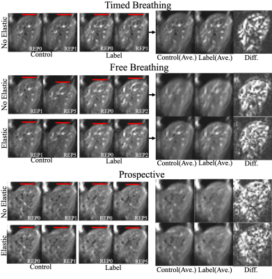 |
Free Breathing 3D ASL Imaging of the Human Liver using Prospective Motion Correction: Preliminary Results
Jörn Huber1, Daniel Hoinkiss1, Meghashree Channaveerappa1, and Matthias Günther1,2
1Fraunhofer MEVIS, Bremen, Germany, 2Faculty 1 (Physics/Electrical Engineering), University of Bremen, Bremen, Germany
Assessment of liver perfusion can yield valuable information about diseases like carcinoma and cirrhosis. Arterial spin labeling enables contrast-free measurements of liver perfusion with the drawback of increased motion sensitivity. Since timed breathing protocols prolong scan times, a novel prospective motion correction technique is presented, adjusting the initial saturation and image readout to the current position of the liver during the breathing cycle. The presented technique is compatible with highly suppressed signals, typically encountered in modern ASL imaging and achieved a significant decrease of cranial-caudal liver translation in acquired 3D GRASE volumes.
|
|
| 15:21 | 0073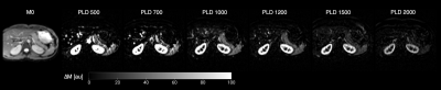 |
Multi-delay pseudo-continuous arterial spin labeling for perfusion quantification in the spleen
Sergio M. Solis-Barquero1,2, Verónica Aramendia-Vidaurreta1,2, Marta Vidorreta3, Rebeca Echeverria-Chasco1,2, Leyre García-Ruiz1,2, Ana Ezponda1,2, Gorka Bastarrika1,2, and María A. Fernández-Seara1,2
1Departament of Radiology, Clínica Universidad de Navarra, Pamplona, Navarra, Spain, 2IdiSNA, Instituto de Investigación Sanitaria de Navarra, Navarra, Spain, 3Siemens Healthineers, Madrid, Spain
The spleen plays an important role in diseases related to portal circulation. Pseudo-continuous arterial spin labeling (PCASL) was used to assess the Splenic Blood Flow (SBF) using six different post labeling delays in five different healthy volunteers. SBF and ATT maps were generated by fitting the one compartment Buxton kinetic model. This study shows the feasibility of using single and multi-delay PCASL to measure blood flow in the spleen. Obtained SBF values are in agreement with previous studies. PCASL enables a reliable measure of splenic blood flow in healthy volunteers.
|
|
| 15:33 | 0074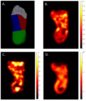 |
Elevated Foot Diffusion and Resting Perfusion in Patients with Diabetic Foot Ulcer: A Pilot and Feasibility Study
David A Reiter1, Jingting Yao2, Scott Edwards1, Marcos Coutinho Schechter1, Maya Fayfman1, Gabriel Santamarina1, Paula Nesbeth1, Vincent Giacalone1, Gerardo Blanco1, Thorsten Feiweier3, Rabindra Tirouvanziam1, Jessica Alvarez1, Benjamine Risk1, and Ravi Rajani1
1Emory University, Atlanta, GA, United States, 2Massachusetts General Hospital, Boston, MA, United States, 3Siemens Healthcare GmbH, Erlangen, Germany
Here we apply intravoxel incoherent motion (IVIM) and fractional Fickian diffusion (FFD) models to multi-b-value diffusion-weighted-MRI to study tissue cellular and resting microvascular properties of the foot of patients with diabetic foot ulcer. We report preliminary results from an ongoing study comparing patients with type 2 diabetes and persistent plantar foot ulcers with healthy age- and BMI-matched individuals. Pseudo-diffusion and mean diffusion coefficients and microvascular volume fraction were elevated in patients, showing large effect sizes in subregions. This approach may prove useful for evaluating patients with ulcers to prognosticate tissue at risk of poor wound healing.
|
|
| 15:45 | 0075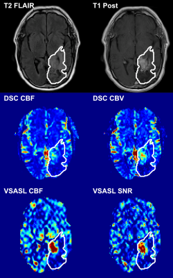 |
Velocity-selective arterial spin labeling perfusion in the evaluation of treated high grade gliomas at 1.5 Tesla
Sebastian Lambrecht1,2, Dapeng Liu1,3, Omar Dzaye4, Matthias Holdhoff4, Peter van Zijl1,3, Qin Qin1,3, and Doris Lin1,3
1Russell H. Morgan Department of Radiology and Radiological Science, The Johns Hopkins University School of Medicine, Baltimore, MD, United States, 2Institute of Neuroradiology, University Hospital LMU Munich, Munich, Germany, 3F. M. Kirby Research Center for Functional Brain Imaging, Kennedy Krieger Institute, Baltimore, MD, United States, 4Department of Oncology, The Johns Hopkins University School of Medicine, Baltimore, MD, United States
Perfusion measures were compared between VSASL and DSC methods at 1.5T in 28 patients with treated high-grade glioma assigned into 2 groups: “tumor” (with detectable enhancing tumor, n=9) and “non-tumor” (without detectable tumor, n=19). All measures (rCBF and tSNR from VSASL, rCBV and rCBF from DSC) showed significant difference between “tumor” and “non-tumor” groups allowing reliable discrimination. In general, there was moderate to excellent agreement and correlation between these measures derived from VSASL vs. DSC. Hence, VSASL has potential to serve as a viable non-invasive alternative to DSC perfusion in the clinical disease surveillance without the need for exogenous contrast.
|
|
| 15:57 | 0076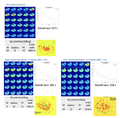 |
Exercise-induced hyperemia ASL and CrCEST at 3T demonstrate individual calf muscle improvement post-revascularization in patients with PAD
Helen Sporkin1, Toral Patel2, Yaqub Betz3, Christopher Kramer2,3, and Craig Meyer1,4
1Biomedical Engineering, University of Virginia, Charlottesville, VA, United States, 2Cardiac Imaging, University of Virginia, Charlottesville, VA, United States, 3Cardiology, University of Virginia, Charlottesville, VA, United States, 4Radiology and Medical Imaging, University of Virginia, Charlottesville, VA, United States
Peripheral arterial disease is a prevalent atherosclerotic disease characterized by atherosclerotic lesions in the limbs. Patients with PAD have been shown to have a delayed phosphocreatine recovery due to chronic ischemia. Patients with PAD may be candidates for revascularization, but outcomes are variable. CrCEST allows for creatine concentrations to be monitored at high spatial resolution, while ASL quantifies perfusion into tissue. We use this combination to revascularization in patients undergoing both endovascular and surgical procedures. Preliminary results link improvement in CrCEST decay curves and improved functional walk scores, while increased perfusion seen on ASL does not necessarily correlate with improvement.
|
|
| 16:09 | 0077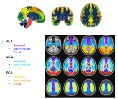 |
Amyloid burden and vascular risk factors correlate with regional cerebral blood flow in a cognitively unimpaired population
Beatriz E. Padrela1, Luigi Lorenzini1, Lyduine E. Collij1, Mara ten Kate1, Anouk den Braber2,3, Jori Tomassen2, Bart N.M. van Beckel1, Pieter Jelle Visser2, Frederik Barkhof1,4, Jan Petr5, and Henk J.M.M. Mutsaerts1
1Department of Radiology and Nuclear Medicine, Amsterdam Neuroscience, Amsterdam University Medical Center, Location VUmc, Amsterdam, Netherlands, 2Department of Neurology, Alzheimer Center, Neuroscience Campus Amsterdam, VU University Medical Center, Amsterdam, the Netherlands, Amsterdam, Netherlands, 3Department of Biological Psychology, Vrije Universiteit Amsterdam, Amsterdam, Netherlands, 4Queen Square Institute of Neurology and Centre for Medical Image Computing (CMIC), University College London, London, United Kingdom, 5Helmholtz-Zentrum Dresden-Rossendorf, Institute of Radiopharmaceutical Cancer Research, Dresden, Germany
Studying the association between cerebral blood flow (CBF), amyloid burden, and vascular risk factors in a cognitively unimpaired elderly population could clarify the role of CBF as a biomarker of cognitive decline. In 196 cognitively unimpaired participants, regional CBF was associated with regional amyloid-PET Centiloid. Vascular risk scores as measured by the Framingham risk score combined with amyloid Centiloid values were associated with increased CBF in vascular territories. Longitudinally, global CBF changes were associated with baseline precuneus amyloid burden.
|
 Back to Meeting Home
 Back to the Program-at-a-Glance
The International Society for Magnetic Resonance in Medicine is accredited by the Accreditation Council for Continuing Medical Education to provide continuing medical education for physicians.
 Back to Meeting Home
 Back to the Program-at-a-Glance
 View the Presentation