Oral Session
Applications of Perfusion & Permeability
Joint Annual Meeting ISMRM-ESMRMB & ISMRT 31st Annual Meeting • 07-12 May 2022 • London, UK

Oral Session
Applications of Perfusion & Permeability
09:15 |
0207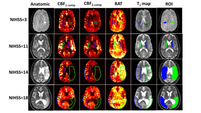 |
Simultaneous hemodynamic and structural imaging of ischemic stroke with MR Fingerprinting ASL
Hongli Fan1,2, Pan Su2, Doris Da May Lin3, Emily B. Goldberg3, Alexandra Walker3, Richard Leigh3, Argye E. Hillis3, and Hanzhang Lu1,2,4
1Department of Biomedical Engineering, Johns Hopkins University School of Medicine, Baltimore, MD, United States, 2The Russell H. Morgan Department of Radiology & Radiological Science, Johns Hopkins University School of Medicine, Baltimore, MD, United States, 3Department of Neurology, Johns Hopkins University School of Medicine, Baltimore, MD, United States, 4F. M. Kirby Research Center for Functional Brain Imaging, Kennedy Krieger Institute, Baltimore, MD, United States
Tissue perfusion and structural imaging provides important information in the evaluation of ischemic stroke. MR-fingerprinting (MRF) arterial spin labeling (ASL) is a novel noninvasive method of ASL perfusion that allows simultaneous estimation of cerebral blood flow (CBF), bolus arrival time (BAT), and tissue T1 map in a single scan of <4 minutes. Here we evaluated the utility of MRF-ASL in ischemic stroke patients in depicting hemodynamic and structural abnormalities, as well as predicting neurological deficits and disability.
|
|
| 09:27 |
0208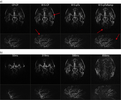 |
Evaluation of a full dynamic density adapted radial 4D-pcASL angiography sequence exploiting B1+-shimming and ramped SPINS excitation at 7T Video Not Available
Christian R. Meixner1, Max Müller1, Sebastian Schmitter2,3, Jürgen Herrler4, Arnd Dörfler4, Michael Uder1, and Armin M. Nagel1,3
1Institute of Radiology, University Hospital Erlangen, Friedrich-Alexander- Universität Erlangen-Nürnberg (FAU), Erlangen, Germany, 2Physikalisch-Technische Bundesanstalt (PTB), Braunschweig und Berlin, Germany, 3Division of Medical Physics in Radiology, German Cancer Research Center (DKFZ), Heidelberg, Germany, 4Department of Neuroradiology, University Hospital Erlangen, Friedrich-Alexander- Universität Erlangen-Nürnberg (FAU), Erlangen, Germany In this work, a whole-brain 4D-pcASL angiography sequence exploiting a radial density-adapted gradient echo readout with golden angle sampling was implemented and evaluated on a 7T system. Various parallel transmission (pTx) strategies were compared to the standard circularly polarized mode: To counteract low transmit efficiency in the labeling, B1+-shimming was used in combination with a full spiral-nonselective pTx-optimized readout train. Further, constant flip angle pTx-excitations in the readout were compared to pTx-pulses that were ramped from 3.5° to 6.5° using a linear scaling factor. This ramped readout strategy outperforms constant flip angles and reveals more of the vascular tree |
|
| 09:39 | 0209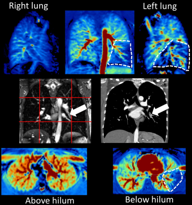 |
A prospective patient study using pseudocontinuous Arterial Spin Labeling to detect pulmonary embolism: preliminary results of an ongoing study
Ferdinand Seith1, Cecilia Zhang1, Manuel Kolb1, Brigitte Gückel1, Rolf Pohmann2, Petros Martirosian3, and Ahmed E. Othman1,4
1Diagnostic and Interventional Radiology, University Hospital of Tübingen, Tübingen, Germany, 2High-Field MR Center, Max Planck Institute for Biological Cybernetics, Tübingen, Germany, 3Section on Experimental Radiology, University Hospital of Tübingen, Tübingen, Germany, 4Neuroradiology, University Hospital of Mainz, Mainz, Germany
We present preliminary results of a prospective study of 96 patients examined with pCASL-MRI of the lung with suspected pulmonary embolism. pCASL-MRI of the lung was acquired in a 1.5T scanner by labeling the pulmonary trunk during systole. Coronal, sagittal and axial images were acquired using a fast bSSFP sequence. Additionally, a mulit-slice coronal bSSFP imaging the whole lung was performed. Two blinded readers evaluated MR-images to detect pulmonary embolism and noted the diagnostic confidence (5=very good). Compared to CT, sensitivity and specificity were 0.971 and 0.951. The median diagnostic confidence was 5 in both readers (kappa 0.739).
|
|
| 09:51 | 0210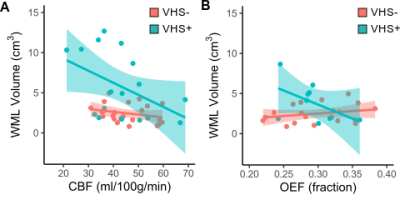 |
Associations between cerebral microvascular hemodynamics and white matter lesion burden in typically aging older adults Video Permission Withheld
Meher R Juttukonda1,2, Kimberly A Stephens1, Yi-Fen Yen1,2, Casey M Howard1, Jonathan R Polimeni1,2, Bruce R Rosen1,2, and David H Salat1,2
1Athinoula A. Martinos Center for Biomedical Imaging, Massachusetts General Hospital, Charlestown, MA, United States, 2Radiology, Harvard Medical School, Boston, MA, United States
Hypoperfusion is frequently considered a cause of age-related white matter lesions (WML). However, reductions in oxygen availability to brain tissue may also be caused by impaired oxygen extraction fraction (OEF). Here, we tested the hypothesis that venous hyperintense signal (VHS) in arterial spin labeling (ASL) MRI is indicative of impaired OEF in typically aging older adults. In participants aged 60–80 years (n=40), we measured VHS with ASL and maximum OEF (OEFmax) with dynamic susceptibility contrast MRI. Lower OEFmax was uniquely associated with higher WML volume in participants with VHS, indicating a potentially distinct cerebrovascular aging pattern in these individuals.
|
|
| 10:03 | 0211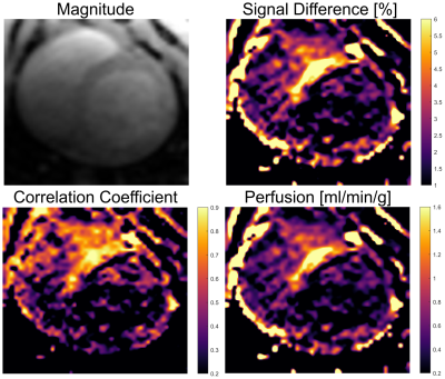 |
First in vivo Myocardial Perfusion Measurements using Intra-arterial Spin Labeling during MR-guided Coronary Interventions in Pigs
Simon Reiss1, Kevin Waescher1, Thomas Lottner1, Ali Caglar Özen1,2, Timo Heidt3, Constantin von zur Mühlen3, and Michael Bock1
1Dept. of Radiology, Medical Physics, Medical Center University of Freiburg, Faculty of Medicine, University of Freiburg, Freiburg, Germany, 2German Consortium for Translational Cancer Research Partner Site Freiburg, German Cancer Research Center (DKFZ), Heidelberg, Germany, 3Dept. of Cardiology and Angiology I, University Hospital Freiburg and Faculty of Medicine, University of Freiburg, Freiburg, Germany
Arterial spin labeling (ASL) provides a method for myocardial perfusion quantification without exogenous contrast agents. In MR-guided coronary catheterizations, perfusion measurements can provide a valuable tool to diagnose myocardial perfusion deficits and monitor the success of the intervention. In these interventions, catheters are usually visualized by active receive RF coils attached to the catheter. Using these active coils in transmit mode would enable intra-arterial spin labeling (iASL) in the coronary arteries with an increased labeling efficiency compared to conventional ASL. Here, we show the first in vivo results using iASL for myocardial perfusion measurements during coronary interventions in pigs.
|
|
10:15 |
0212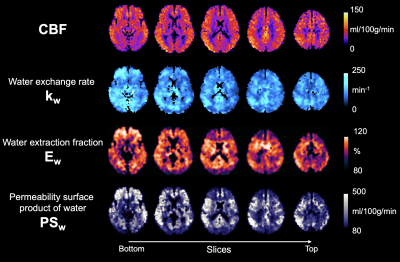 |
BBB water exchange rate and permeability mapping with multi-delay diffusion weighted pseudo-continuous arterial spin labeling
Xingfeng Shao1 and Danny JJ Wang1
1Laboratory of FMRI Technology (LOFT), Mark & Mary Stevens Neuroimaging and Informatics Institute, Keck School of Medicine, University of Southern California, Los Angeles, CA, United States
We proposed a new sequence which improves the SNR of diffusion weighted ASL signals, and simultaneously estimates kw, water extraction ratio (Ew) and PSw using a multi-delay approach with PLDs ranging from 1500 to 2700 msec. Whole brain average CBF = 67.8±6.7 ml/100g/min, kw = 103.7±16.6 min-1, Ew = 94.8±1.3% and PSw = 202.2±41.3 ml/100g/min. These results match the recent literatures on kw and PSw studies. Ew ~95% suggests two long PLDs of 2400 and 2700 msec are sufficient to allow complete water extraction.
|
|
| 10:27 | 0213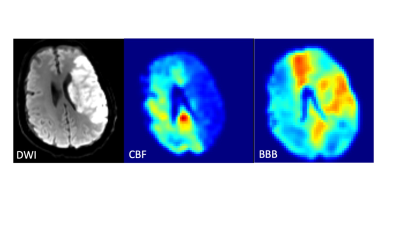 |
Use of pCASL MRI to quantify Cerebral Blood Flow and Blood-Brain Barrier Permeability in Patients with Ischemic Stroke Video Not Available
Nikolaos Mouchtouris1, Mahdi Alizadeh2, Adam Flanders2, Chengyuan Wu1, Feroze Mohamed2, Reid Gooch1, Stavropoula Tjoumakaris1, and Pascal Jabbour1
1Neurosurgery, THOMAS JEFFERSON UNIVERSITY HOSPITAL, Philadelphia, PA, United States, 2Radiology, THOMAS JEFFERSON UNIVERSITY HOSPITAL, Philadelphia, PA, United States
In this pilot study, we apply diffusion-prepared pseudo-continuous arterial spin labeling (DP-pCASL) non-contrast MRI sequence to patients with anterior circulation ischemic strokes and determine the cerebral blood flow (CBF) and blood-brain barrier (BBB) permeability in the affected vascular territories. When comparing their imaging findings to 24 control hemispheres (12 healthy volunteers), we demonstrated that both the CBF and BBB of the affected MCA territory was significantly lower than the MCA territories in healthy volunteers. Additionally, the BBB permeability in the affected territory appears to be elevated in acute ischemic strokes but decreases in subacute strokes.
|
 Back to Meeting Home
 Back to the Program-at-a-Glance
The International Society for Magnetic Resonance in Medicine is accredited by the Accreditation Council for Continuing Medical Education to provide continuing medical education for physicians.
 Back to Meeting Home
 Back to the Program-at-a-Glance
 View the Presentation