Oral Session
Flow from Head to Toe
Joint Annual Meeting ISMRM-ESMRMB & ISMRT 31st Annual Meeting • 07-12 May 2022 • London, UK

09:15 |
0214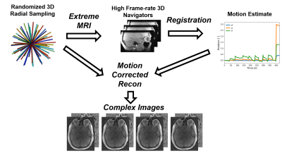 |
Motion-Corrected 4D-Flow for Neurovascular Applications
Leonardo A Rivera-Rivera1, Steven Kecskemeti1, Mu-Lan Jen1, Zach Miller1, Sterling C Johnson1, Laura Eisenmenger1, and Kevin M Johnson1
1University of Wisconsin, Madison, Madison, WI, United States Accurate and precise assessment of neurovascular flow in the brain requires high image fidelity which can be difficult to obtain in populations with high tendencies to move, such as geriatrics and pediatrics. This study presents a 3D radial sampling and multi-scale low rank image reconstruction for self navigated motion correction of 4D-Flow with validations in phantom and invivo studies. |
|
| 09:27 |
0215 |
Free-running 3D anatomical and flow MRI using Synchronization of Neighboring Acquisitions by Physiological Signals (SyNAPS)
Mariana B. L. Falcão1, Adèle L.C. Mackowiak1, Simone Rumac1, Mario Bacher1,2, Giulia Rossi1, Milan Prša3, Estelle Tenisch1, Tobias Rutz4, Jessica Bastiaansen1,5,6, Ruud Van Heeswijk1, Peter Speier2, Michael Markl7,8, Matthias Stuber1,9, and Christopher W. Roy1
1Department of Diagnostic and Interventional Radiology, University Hospital (CHUV) and University of Lausanne (UNIL), Lausanne, Switzerland, 2Siemens Healthcare GmbH, Erlangen, Germany, 3Woman- Mother- Child Department, University Hospital (CHUV) and University of Lausanne (UNIL), Lausanne, Switzerland, 4Service of Cardiology, Centre de Resonance Magnétique Cardiaque (CRMC), University Hospital (CHUV) and University of Lausanne (UNIL), Lausanne, Switzerland, 5Department of Diagnostic, Interventional and Pediatric Radiology (DIPR), University hospital Bern (Inselspital), Bern, Switzerland, 6Translational Imaging Center, sitem-insel, Bern, Switzerland, 7Department of Radiology, Feinberg School of Medicine, Northwestern University, Chicago, IL, United States, 8Department of Biomedical Engineering, Northwestern University, Chicago, IL, United States, 9Center for Biomedical Imaging (CIBM), Lausanne, Switzerland
We introduce a novel method for combining multiple free-running MRI acquisitions together, through the use of cardiac and respiratory signal extraction with Pilot Tone navigation called Synchronization of Neighboring Acquisitions by Physiological Signals (SyNAPS). We demonstrate the initial feasibility and utility of SyNAPS on a setup for joint reconstruction of back-to-back dynamic anatomical and flow MRI acquisitions, here named 4D flow SyNAPS. Overall, 4D flow SyNAPS enabled an improved structural visualization, when compared to the magnitude images from free-running 4D flow datasets alone, and the resulting flow measurements showed better agreement with reference 2D flow acquisitions.
|
|
| 09:39 |
0216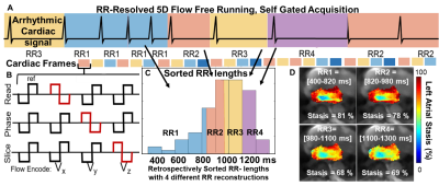 |
RR-Resolved 5D flow for Decoding the Impact of Cardiac Rhythm on Left Atrial Flow Dynamics in Atrial Fibrillation and Stroke
Justin Baraboo1, Elizabeth Weiss1, Maurice Pradella1, Liliana Ma1, Julia Hwang1, Suvai Gunasekaran1, Mariana Falcão2, Christopher W Roy2, Matthias Stuber2, Rod Passman1, Daniel Kim1, and Michael Markl1
1Northwestern University, Chicago, IL, United States, 2University of Lausanne, Lausanne, Switzerland
RR-resolved 5D flow is a novel MRI technique that can acquire time-resolved 3D hemodynamics for varying RR-interval durations during atrial fibrillation (AF) (5D = 3D+cardiac time+RR-interval duration). The purpose of this study was to apply RR-resolved 5D flow MRI in a cohort of AF patients with and without a prior history of stroke. We saw significantly lowered left atrial peak velocities and trends toward higher atrial blood stasis in AF patients with previous stroke history vs no previous stroke history who underwent arrhythmia during MRI acquisition. RR-resolved 5D flow may be a promising approach for cardiovascular imaging in arrhythmic patients.
|
|
| 09:51 | 0217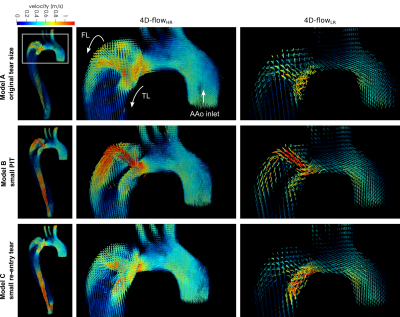 |
Impact of 4D-flow MRI spatial resolution on quantitative hemodynamics in 3D-printed aortic dissection models with varying tear size
Judith Zimmermann1,2, Kathrin Bäumler1, Tyler E Cork1,3, Michael Loecher1,4, Alison L Marsden3,5, Dominik Fleischmann1, and Daniel B Ennis1,4
1Department of Radiology, Stanford University, Stanford, CA, United States, 2Department of Computer Science, Technical University of Munich, Garching, Germany, 3Department of Bioengineering, Stanford University, Stanford, CA, United States, 4Division of Radiology, Veterans Affairs Health Care System, Palo Alto, CA, United States, 5Department of Pediatrics, Stanford University, Stanford, CA, United States
Understanding flow dynamics in type-B aortic dissection (TBAD) is of high clinical interest to predict complications and individualize treatment. Here, we sought to evaluate 4D-flow MRI at high and low spatial image resolution, in comparison to high resolution, high signal-to-noise 2D phase contrast MRI. Further, we compare TBAD hemodynamics and flow alterations owing to changes in tear size. We leverage novel 3D printing technology to manufacture three compliant TBAD models with altered tear morphology and perform pressure- and flow-controlled MRI with an advanced in vitro flow setup.
|
|
10:03 |
0218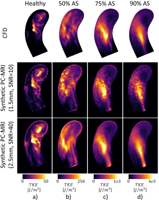 |
Synthesis of 4D Flow Tensor MRI of Patient-Specific Turbulent Flow
Pietro Dirix1, Stefano Buoso1, Eva Peper1, and Sebastian Kozerke1
1Institute for Biomedical Engineering, University and ETH Zürich, Zürich, Switzerland
We present a pipeline to synthesize patient-specific pulsatile turbulent 4D flow MRI datasets of the aorta. Aortic motion and inflow are extracted from in-vivo 2D cine and time-resolved 2D phase-contrast data. Computational fluid dynamics is used to obtain 4D velocity and turbulence fields to simulate MR signals using multipoint 4D flow tensor MRI protocols, which are reconstructed into velocity and turbulence maps with a Bayesian approach. As a result, realistic paired data of ground truth and their projection into MR images enable assessing accuracy and precision of encoding and inference, training of inference machines and, ultimately, deriving optimal experimental designs.
|
|
| 10:15 | 0219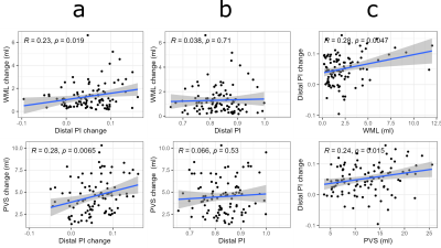 |
Longitudinal associations between 4D flow MRI intracranial pulsatility, white matter lesions, and perivascular space dilation across 5 years
Tomas Vikner1, Nina Karalija1,2, Anders Eklund1,2, Jan Malm3, Lars Nyberg1,2,4, and Anders Wåhlin1,2,5
1Department of Radiation Sciences, Umeå University, Umeå, Sweden, 2Umeå Center for Functional Brain Imaging (UFBI), Umeå University, Umeå, Sweden, 3Department of Clinical Science, Neurosciences, Umeå University, Umeå, Sweden, 4Department of Integrative Medical Biology (IMB), Umeå University, Umeå, Sweden, 5Department of Applied Physics and Electronics, Umeå University, Umeå, Sweden
Small vessel disease (SVD) is associated with elevated vascular stiffness and pulsatility, but longitudinal studies are lacking. Using a 4D flow MRI approach sensitive to age-differences in pulsatility in large and small cerebral arteries, we evaluated 5-year changes in pulsatility in relation to changes in white matter lesions (WML) and perivascular spaces (PVS), radiological markers of SVD. Pulsatility change correlated with WML and PVS volume change, but pulsatility at baseline did not predict WML and PVS progression. However, WML and PVS volumes at baseline predicted 5-year pulsatility change, suggesting that microvascular dysfunction associated with SVD accelerates pulsatility increases.
|
|
| 10:27 | 0220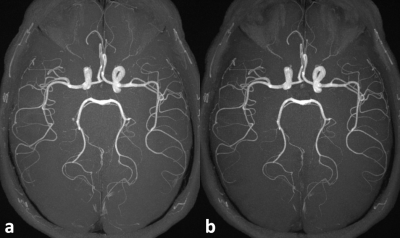 |
Improved Agreement of Cerebral Arterial Blood Flow Velocity Measures Using 3D Quantitative QISS MRA
Ioannis Koktzoglou1,2, Rong Huang1, and Robert R Edelman1,3
1Radiology, NorthShore University HealthSystem, Evanston, IL, United States, 2Radiology, Pritzker School of Medicine, University of Chicago, Chicago, IL, United States, 3Radiology, Northwestern University Feinberg School of Medicine, Chicago, IL, United States
Quantitative time of flight (qTOF) is a recently described 3D magnetic resonance angiography (MRA) technique for simultaneous luminal and hemodynamic evaluation of the intracranial arteries, that provides higher contrast-to-noise ratio efficiency than 3D phase contrast (PC) MRA and reduces in-plane flow displacement artifacts visible on standard 3D TOF MRA. We hypothesized that the use of a quiescent interval slice-selective-based data acquisition strategy (qQISS) that boosts arterial-to-background contrast might improve the quantitation of intracranial arterial flow velocity. Compared to qTOF MRA, we found that qQISS MRA improved agreement with PC MRA for measuring intracranial blood velocity.
|
 Back to Meeting Home
 Back to the Program-at-a-Glance
The International Society for Magnetic Resonance in Medicine is accredited by the Accreditation Council for Continuing Medical Education to provide continuing medical education for physicians.
 Back to Meeting Home
 Back to the Program-at-a-Glance
 View the Presentation