Oral Session
MR Angiography
Joint Annual Meeting ISMRM-ESMRMB & ISMRT 31st Annual Meeting • 07-12 May 2022 • London, UK

16:45 |
0328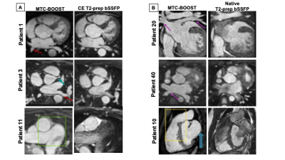 |
Gadolinium-free Bright and Black-Blood 3D whole-heart MRI for efficient anatomical assessment in patients with Congenital Heart Disease
Anastasia Fotaki1, Kuberan Pushparajah1, Reza Hajhosseiny1, Alina Schneider1, Karl Kunze1,2, Radhouene Neji1,3, Harith Alam4, Alessandra Frigiola4, René Botnar1,5, and Claudia Prieto1,5
1Biomedical Engineering, King's College London, London, United Kingdom, 2MR Research Collaborations, Siemens Healthcare Limited, Frimley, United Kingdom, 3MR Research Collaborations, Siemens Healthcare Limited, London, United Kingdom, 4Guy’s and St Thomas’s Hospital, London, United Kingdom, 5Escuela de Ingeniería, Pontificia Universidad Católica de Chile, Santiago, Chile
A recently developed sequence, the Magnetisation Transfer Contrast Bright Blood phase SensiTive (MTC-BOOST), enables free-breathing simultaneous acquisition of a bright-blood and black-blood 3D whole-heart dataset. We sought to compare the acquisition time, image quality and diagnostic performance of the proposed MTC-BOOST against the native clinical T2-prep bSSFP and the contrast-enhanced (CE) T2-prep bSSFP sequence in a cohort of patients with Congenital Heart Disease. Our study showed that MTC-BOOST offers good delineation of the intracardiac and vascular anatomy that is comparable to the clinical CE T2prep bSSFP and superior to the non-CE T2-prep bSSFP while offering a shorter acquisition time.
|
|
| 16:57 | 0329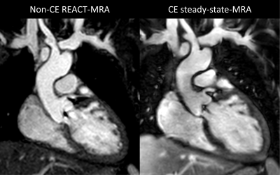 |
Non-contrast-enhanced free-breathing thoracic MRA using gated REACT at 3T in small children with congenital heart disease Video Permission Withheld
Alexander Isaak1, Narine Mesropyan1, Christopher Hart2, Dmitrij Kravchenko1, Christoph Endler1, Leon M. Bischoff1, Shuo Zhang3, Christoph Katemann3, Oliver Weber3, Daniel Kuetting1, Ulrike Attenberger1, Darius Dabir1, and Julian A. Luetkens1
1Department of Diagnostic and Interventional Radiology, University Hospital Bonn, Bonn, Germany, 2Department of Diagnostic and Interventional Radiology & Department of Pediatric Cardiology, University Hospital Bonn, Bonn, Germany, 3Philips GmbH Market DACH, Hamburg, Germany
Application of high-resolution 3D MR angiography (MRA) in small children with congenital heart disease (CHD) is challenging and generally requires contrast agent administration. In a cohort of pediatric CHD patients (median age: 4 years), non-contrast-enhanced free-breathing gated 3D mDixon REACT-MRA provided comparable overall image quality to contrast-enhanced free-breathing 3D mDixon steady-state MRA for assessment of the thoracic vasculature. REACT-MRA allowed for accurate and reliable vessel size measurements. Although fat-water separation artifacts were observed, they could be extenuated by reconstruction of in- and out-of-phase images. Gated REACT-MRA allows for a contrast-free assessment of the thoracic vasculature in small children with CHD.
|
|
| 17:09 | 0330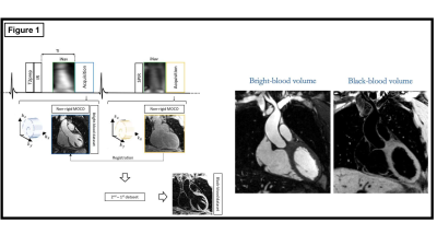 |
Feasibility of simultaneous non-contrast coronary MR angiography and vulnerable plaque imaging in acute myocardial infarction
Reza Hajhosseiny1,2, Adam Hartley2, Graham Cole2, Camila Munoz1, Amarjit Sethi2, Rasha Al-Lamee2, Deepa Gopalan2, Ben Ariff2, Raffi Kaprielian2, Karl Kunze3,4, Radhouene Neji3,4, Claudia Prieto1, Ramzi Khamis2, and René M Botnar1
1Biomedical Engineering Department, School of Biomedical Engineering and Imaging Sciences, King's College London, London, United Kingdom, 2National Heart and Lung Institute, Imperial College London, London, United Kingdom, 3MR Research Collaborations, Siemens Healthcare Limited, Frimley, United Kingdom, 4School of Biomedical Engineering and Imaging Sciences, King's College London, London, United Kingdom
CMR plaque imaging has the potential to predict future coronary events, but is limited by low spatial-resolution, misregistration artefacts, respiratory motion related image quality degradation and unpredictable acquisition times. Here we clinically evaluate the feasibility of a novel 3D free-breathing, non-rigid motion corrected, non-contrast CMR sequence (iT2prep-BOOST) that enables simultaneous, high-resolution visualisation of the coronary arteries and high-risk plaque features on co-registered bright and black blood images in patients presenting with acute myocardial infarction. We found that iT2prep-BOOST can simultaneously visualise coronary artery stenosis as well as culprit and bystander coronary plaque, as validated against invasive coronary angiography and OCT.
|
|
| 17:21 | 0331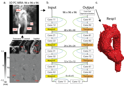 |
A 3D Dense-U-Net for fully automated 5D flow MRI segmentation
Liliana E. Ma1,2, Haben Berhane1,2, Justin Baraboo1,2, Michael Sugimura3, Christopher W. Roy4, Mariana Falcão4, Jérôme Yerly4, Matthias Stuber4,5, and Michael Markl1,2
1Department of Radiology, Northwestern University, Feinberg School of Medicine, Chicago, IL, United States, 2Department of Biomedical Engineering, Northwestern University, Evanston, IL, United States, 3Self-employed, Chicago, IL, United States, 4Department of Radiology, Lausanne University Hospital (CHUV) and University of Lausanne (UNIL), Lausanne, Switzerland, 5Center for Biomedical Imaging, Lausanne, Switzerland
Recently, a free-running 5D flow framework was introduced and validated. However, some 5D flow MRI is based on 3D radial imaging, which is limited by reduced SNR that can result in challenges with 3D segmentation. A number of previous studies have investigated automatic segmentation for 4D flow MRI, however these have been traditionally optimized for Cartesian datasets, which are typically acquired over much smaller spatial matrices and cover only one respiratory position. The purpose of this study was thus to adapt and expand a deep-learning framework to cardiac 5D flow MRI data for automatic segmentation of the thoracic aorta.
|
|
| 17:33 | 0332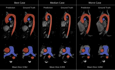 |
Automatic Segmentation of the Great Arteries for Robust Hemodynamic Assessment
Javier Montalt-Tordera1, Endrit Pajaziti1, Rod Jones2, Jennifer Steeden1, Silvia Schievano1, and Vivek Muthurangu1
1University College London, London, United Kingdom, 2Great Ormond Street Hospital, London, United Kingdom
Computational fluid dynamics (CFD) are useful in the assessment of blood flow conditions in patients with congenital heart disease. A necessary, time-consuming step in the creation of CFD models is the segmentation of the anatomy of interest. In this work, a neural network was trained to segment the aorta and the pulmonary arteries in 3D MRI, and its performance was evaluated in the context of a CFD application. The network performs well in terms of Dice score and is shown to lead to accurate pressure and flow velocity fields, with errors at the level of inter-observer variability.
|
|
| 17:45 |
0333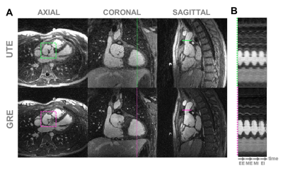 |
Free-running contrast-enhanced ultra-short TE (UTE) for cardiac and respiratory motion-resolved flow artifact-free 5D whole-heart MRI
Giulia M.C. Rossi1, Ludovica Romanin1,2, Mariana B.L. Falcão1, Bastien Milani1, Davide Piccini1,2, Jérôme Yerly1,3, Jürg Schwitter4,5,6, Milan Prša7, Tobias Rutz8, Estelle Tenisch1, Matthias Stuber1,3, and Christopher W. Roy1
1Department of Radiology, Lausanne University Hospital (CHUV) and University of Lausanne (UNIL), Lausanne, Switzerland, 2Advanced Clinical Imaging Technology (ACIT), Siemens Healthcare AG, Lausanne, Switzerland, 3CIBM Center for Biomedical Imaging, Lausanne, Switzerland, 4Division of Cardiology, Lausanne University Hospital (CHUV), Lausanne, Switzerland, 5Director CMR-Center, Lausanne University Hospital (CHUV), Lausanne, Switzerland, 6Faculty of Biology and Medicine, University of Lausanne (UNIL), Lausanne, Switzerland, 7Division of Pediatric Cardiology, Woman-Mother-Child Department, Lausanne University Hospital (CHUV) and University of Lausanne (UNIL), Lausanne, Switzerland, 8Service of Cardiology, Heart and Vessel Department, Lausanne University Hospital (CHUV) and University of Lausanne (UNIL), Lausanne, Switzerland Free-running whole-heart MRI can suffer from flow artifacts. Despite the efficiency of 3D radial UTE in minimizing the latter, its integration as a free-running sequence has so far been challenging due to poor-quality self-gating, which has necessitated an inefficient dual-echo approach. In this work we show that self-gating signals from a single-echo ferumoxytol-enhanced free-running 3D radial UTE sequence are comparable to the dual-echo approach, allowing to significantly improve scanning efficiency and produce dynamic images that are free from flow artifacts. |
|
| 17:57 | 0334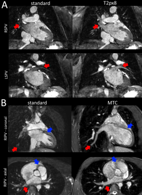 |
Pulmonary vein MRA with reduced sensitivity to off-resonance using optimized contrast preparation
Mohamed Nagiub1, Madhusudan Ganigara1, Bharti Sharma1, René Botnar2, Tarique Hussain1, and Joshua S Greer1
1Pediatrics, UT Southwestern Medical Center, Dallas, TX, United States, 2Biomedical Engineering, King's College London, London, United Kingdom
3D bSSFP is the standard for anatomical evaluation of congenital heart disease, but provides suboptimal visualization of the pulmonary veins due to off resonance and flow. Solutions have been proposed to reduce the sensitivity of the bSSFP acquisition to these factors with limited success. However, the T2prep pulse that is routinely applied for bSSFP imaging also likely contributes to pulmonary vein signal loss. In this study, we compare multiple contrast preparation schemes for 3D bSSFP imaging which minimize sensitivity of off-resonance to improve pulmonary vein visualization.
|
 Back to Meeting Home
 Back to the Program-at-a-Glance
The International Society for Magnetic Resonance in Medicine is accredited by the Accreditation Council for Continuing Medical Education to provide continuing medical education for physicians.
 Back to Meeting Home
 Back to the Program-at-a-Glance
 View the Presentation