Oral Session
Vessel Wall & Function
Joint Annual Meeting ISMRM-ESMRMB & ISMRT 31st Annual Meeting • 07-12 May 2022 • London, UK

| 09:27 | 0007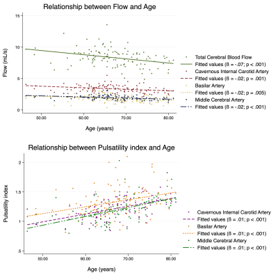 |
Defining Normative Cerebral Hemodynamics in Cognitively Healthy Older Adults with 4D Flow MRI
Grant S Roberts1, Anthony Peret2, Leonardo Rivera-Rivera1,3, Karly A Cody3, Howard A Rowley2, Oliver Wieben1,2, Sterling C Johnson3, Kevin M Johnson1,2, and Laura B Eisenmenger2
1Dept of Medical Physics, University of Wisconsin - Madison, Madison, WI, United States, 2Dept of Radiology, University of Wisconsin - Madison, Madison, WI, United States, 3Dept of Medicine, University of Wisconsin - Madison, Madison, WI, United States
It has been shown that vascular disease is strongly associated with Alzheimer’s disease (AD). It is thus important to establish normative cerebrovascular hemodynamics in aging populations. In this study, we comprehensively assess macrovascular hemodynamics using 4D flow MRI to obtain flow rates and pulsatility indices in 110 cognitively healthy, older adults and correlate these measures with age, sex, atherosclerotic cardiovascular disease (ASCVD) risk scores, and APOE genotypes. We found a (1) negative correlation between flow vs. age and flow vs. ASCVD, (2) a positive correlation between pulsatility vs. age and pulsatility vs. ASCVD, and (3) no correlations with APOE genotypes.
|
|
| 09:39 | 0008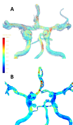 |
Wall shear stress and velocity pulsatility in the parent artery of an unruptured intracranial aneurysm - a 7T 4D flow MRI study.
Rick J. van Tuijl1, Ynte M. Ruigrok2, Kimberley M. Timmins1, Birgitta K Velthuis1, Pim van Ooij1, Jaco J.M. Zwanenburg1, and Irene C. van der Schaaf1
1Radiology, UMC Utrecht, Utrecht, Netherlands, 2Neurology, UMC Utrecht, Utrecht, Netherlands The influence of maximum wall shear stress (WSSMAX) and velocity pulsatility (vPI) are extensively investigated in aortic aneurysms, but its influence along the Circle of Willis (CoW) in patients with unruptured intracranial aneurysms (UIA) is unknown. Thirteen patients with an UIA were scanned at 7T-MRI using 4D phase-contrast MRI, comparing hemodynamic parameters between the parent artery of the UIA and the corresponding contralateral artery. The parent vessel of the UIA showed significantly higher WSSMAX, blood-flow and velocity, and lower vPI compared to the unaffected contralateral side. This study supports a hemodynamic role in the aetiology of aneurysm formation. |
|
09:51 |
0009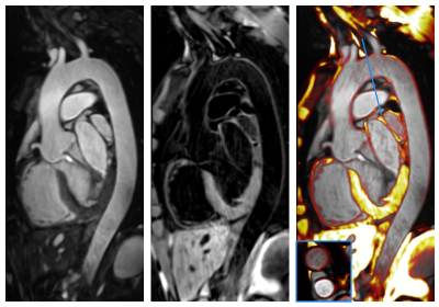 |
Simultaneous lumen and vessel wall imaging with iT2Prep-BOOST for an efficient comprehensive assessment of aortic disease
Camila Munoz1, Anastasia Fotaki1, Reza Hajhosseiny1, Karl P Kunze2, Radhouene Neji1,2, René M Botnar1, and Claudia Prieto1
1School of Biomedical Engineering and Imaging Sciences, King's College London, London, United Kingdom, 2MR Research Collaborations, Siemens Healthcare Limited, Frimley, United Kingdom
Bright- and black-blood imaging are key contrasts for the assessment of lumen and vessel wall integrity in aortic MRI. These contrasts are usually acquired separately, resulting in long and inefficient examinations. Here we extend a previously introduced motion-compensated interleaved acquisition (iT2Prep-BOOST) to produce high-quality co-registered 3D bright- and black-blood images from a short predictable scan of ~8min. The proposed iT2Prep-BOOST approach resulted in bright-blood images with improved contrast compared to conventional bSFFP images, and black-blood images with increased volumetric coverage and resolution compared to conventional HASTE images, showing promise for a comprehensive assessment of aortic disease from a single scan.
|
|
| 10:03 | 0010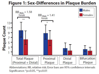 |
Sex-Differences in Intracranial Plaque Burden in Hypertensive Patients with Acute Ischemic Stroke: A Vessel Wall MR Imaging Study
Jae W. Song1, Jiayu Xiao2, Steven Y Cen2, Xiao Liu3, Fang Wu4, Konrad Schlick5, Qi Yang3, Shlee S Song5, and Zhaoyang Fan2
1University of Pennsylvania, Philadelphia, PA, United States, 2University of Southern California, Los Angeles, CA, United States, 3Chaoyang Hospital, Beijing, China, 4Xuanwu Hospital, Beijing, China, 5Cedars-Sinai Medical Center, Los Angeles, CA, United States
Hypertension is associated with intracranial atherosclerosis (ICAS) and is a leading cause of acute ischemic stroke (AIS). Given sex-differences in the severity of hypertension, we hypothesized sex-differences in ICAS burden among hypertensive patients with AIS. Results show males have significantly higher adjusted total plaque burdens than females. Subgroup analyses show treated hypertensive males with AIS have higher total proximal and bifurcation plaques than treated females. Untreated hypertensive females have significantly higher total proximal and bifurcation plaques compared to untreated males. The results suggest attention to differential hypertension management by sex may be warranted to reduce ICAS burden and AIS risk.
|
|
| 10:15 | 0011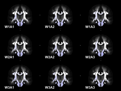 |
Regional diffusion imaging changes related to obesity, after adjusting for other risk factors: the UK Biobank study
Karolina Wartolowska1 and Alastair JS Webb1
1University of Oxford, Oxford, United Kingdom
Small vessel disease is a common cause of white matter damage in older people. It is strongly associated with age, hypertension, diabetes, and obesity. We have investigated the associations between body mass index (BMI) and diffusion changes (adjusted for other risk factors) in data from UK Biobank. BMI was more strongly related to diffusion changes in inferior brain regions, depending on whether the white matter tracts were within the anterior or posterior circulation. Obesity affects white matter integrity, beyond the effect of cardiovascular risk factors.
|
|
| 10:27 | 0012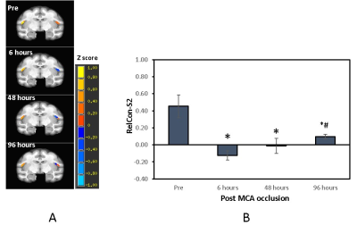 |
Temporal correlation of functional connectivity with cerebral blood flow and T2 relaxation time in the monkey brain following ischemic stroke
Chun-Xia Li1, Frank Tong2, Doty Kempf1, Leonard Howell1, and Xiaodong Zhang1
1Emory University, Atlanta, GA, United States, 2Department of Radiology, Emory University, Atlanta, GA, United States Somatosensory deficits are seen in most stroke patients and can improve during recovery from stroke as reported previously. However, the underlining mechanism of the recovery remains poorly understood. The nonhuman primate (NHP) model of stroke has demonstrated similar functional connectivity evolution as seen in stroke patients and the secondary somatosensory cortex (S2) in NHPs is recognized as a direct extrapolation of somatosensory cortex in human. In the present study, the progressive function alteration of S2 and its relationship with the cerebral blood flow (CBF) and T2 changes in the monkey brain during acute stroke were examined and evaluated. |
|
| 10:39 | 0013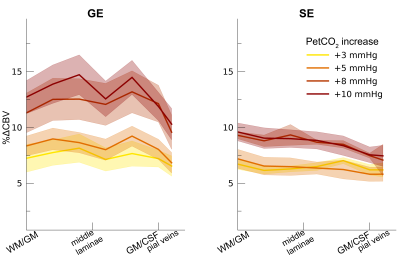 |
How vascular properties affect the BOLD contrast across cortical depth.
Wouter Schellekens1, Alex Bhogal1, Emiel Roefs1, Mario Báez-Yáñez1, Jeroen Siero1, and Natalia Petridou1
1UMC Utrecht, Utrecht, Netherlands
In the current study, we quantify vascular properties in relation to laminar BOLD fMRI signals for differently sized vascular compartments across cortical depth. Using hypercapnic and hyperoxic breathing conditions, while measuring from macro- and micro-vascular compartments, we estimate effects of dilation capacity, theoretical maximum signal intensity, and relative change in cerebral blood volume on laminar BOLD contrasts. We show that enlarged signals for larger pial veins are mainly caused by their capacity for dilation. BOLD signal differences between macro- and micro-vascular compartments are not likely caused by differences in theoretical maximum signal intensity, or relative changes in cerebral blood volume.
|
 Back to Meeting Home
 Back to the Program-at-a-Glance
The International Society for Magnetic Resonance in Medicine is accredited by the Accreditation Council for Continuing Medical Education to provide continuing medical education for physicians.
 Back to Meeting Home
 Back to the Program-at-a-Glance
 View the Presentation