Oral Session
Perfusion Techniques
Joint Annual Meeting ISMRM-ESMRMB & ISMRT 31st Annual Meeting • 07-12 May 2022 • London, UK

| 16:45 | 0335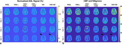 |
Dual-module velocity-selective arterial spin labeling (dm-VSASL) using velocity-selective saturation and inversion pulses
Jia Guo1,2
1Bioengineering, University of California Riverside, Riverside, CA, United States, 2Center for Advanced Neuroimaging, University of California Riverside, Riverside, CA, United States
Recently velocity-selective saturation (VSS) based dual-module (dm-) preparation and velocity-selective inversion (VSI) based preparation have demonstrated improved SNR efficiency in velocity-selective arterial spin labeling (VSASL) than conventional single-module VSS based preparation. In this study, a novel strategy was developed to allow dm-VSI labeling to further improve the SNR efficiency. Implementation of dm-VSASL were evaluated for VSS and/or VSI combinations. The theoretical performance was compared via ASL signal modeling, and then validated by in vivo human experiments with an increase of 6.6%. Dm-VSASL is capable of providing further improved SNR performance, with potentially better suppression of diffusion attenuation/eddy current artifacts.
|
|
| 16:57 | 0336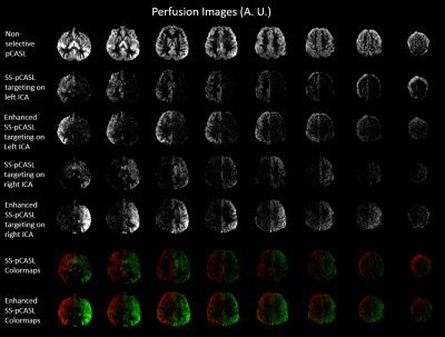 |
Enhanced superselective pseudo-Continuous Arterial Spin Labeling using parallel transmission with B1 phase shimming at 7T
Chenyang Zhao1, Kai Wang1, and Danny JJ Wang1,2
1Laboratory of FMRI Technology (LOFT), Mark & Mary Stevens Neuroimaging and Informatics Institute, Keck School of Medicine, University of Southern California, Los Angeles, CA, United States, 2Department of Neurology, Keck School of Medicine, University of Southern California, Los Angeles, CA, United States
Superselective pseudo-Continuous Arterial Spin Labeling (SS-pCASL) suffers from low labeling efficiency due to B1 inhomogeneity at 7T. An enhanced SS-pCASL using parallel transmission with B1 phase shimming was proposed. B1 phase shimming algorithm was designed to penalize low and high B1 amplitude in the labeled and unlabeled ICA, respectively. B1 amplitude was increased by 40% for the labeled ICA and decreased by 90% for the unlabeled ICA. Simulation and in-vivo results demonstrated that enhanced SS-pCASL improved labeling efficiency for the labeled ICA and suppressed labeling oscillation for the unlabeled ICA.
|
|
| 17:09 | 0337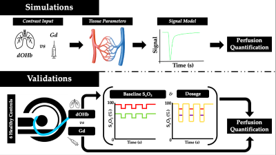 |
Deoxyhemoglobin versus Gadolinium as Contrast in Dynamic Susceptibility Perfusion Imaging: Simulations and Scan Validations
Jacob Benjamin Schulman1, Ece Su Sayin2, Julien Poublanc3, Angelica Manalac1, Joseph A Fisher4, Olivia Sobczyk4, James Duffin4, Harrison Levine2, David J Mikulis3, and Kamil Uludag5
1Medical Biophysics, University of Toronto, Toronto, ON, Canada, 2Physiology, University of Toronto, Toronto, ON, Canada, 3Joint Department of Medical Imaging and the Functional Neuroimaging Lab, University Health Network, Toronto, ON, Canada, 4Department of Anaesthesia and Pain Management, University Health Network, Toronto, ON, Canada, 5Techna Institute & Koerner Scientist in MR Imaging, University Health Network, Toronto, ON, Canada
DSC-MRI requires bolus administration of gadolinium, an exogenous contrast agent with notable limitations. We used simulations to compare gadolinium with a deoxyhemoglobin bolus generated in the lungs as perfusion contrast and verified the findings in healthy human subjects. We found that the calculated cerebral blood volume is dependent on the contrast agent, the magnitude of deoxyhemoglobin concentration increase (dosage), and the baseline oxygen saturation. We also found a reduction of macrovascular contamination with deoxyhemoglobin contrast. This work reveals novel insights for the use of deoxyhemoglobin, an endogenous and safe contrast agent for determining perfusion in the human brain.
|
|
| 17:21 | 0338 |
Subject-specific optimization of background suppression for arterial spin labeling MRI using a real-time feedback loop on the scanner
Kirsten Koolstra1, Marius Staring1, Paul de Bruin2, and Matthias J P van Osch1
1Leiden University Medical Center, Leiden, Netherlands, 2Philips, Best, Netherlands Background suppression (BGS) in arterial spin labeling (ASL) leads to perfusion images with a higher temporal signal-to-noise ratio (tSNR) compared to ASL without BGS. The optimal inversion times (TIs), and therefore the quality of the BGS, depend on the T1 relaxation times of the underlying tissue and on inhomogeneities of the scanner’s magnetic fields (B0, B1+). In this work, we designed and implemented a feedback mechanism that optimized the quality of background suppression in real time on the scanner. The results show an increased tSNR for the subject-specific optimization of BGS compared to standard BGS in 12 healthy volunteers. |
|
17:33 |
0339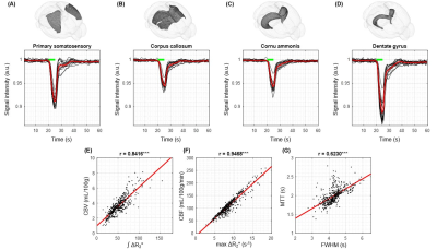 |
Development of perfusion technique with dynamic BOLD MRI and transient hypoxia in mouse
THI THUY LE1, DONG KYU LEE2, GEUN HO IM2, and SEONG GI KIM1
1Department of Biomedical Engineering, Sungkyunkwan University, Suwon, Republic of Korea, Center for Neuroscience Imaging Research (CNIR), Institute for Basic Science (IBS), Suwon, Republic of Korea, Suwon, Korea, Republic of, 2Center for Neuroscience Imaging Research (CNIR), Institute for Basic Science (IBS), Suwon, Republic of Korea, Suwon, Korea, Republic of
Tissue perfusion can be measured by arterial spin labeling and dynamic susceptibility contrast (DSC) method. However, these methods cannot be easily applied to mouse. Alternatively, a bolus of paramagnetic deoxyhemoglobin induced by transient hypoxia may be used as an endogenous blood pool contrast agent. Dynamic BOLD MRI with hypoxic challenge was adopted in mice. A stimulus duration was determined, and its impact to animal physiology was investigated. With corrected arterial input function, quantitative CBF and CBV were determined. Overall, we successfully developed a technique with BOLD MRI and transient hypoxia.
|
|
| 17:45 | 0340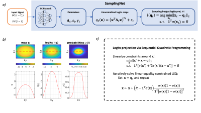 |
Learning Time-Adaptive Data-Driven Sampling Pattern for Accelerated 3D Myocardial Perfusion Imaging
Valery Vishnevskiy1, Tobias Hoh1, and Sebastian Kozerke1
1University and ETH Zurich, Zurich, Switzerland
We investigated potential benefits of optimizing accelerated 3D myocardial perfusion MRI by time-adaptive data-driven k-t Cartesian sampling during the scan. To this end, the sampling mask for each following acquisition window is inferred by a neural network on the fly using the acquired history of MR signal projections. It is demonstrated that time-adaptive data-driven sampling reduces reconstruction errors by 25% along with a reduction of signal underestimation during the contrast bolus passage when compared to a predefined fixed sampling pattern.
|
|
| 17:57 | 0341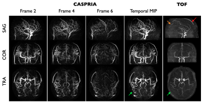 |
Combined Angiographic, Structural and Perfusion Radial Imaging using Arterial Spin Labeling
Thomas W Okell1, Joseph G Woods2, and Mark Chiew1
1Wellcome Centre for Integrative Neuroimaging (FMRIB), Nuffield Department of Clinical Neurosciences, University of Oxford, Oxford, United Kingdom, 2CFMRI, Department of Radiology, University of California San Diego, San Diego, CA, United States A Combined Angiographic, Structural and Perfusion Radial Imaging using Arterial Spin Labeling (CASPRIA) pulse sequence is presented which allows the simultaneous acquisition of non-contrast dynamic angiograms, quantitative perfusion maps and multi-contrast T1-weighted structural images within a single six-minute scan. Compared to conventional imaging methods, which took 70% longer to acquire, CASPRIA yielded comparable quantitative perfusion estimates, dynamic (rather than static) angiography with improved distal vessel visibility and structural images with greater contrast flexibility. With further work, the estimation of quantitative tissue T1 values could also be possible. |
 Back to Meeting Home
 Back to the Program-at-a-Glance
The International Society for Magnetic Resonance in Medicine is accredited by the Accreditation Council for Continuing Medical Education to provide continuing medical education for physicians.
 Back to Meeting Home
 Back to the Program-at-a-Glance
 View the Presentation