Oral Session
Cartilage
Joint Annual Meeting ISMRM-ESMRMB & ISMRT 31st Annual Meeting • 07-12 May 2022 • London, UK

| 16:45 | 0342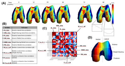 |
The Knee Connectome: Studying Localized Synchrony in 8-year Cartilage Changes in Thickness and T2
Jennifer Anne Cummings1, Kenneth Gao1, Vincent Chen2, Alejandro Morales Martinez2, Sharmila Majumdar1, and Valentina Pedoia1
1University of California, San Francisco, San Francsico, CA, United States, 2University of California, San Francisco, San Francisco, CA, United States
The trajectories of femur cartilage thickness and T2 relaxation time over an 8 year period were analyzed for 787 subjects from the Osteoarthritis Initiative dataset. Biomarkers were automatically extracted and used to generate time series for 27 regions of interest within the cartilage map to create a connectivity matrix (connectome). Statistically significant differences in thickness and T2 trajectories were found to correlate with demographic and clinical factors such as sex, BMI, Radiographic OA and presence of knee pain.
|
|
| 16:57 | 0343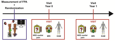 |
T1ρ can detect microstructural changes in cartilage induced by conservative interventions: a one-year randomized controlled trial
Valentina Mazzoli1,2, Scott Uhlrich2,3, Feliks Kogan1, Amy Silder2,4, Andrea Finlay5, Scott Delp 3,4, Gary Beaupre4,6, Julie Kolesar4,6, and Garry Gold1
1Radiology, Stanford University, Stanford, CA, United States, 2Musculoskeletal Research Laboratory, VA Palo Alto Healthcare System, Palo Alto, CA, United States, 3Mechanical Engineering, Stanford University, Stanford, CA, United States, 4Bioengineering, Stanford University, Stanford, CA, United States, 5Center for Innovation and Implementation, VA Palo Alto Healtcare System, Palo Alto, CA, United States, 6Musculoskeletal Research Laboratory, VA Palo Alto Healtcare System, Palo Alto, CA, United States
We performed a single-blind randomized clinical trial in subjects with medial knee OA to evaluate the effect of a gait modification. Primary outcomes were pain and KAM (surrogate measure of medial knee loading). We also utilized cartilage T2 and T1ρ to assess (micro)structural changes induced by the treatment. In the Intervention group, we observed a greater reduction in pain and KAM (reduced loading), and less increase in T1ρ in the medial femoral compartment, potentially indicating slowed cartilage degeneration. Our study shows that quantitative cartilage MRI is a promising outcome measure in clinical trial assessing non-surgical treatment for knee OA.
|
|
| 17:09 | 0344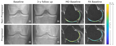 |
DTI of articular cartilage can predict 3-year radiographic progression in OA patients at early stage of disease
Jose G Raya1, Alejandra Duarte2, Richard Kijowski1, Svetlana Krasnokutsky-Samuels1, Jenny T Bencardino3, and Jonathan Samuels1
1NYU Langone Medical Center, New York, NY, United States, 2Fundacion Santa Fe de Bogota, Bogota, Colombia, 3Penn Medicine, Philadelphia, PA, United States
The goal of this study is to validate DTI of articular cartilage at 3T as a biomarker for OA progression in a population at early stages of the disease and high likelihood of short-term progression. The goal of this study is to validate DTI of articular cartilage at 3T as a biomarker for OA progression in a population at early stages of the disease and high likelihood of short-term progression. We show that DTI has potential for prognosis of 3-year radiographic changes in a population with early stages of disease.
|
|
17:21 |
0345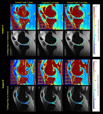 |
Deep Learning Predicts Synthetic T1rho Maps from T2 Maps for Knee MRI
Michelle W. Tong1,2, Aniket A. Tolpadi1,2, Alex Beltran1,2, Sharmila Majumdar2, and Valentina Pedoia 2
1Department of Bioengineering, University of California Berkeley, Berkeley, CA, United States, 2Department of Radiology and Biomedical Imaging, University of California San Francsico, San Francisco, CA, United States
This study explores the use a deep learning network to generate T1rho maps from T2 maps for knee MRI. T1rho maps have clinical value in the diagnosis of osteoarthritis in addition to T2 maps while T2 maps are more widely adopted and available in clinical datasets. This study found synthetic T1rho maps images maintain excellent fidelity for data collected in a research setting while performance is reduced for data collected in a clinical setting. This work elucidates the promise of deep learning in accelerating imaging protocols through domain adaptation as opposed to more common reconstruction approaches.
|
|
| 17:33 | 0346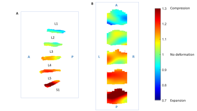 |
Deformation-field magnetic resonance imaging for non-invasive evaluation of disc deformation in vivo
Frida Johansson1,2, Zainab Sirat1, Hanna Hebelka1,3, Helena Brisby1,4, Fredrik Nordström1,2, and Kerstin Lagerstrand1,2
1Institute of Clinical Sciences, University of Gothenburg, Gothenburg, Sweden, 2Medical Physics and Biomedical Engineering, Sahlgrenska University Hospital, Gothenburg, Sweden, 3Department of Radiology, Sahlgrenska University Hospital, Gothenburg, Sweden, 4Department of Orthopaedics, Sahlgrenska University Hospital, Gothenburg, Sweden
In vivo evaluation of the intervertebral disc (IVD) during spinal loading may yield greater insight into the biomechanical properties of the IVD. This study aimed to investigate how the lumbar IVD deforms during loading, quantified with a novel non-invasive method utilizing MRI and image registration. Findings showed that the intradiscal deformation depends not only on disc degeneration but also on the lumbar spine level. This highlights the need for tools that can evaluate the mechanical properties of the disc in vivo. The proposed method offers a possibility to depict and track biomechanical changes non-invasively while characterizing disc structures in detail.
|
|
| 17:45 | 0347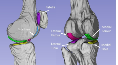 |
Bi-exponential UTE-T2* Evaluation of Longitudinal Changes in Knee Cartilage Over 2 Years Following Anterior Cruciate Ligament Reconstruction
Ashley A Williams1,2, Yongxian Qian3,4, and Constance R Chu1,2
1Orthopaedic Surgery, Stanford University, Stanford, CA, United States, 2Veterans Affairs Palo Alto Health Care System, Palo Alto, CA, United States, 3Department of Radiology, New York University Grossman School of Medicine, New York, NY, United States, 4Center for Biomedical Imaging, NYU Langone Health, New York, NY, United States
This work evaluates mono- and bi-exponential UTE-T2* relaxation in 6 tread mark regions of knee cartilage of 18 participants with ACL-injury prior to reconstruction surgery and in 15/18 participants 2 years after surgery. Bi-exponential T2*short had a smaller fractional contribution than T2*long in all regions examined. However, T2*short accounted for a higher fraction of total signal in deep cartilage layers compared to superficial. While both mono- and bi-exponential UTE-T2* analyses showed significant longitudinal changes, bi-exponential analyses did not exceed the sensitivity of mono-exponential UTE-T2* for detection of knee cartilage compositional changes over the first two years following ACL reconstruction.
|
|
| 17:57 | 0348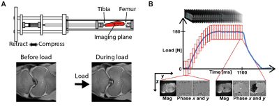 |
High Frame Rate Strain and Viscoelastic Analysis on Bovine Tibiofemoral Joints During Compression
Woowon Lee1, Emily Y Miller2, Callan M Luetkemeyer1, and Corey P Neu1,2
1Paul M. Rady Department of Mechanical Engineering, University of Colorado, Boulder, CO, United States, 2Biomedical Engineering Program, University of Colorado, Boulder, CO, United States
Osteoarthritis (OA) is a degenerative process in cartilage mainly occurring in knee. OA affects the structure and function of cartilage thus, measuring the mechanical properties of cartilage is crucial. We use spiral DENSE MRI to quantify the mechanics of intact and defected bovine articular cartilage. The samples underwent cyclic compressive load and 27 frames are captured while the joint was compressed. Displacement and strain gradually increased over time and the defected cartilage showed higher strain in the tibiofemoral contact areas. We also found creep response in the defected cartilage which may be a potential biomarker for OA detection.
|
|
| 18:09 | 0349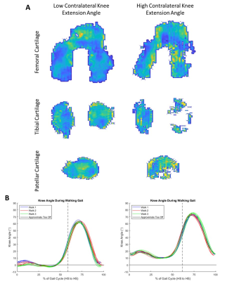 |
Cartilage T2 Relaxation Times are Related to Walking Asymmetries in Osteoarthritic Knees
Amelia Kruse1, Austin Carcia1, Adam Bradshaw1, Charles Ho1, Johnny Huard1, Scott Tashman1, and Lauren Watkins1
1Steadman Philippon Research Institute, Vail, CO, United States
In this exploratory study, we examine relationships between joint function and cartilage composition in individuals with knee osteoarthritis (OA). MRI T2 relaxation times were used to assess articular cartilage and joint kinematics were recorded over walking gait cycles using video-motion analysis. There were significant correlations between cartilage T2 relaxation times and knee flexion, extension, and swing-stance ratio in both affected and contralateral legs. MRI measures of cartilage quality and joint function provide complimentary information that may be useful in measuring disease progression and intervention effectiveness using MRI.
|
 Back to Meeting Home
 Back to the Program-at-a-Glance
The International Society for Magnetic Resonance in Medicine is accredited by the Accreditation Council for Continuing Medical Education to provide continuing medical education for physicians.
 Back to Meeting Home
 Back to the Program-at-a-Glance
 View the Presentation