Oral Session
Joints
Joint Annual Meeting ISMRM-ESMRMB & ISMRT 31st Annual Meeting • 07-12 May 2022 • London, UK

| 14:45 | 0093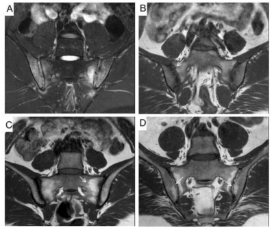 |
Evaluation inflammatory and chronic structural sacroiliac joint changes in Axial Spondyloarthritis using semiquantitative MRI scoring system
yimeng zhang1, zikang guo1, and xinwei lei2
1First Central Clinical College, Tianjin Medical University, Tianjin, Tianjin, China, 2Tianjin First Central Hospital, Tianjin, Tianjin, China
There are many clinical studies investigating the differences between non-radiographic axial spondyloarthritis and ankylosing spondylitis(AS), but few focus on radiology. Spondyloarthritis Research Consortium of Canada (SPARCC) and Sacroiliac Joint Structural Score (SSS) scoring systems can assess a broad spectrum of inflammatory and structural lesions. Our results indicate SPARCC scores were significantly improved after treatment. A significant increase in SSS scores for fat metaplasia and backfill was noted. Therefore, using semiquantitative MRI scoring methods can assess abnormal lesions and treatment response. Higher structural damages were seen in AS patients. SPARCC can assess severity of disease pre-treatment and treatment effectiveness.
|
|
| 14:57 | 0094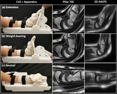 |
Extended and weight bearing wrist MRI using a positioning apparatus and flexible glove coil to explore the cause of dorsal pain
Bili Wang1,2, Jerzy Walczyk1,2, Mohammed Ahmed1, Madeline Rocks3, Stuart Elkowitz3, Louis Catalano3, Christopher Burke1, Steven Daniels1, and Ryan Brown1,2
1Bernard and Irene Schwartz Center for Biomedical Imaging, Department of Radiology, New York University Grossman School of Medicine, New York, NY, United States, 2Center for Advanced Imaging Innovation and Research (CAI2R), Department of Radiology, New York University Grossman School of Medicine, New York, NY, United States, 3Department of Orthopedic Surgery, New York University Grossman School of Medicine, New York, NY, United States
Dorsal wrist pain during extension or weight bearing is a common symptom, the etiology of which often remains unclear. Clinical MRI often fails to reveal causative pathology in part because it is performed in the neutral position. We built an apparatus to guide motion and used a flexible coil to capture signal in a range of positions that better match the conditions during which pain is reported. Results in nine asymptomatic volunteers show excellent tissue structure delineation, strong measurement agreement among readers, and increases in dorsal capsule thickness and radiocapitate, radiolunate, capitolunate and extensor tendon angulation during weight bearing.
|
|
| 15:09 | 0095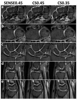 |
Accelerated High-Resolution 3D Isotropic Wrist MRI at 7 Tesla: Compressed Sensing vs. SENSE
Bobby Runderkamp1, Matthan Caan2, Wietske van der Zwaag3, Robert Hemke1, Mario Maas1, Mads Andersen4,5, Gustav Strijkers2, Karin Markenroth Bloch4, and Aart Nederveen1
1Radiology & Nuclear Medicine, Amsterdam UMC, Amsterdam, Netherlands, 2Biomedical Engineering and Physics, Amsterdam UMC, Amsterdam, Netherlands, 3Spinoza Center for Neuroimaging, Amsterdam, Netherlands, 4Lund University Bioimaging Center, Lund, Sweden, 5Philips Healthcare, Copenhagen, Denmark
Imaging of the intricate arrangement of small structures in the wrist benefits from high resolution imaging, which the increased SNR at 7 Tesla can enable. However, the increased resolution requires longer scan times, decreasing patient comfort and increasing risk of motion artifacts. In this work, we evaluated compressed sensing (CS) acceleration of a clinical wrist 7T MRI protocol, 0.45mm isotropic resolution, in comparison to SENSE-acceleration. We show that CS-acceleration can produce adequate to good image quality in significantly shorter scan times, not achievable by increasing SENSE-acceleration.
|
|
| 15:21 | 0096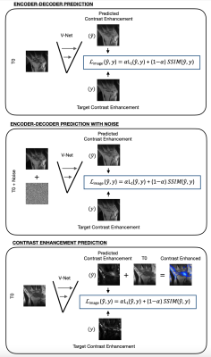 |
Gadolinium Free Synthetic Inflammation Imaging
Hari Umesh1, Francesco Caliva2, Jing Liu2, Xiaojuan Li3, Thomas Link2, Sharmila Majumdar2, and Valentina Pedoia2
1University of Illinois at Urbana-Champaign, Champaign, IL, United States, 2UCSF, San Francisco, CA, United States, 3Cleveland Clinic, Cleveland, OH, United States
Traditional MRI imaging involves injecting gadolinium-diethylenetriamine pentaacetic acid (Gd-DTPA) , a pure substance that induces contrast to highlight inflammation and blood vessels in MRI images, proven to increase accuracy in radiologic diagnosis. We present a 2D V-net image contrast generation model that accurately predicts Gd-DTPA induced contrast from corresponding images without Gd-DTPA induced contrast.
|
|
15:33 |
0097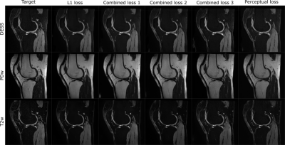 |
Deep-Learning-based contrast synthesis from MRF parameter maps in the knee
Olli Nykänen1,2, Antti Isosalo1, Satu I Inkinen1, Victor Casula1,3, Mika Nevalainen1,3,4, Riccardo Lattanzi5, Martijn Cloos6, Mikko J Nissi2, and Miika T Nieminen1,3,4
1Research Unit of Medical Imaging, Physics and Technology,, University of Oulu, Oulu, Finland, 2Department of Applied Physics, University of Eastern Finland, Kuopio, Finland, 3Medical Research Center, University of Oulu and Oulu University Hospital, Oulu, Finland, 4Department of Diagnostic Radiology, Oulu University Hospital, Oulu, Finland, 5Center for Advanced Imaging Innovation and Research (CAI2R) and Bernard and Irene Schwartz Center for Biomedical Imaging, Department of Radiology, New York University Grossman School of Medicine, New York, NY, United States, 6Centre for Advanced Imaging, Queensland University, Brisbane, Australia
In this study, deep convolutional neural networks (DCNN) are used to synthesize contrast-weighted magnetic resonance (MR) images from quantitative parameter maps of the knee joint obtained with magnetic resonance fingerprinting (MRF). Training of the neural networks was performed using data from 142 patients, for which both standard MR images and quantitative MRF maps of the knee were available. The study demonstrates that synthesizing contrast-weighted images from MRF-parameter maps is possible utilizing DCNNs. Furthermore, the study indicates a need to tune up the dictionary used in MRF so that the parameters expected from the target anatomy are well-covered.
|
|
| 15:45 | 0098 |
Improving diagnostic confidence using a Deep-Learning Reconstructed Fast Motion-Robust PROPELLER protocol for Shoulder Imaging
Laura Carretero1,2, Xinzeng Wang3, Eugenia Sánchez4, Satish Nagrani4, Daniel Litwiller5, Pablo García-Polo1, Maggie Fung6, and Mario Padrón4
1GE Healthcare, Madrid, Spain, 2Rey Juan Carlos University, Madrid, Spain, 3GE Healthcare, Houston, TX, United States, 4Clínica Cemtro, Madrid, Spain, 5GE Healthcare, Colorado, CO, United States, 6GE Healthcare, New York, NY, United States PROPELLER imaging is the choice for motion-prone anatomies, to generate good diagnostic quality images even for challenging patients. However, it is associated with prolonged scan times. In this study we evaluate the application of a new DL-based reconstruction algorithm to enhance a fast PROPELLER shoulder protocol with the goal of providing consistent diagnostic confidence and an overall improved image quality compared to conventional routine protocols. Our study demonstrates the proposed under 10min shoulder MRI protocols are interchangeable and have the same diagnostic confidence with better SNR and lesion conspicuity than routine MRI protocols for shoulder post-contrast and instability exams. |
|
| 15:57 | 0099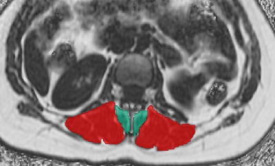 |
The role of facet joint arthropathy in chronic low back pain and its association with adjacent paraspinal muscle composition Video Permission Withheld
Nico Sollmann1,2,3, Noah B. Bonnheim4, Gabby B. Joseph1, Ann A. Lazar5, Ravi Chachad1, Jiamin Zhou1, Jeannie F. Bailey4, Xiaojie Guo4, Thomas M. Link1, Aaron J. Fields4, and Roland Krug1
1Department of Radiology and Biomedical Imaging, University of California San Francisco, San Francisco, CA, United States, 2Department of Diagnostic and Interventional Radiology, University Hospital Ulm, Ulm, Germany, 3Department of Diagnostic and Interventional Neuroradiology, Technical University of Munich, Munich, Germany, 4Department of Orthopaedic Surgery, University of California San Francisco, San Francisco, CA, United States, 5Department of Epidemiology and Biostatistics, University of California San Francisco, San Francisco, CA, United States
Low back pain (LBP) is a global health burden, but patient phenotyping based on imaging that would facilitate timely and effective treatment regimens lacks behind. One issue is that associations between different structures at the degenerative lumbar spine are not yet well characterized. In this study we revealed that facet joint arthropathy (FJA) at level L4/L5 is associated with the fat fraction (FF) of adjacent paraspinal musculature (PSM) as derived from chemical shift encoding-based water-fat MRI (CSE-MRI), as well as with Modic-type endplate changes, endplate defects, and intervertebral degenerative disk disease (DDD) in patients with chronic LBP.
|
|
| 16:09 | 0100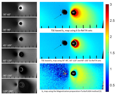 |
Turbo-spin echo based B1+ mapping in the presence of metallic hardware Video Permission Withheld
Iman Khodarahmi1, Mahesh B Keerthivasan2, and Jan Fritz1
1Radiology, New York University School of Medicine, New York, NY, United States, 2Siemens Medical Solutions USA Inc, Malvern, PA, United States
A robust B1+ mapping technique in the presence of metallic hardware is still unavailable. Our proposed B1+ quantification technique relies on turbo-spin echo or SEMAC acquisitions to decrease metal-related susceptibility artifacts while resolving B1+ values from signal variations at various sets of excitation and refocusing flip angles. Apriori knowledge of signal evolution is obtained by simulating the Bloch equations at each B1+ value. Phantom validation showed promising results, particularly at areas close to the metal surface, which are invisible with other mapping techniques.
|
 Back to Meeting Home
 Back to the Program-at-a-Glance
The International Society for Magnetic Resonance in Medicine is accredited by the Accreditation Council for Continuing Medical Education to provide continuing medical education for physicians.
 Back to Meeting Home
 Back to the Program-at-a-Glance
 View the Presentation