Oral Session
Novel MR Technology
Joint Annual Meeting ISMRM-ESMRMB & ISMRT 31st Annual Meeting • 07-12 May 2022 • London, UK

| 09:15 | 0405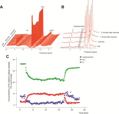 |
Skeletal muscle acetylcarnitine during submaximal exercise and recovery: Interleaved 1H/31P MRS 7T pilot study
Radka Klepochová1,2, Fabian Niess2, Martin Meyerspeer3, Siegfried Trattnig2,4, Michael Krebs1, Alexandra Kautzky-Willer1, Michael Leutner1, and Martin Krššák1,2
1Division of Endocrinology and Metabolism, Department of Internal Medicine III, Medical University of Vienna, Vienna, Austria, 2High-Field MR Center, Department of Biomedical Imaging and Image-Guided Therapy, Medical University of Vienna, Vienna, Austria, 3High-Field MR Center, Center for Medical Physics and Biomedical Engineering, Medical University of Vienna, Vienna, Austria, 4Christian Doppler Laboratory for Clinical Molecular MR Imaging (MOLIMA), Christian Doppler Laboratory, Vienna, Austria
The study aimed to explore the behavior of skeletal muscle acetylcarnitine and phosphocreatine during submaximal plantar flexion exercise using interleaved 1H/31P MRS at 7T. Acetylcarnitine decreased to steady state during the exercise, increased back to initial levels in early recovery period and started to decline during later phase of recovery. Studies including more volunteers and patients with broader range of metabolic conditions and physical fitness will be necessary for detailed quantitative analysis.
|
|
| 09:27 | 0406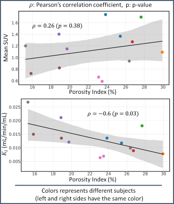 |
Exploratory study of relationships between [18F] Sodium fluoride PET metabolic bone measures and MRI bone porosity index in the tibial tuberosity
Marco Barbieri*1, Lauren Watkins*1, Bryan Haddock2, Garry E. Gold1,3, and Feliks Kogan1
1Radiology, Stanford University, Stanford, CA, United States, 2Department of Clinical Physiology, Nuclear Medicine and PET, Rigshospitalet, Copenhagen University Hospital, Copenhagen, Denmark, 3Bioengineering, Stanford University, Stanford, CA, United States
Molecular information derived from dynamic [18F]NaF PET imaging holds promise to study bone remodeling in bone and joint disorders. The porosity index (PI), based on ultra-short echo-time (uTE) MRI, has been proposed to study bone porosity in clinically valuable acquisition times. We explored the association between bone metabolic and porosity information in the tibial tuberosity in a cohort of 10 subjects with knee OA using hybrid PET-MRI imaging. We found a moderate negative correlation between the metabolic bone remodeling (Ki) and the porosity index. These promising results may highlight a new tool to unveil unknown pathways in musculoskeletal disease pathophysiology.
|
|
| 09:39 | 0407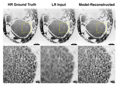 |
Deep learning super-resolution of MR images of the distal tibia improves image quality and assessment of bone microstructure
Trevor Chan1,2, Nada Kamona1,2, Brian-Tinh Vu1,2, Felix Wehrli1,2, and Chamith Rajapakse1,2
1Radiology, University of Pennsylvania, Philadelphia, PA, United States, 2Bioengineering, University of Pennsylvania, Philadelphia, PA, United States
We apply a probabilistic deep learning model to perform image super-resolution on magnetic resonance (MR) images. Our results show that the model is capable of high performance in MR; we upsample low resolution images of the distal tibia to 2x initial spatial resolution–equivalent to capturing 4x fewer samples in K-space–with the goal of reconstructing details in the trabecular architecture. We validate our results by comparing trabecular bone microstructure metrics across high-resolution ground truth, model-reconstructed, and low-resolution input images. By drastically reducing scan time for high-resolution imaging, these methods have the potential to make MRI assessment of bone strength clinically viable.
|
|
| 09:51 | 0408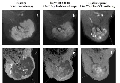 |
Early Chemotherapeutic Response Evaluation In Ewing Sarcoma Using Diffusion Weighted MRI Video Permission Withheld
Esha Baidya Kayal1, Jayendra Tiru Alampally2, Raju Sharma3, Sameer Bakhshi3, Amit Mehndiratta1,3, Rakesh Kumar3, Chandrashekhara SH3, Manisha Jana3, Ashu Seith Bhalla3, Mehar Chand Sharma3, Asit Ranjan Mridha3, Sreenivas Vishnubhatla3, and Devasenathipathy Kandasamy3
1Indian Institute of Technology Delhi, Delhi, India, 2Krishna Imaging & Diagnostics, Telengana, India, 3All India Institute of Medical Sciences Delhi, Delhi, India The objective of this prospective study was to assess the role of diffusion weighted MRI for early evaluation of chemotherapy response in patients with Ewing sarcoma with RECIST 1.1 criteria correlation. Absolute apparent diffusion coefficient (ADC), normalized ADC (nADC) and tumor diameter at baseline and after 1st cycle of chemotherapy were assessed for early detection of chemotherapy response in 16 patients. Results showed, baseline nADC and after 1st chemotherapy cycle, relative percentage change (Δ%) in nADC & tumor-size were significant and observed to be useful markers of early chemotherapeutic response (AUC=0.700, 0.750, 0.938) in Ewing sarcoma. |
|
| 10:03 | 0409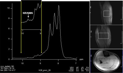 |
Detection of a new resonance in human calf muscle in vivo at 7.0T using the down-field MRS
Ravi Prakash Reddy Nanga1, Mark Elliott1, Neil Wilson1, Sophia Swago2, Walter Witschey1, and Ravinder Reddy1
1Radiology, Center for Advance Metabolic Imaging in Precision Medicine, Perelman School of Medicine at The University of Pennsylvania, Philadelphia, PA, United States, 2Bioengineering, University of Pennsylvania, Philadelphia, PA, United States
We have detected a resonance that was not reported previously, with a chemical shift of ~9.7ppm occurring in down-field MRS (DFMRS) from the human calf muscle in vivo. Based on phantom data, we speculate that the contribution to this broad peak might be from nicotinamide riboside (NR) and nicotinamide mononucleotide (NMN). This is for the first time that this peak is observed and reported from the down-field spectra. It seems to be very specific to calf muscle and has not been identified from the down-field spectra of human brain in vivo, suggesting that it is a feature of muscle metabolism.
|
|
| 10:15 | 0410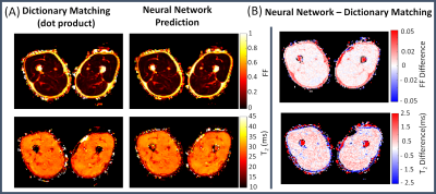 |
A Neural Network Application for Fast Simultaneous Muscle T2-Water and Fat Fraction Mapping from Multi-Spin-Echo Acquisitions
Marco Barbieri1, Melissa T. Hooijmans2, Garry E. Gold1,3, Feliks Kogan1, and Valentina Mazzoli1
1Radiology, Stanford University, Stanford, CA, United States, 2Radiology and Nuclear Medicine, Amsterdam University Medical Center, Amsterdam, Netherlands, 3Bioengineering, Stanford University, Stanford, CA, United States
Muscle T2 relaxometry can be used to monitor disease activity in neuromuscular disorders. Dictionary matching of multi-echo-spin-echo (MESE) data is the gold-standard method to estimate the T2 of the myocitic component (T2-water) because of its ability to correct for multiple confounding factors, but suffers from a high computational burden. This work proposes a neural network (NN) approach for fast muscle T2-water mapping with subject-specific T2-fat calibration to overcome computational limitations of the dictionary method. The method was validated in-vivo against the standard dictionary approach. The NN application outperformed the dictionary approach in computational resources (x140 faster) while retaining quantitative accuracy.
|
|
| 10:27 | 0411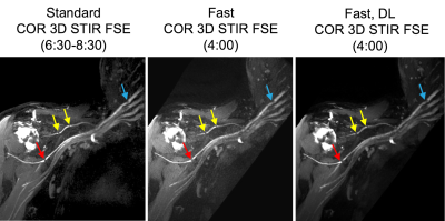 |
Deep Learning Reconstruction-Enabled 2D and 3D MR Neurography
Ek T. Tan1, Yan Wen2, Kang Wang2, Jake A. Fiore1, R. Marc Lebel2, Suryanarayanan Kaushik2, Maggie Fung2, and Darryl B. Sneag1
1Hospital for Special Surgery, New York, NY, United States, 2GE Healthcare, Chicago, IL, United States
Deep learning reconstruction (DLRecon) was applied to shorter acquisition 2D Dixon and 3D short-tau-inversion recovery (STIR) brachial plexus MR neurography (MRN) sequences and were compared both qualitatively and quantitatively to standard clinical sequences. DLRecon 2D and 3D images demonstrated similar quality to standard images. DLRecon may facilitate implementation of shorter MRN protocols, thereby helping streamline MRN’s incorporation into busy clinical practices and providing the option of acquiring additional imaging planes.
|
|
| 10:39 | 0412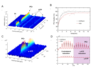 |
Effects of Aging and Muscle Degeneration on Metabolic Response to High-Intensity Muscle Contraction in Mice by Phosphorus-31 MRS
Kihwan Kim1, Yuran Zhu2, Yudu Li3, Zhi-Pei Liang 3,4, and Xin Yu2,5
1Biomedical Engineering, Case Western Reserve University, Cleveland, OH, United States, 2Biomedical Engineering, Case Western Reserve University, cleveland, OH, United States, 3Electrical and Computer Engineering, University of Illinois at Urbana-Champaign, Urbana, IL, United States, 4Beckman Institute for advanced Science and Technology, University of Illinois at Urbana-Champaign, Urbana, IL, United States, 5Case Center for Imaging Research, Case Western Reserve University, cleveland, OH, United States
In this study, 31P-MRS was employed to evaluate the effects of aging and muscle degeneration on metabolic response to stimulation-induced, high-intensity muscle contraction in two age groups of C57BL/6 and mdx mice, a mouse model of moderate muscular dystrophy. Significant, age-dependent differences in mitochondrial oxidative capacity and creatine kinase activity after muscle stimulation were observed between C57BL/6 and mdx mice, suggesting a positive metabolic response to muscle contraction that can be modulated by aging and muscle degeneration.
|
 Back to Meeting Home
 Back to the Program-at-a-Glance
The International Society for Magnetic Resonance in Medicine is accredited by the Accreditation Council for Continuing Medical Education to provide continuing medical education for physicians.
 Back to Meeting Home
 Back to the Program-at-a-Glance
 View the Presentation