Oral Session
Epilepsy: From Animal Models to AI
Joint Annual Meeting ISMRM-ESMRMB & ISMRT 31st Annual Meeting • 07-12 May 2022 • London, UK

Oral Session
Epilepsy: From Animal Models to AI
| 09:15 | 0585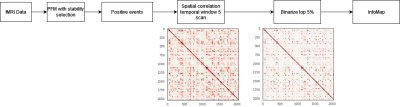 |
Automatic detection of spatio-temporal patterns of interictal epileptic activity with fMRI
Cristina Tobías1, Eneko Uruñuela1, Vicente Ferrer-Gallardo1, Hannah Goldberg2, Christine Engelman2, Mark Lowe2, Stephen E. Jones2, and César Caballero-Gaudes1
1Signal Processing in Neuroimaging, Basque Center on Cognition Brain and Language, San Sebastián, Spain, 2Imaging Institute, Cleveland Clinic, OH, USA., Cleveland, OH, United States
We propose a novel methodology to identify spatio-temporal patterns of interictal epileptic activity in refractory epileptic patients using only BOLD functional MRI. It is based on clustering the activity-inducing signal deconvolved from the fMRI data with the Infomap algorithm. The proposed approach was validated on data acquired during a finger tapping task, and evaluated in four epileptic patients scanned at 7T. This method obtained activation maps that were concordant with the results obtained with EEG-informed GLM and MEG-based dipole source localization in 3 out of 4 patients, demonstrating its potential for mapping epileptic activity based only on fMRI data.
|
|
09:27 |
0586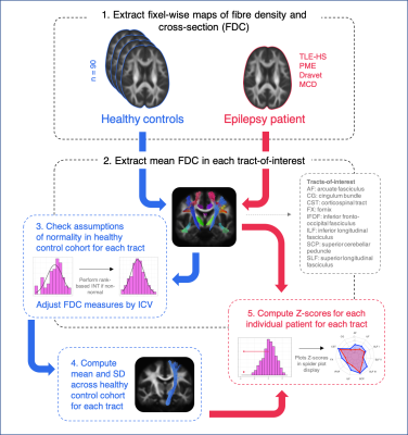 |
Detecting tract-specific abnormalities in individual patients with epilepsy using fixel-based analysis
Remika Mito1, Mangor Pedersen2, Robert Smith1,3, Donna Parker1, Leonid Churilov4, David Vaughan1,3,5, and Graeme Jackson1,3,5
1Florey Institute of Neuroscience and Mental Health, Melbourne, VIC, Australia, 2Department of Psychology and Neuroscience, Auckland University of Technology, Auckland, New Zealand, 3Florey Department of Neuroscience and Mental Health, University of Melbourne, Melbourne, VIC, Australia, 4Melbourne Medical School, University of Melbourne, Melbourne, VIC, Australia, 5Department of Neurology, Austin Health, Melbourne, VIC, Australia
Fibre tract-specific abnormalities have been identified in various neurological disorders and conditions using fixel-based analysis. However, for this technique to be useful in clinical practice, such abnormalities should be detectable in individual patients. In this work, we apply the fixel-based analysis framework to detect fibre density and cross-sectional changes in individual patients with varying epilepsy diagnoses. Abnormalities could be detected in individual epilepsy patients, both at the tract level, and at the fixel-level, with different patterns of tract-based abnormality observed across the different epilepsy syndromes. This information could be clinically valuable when examining individual patients.
|
|
| 09:39 |
0587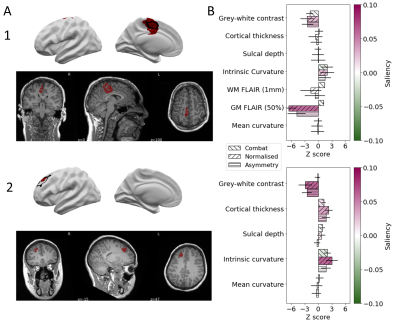 |
Automated lesion prediction and characterisation of focal cortical abnormalities: a MELD study
Konrad Wagstyl1, Mathilde Ripart2, MELD Consortium3, Hannah Spitzer4, Torsten Baldeweg2, and Sophie Adler2
1Wellcome Centre for Human Neuroimaging, UCL, London, United Kingdom, 2Great Ormond Street Institute for Child Health, UCL, London, United Kingdom, 3UCL, London, United Kingdom, 4Institute of Computational Biology, Helmholtz Zentrum München, Munich, Germany
Identification of subtle epilepsy-causing focal cortical dysplasias (FCD) on MRI remains an outstanding challenge during presurgical assessment of patients. The Multi-centre Epilepsy Lesion Detection (MELD) project created an MRI lesion detection algorithm using a large, heterogenous cohort. The algorithm had a sensitivity of 67%, performing well on unseen test sites. Analysis of MRI lesions revealed distinct subgroups, with differing histopathological subtypes, imaging features and detection rates. Individual patient reports highlight a predicted lesion’s location, imaging features and their relative saliency to the classifier. This tool has the potential to improve presurgical lesion identification on MRI from patients with epileptogenic FCD.
|
|
| 09:51 | 0588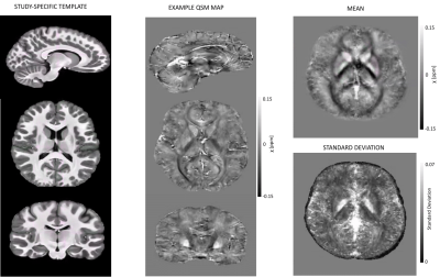 |
Ultra-high field normative quantitative susceptibility mapping (QSM) in children and cortical pathology in drug-resistant focal epilepsy
Chiara Casella1,2, Katy Vecchiato1,2,3, Ayse Sila Dokumaci2,4, Philippa Bridgen2,4, Shaihan Malik2,4, Joseph V Hajnal2,4, Sharon Giles2,4, Jan Sedlacik1,2,4,5, Karin Shmueli2,6, Tom Wilkinson2,4, Raphael Tomi-Tricot2,4,7, David W Carmichael2,4, and Jonathan O'Muircheartaigh1,2,3,8
1Centre for the Developing Brain, School of Biomedical Engineering and Imaging Sciences, King's College London, London, United Kingdom, 2London Collaborative Ultra high field System (LoCUS), London, United Kingdom, 3Department for Forensic and Neurodevelopmental Sciences, Institute of Psychiatry, Psychology and Neuroscience, King's College London, London, United Kingdom, 4School of Biomedical Engineering and Imaging Sciences, King's College London, London, United Kingdom, 5Radiology Department, Great Ormond Street Hospital for Children, London, United Kingdom, 6Department of Medical Physics and Biomedical Engineering, University College London, London, United Kingdom, 7MR Research Collaborations, Siemens Healthcare Limited, Frimley, United Kingdom, 8MRC Centre for Neurodevelopmental Disorders, London, United Kingdom
We developed an analysis pipeline to explore normative QSM values in children and adolescents at 7T, and assessed the feasibility of this approach for detecting tissue-alterations in paediatric drug-resistant epilepsy. Normative values consistent with previous QSM studies were reported for deep brain regions, and normative cortical susceptibility values were described for the first time in children and adolescents at 7T. Finally, sensitivity to susceptibility changes in epileptogenic lesions was demonstrated using the analysis pipeline.
|
|
| 10:03 | 0589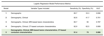 |
Multimodal Lesion Phenotyping Improves Seizure Outcome Classification after Traumatic Brain Injury: An EpiBioS4Rx Study
Rachael Garner1, Alexis Bennett1, Akul Sharma1, Michael Douglas Morris2, Marianna La Rocca1,3, Giuseppe Barisano1, Ruskin Cua4, Paul Vespa2, Arthur W Toga1, and Dominique Duncan1
1USC Stevens Neuroimaging and Informatics Institute, Keck School of Medicine, University of Southern California, Los Angeles, CA, United States, 2David Geffen School of Medicine, University of California, Los Angeles, Los Angeles, CA, United States, 3Dipartimento Interateneo di Fisica, Università di Bari, Bari, Italy, 4USC Department of Radiology, Keck School of Medicine of USC, University of Southern California, Los Angeles, CA, United States
Posttraumatic epilepsy (PTE), or recurrent seizures after traumatic brain injury (TBI), is a debilitating complication of TBI. We present a multimodal approach to classify seizure outcomes using computed tomography (CT) and magnetic resonance imaging (MRI) features that characterize lesion phenotypes. Five logistic regression models to predict seizure outcome are presented, using patient demographics and clinical information in conjunction with CT and MRI variables that describe lesion characteristics including contusion type and location as well as whole-brain lesion volumetrics. The optimal model utilized all four categories of features, yielding 91.4% sensitivity, 75% specificity, and 0.886 area under the curve performance.
|
|
| 10:15 | 0590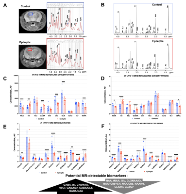 |
Non-invasive metabolic biomarkers of interneuron cell transplantation therapy in an epilepsy model
Marina Radoul1,2, Kai Qiao1,2, Donghyun Hong2, Mansi B. Parekh3, Philip Hampel3, Eric Steven Sevilla3, David Traver3, Hannah Kim3, Marina Bershteyn3, Yves Maury3, Catherine Priest3, Cory R. Nicholas3, and Myriam M. Chaumeil1,2
1Department of Physical Therapy and Rehabilitation Science, University of California San Francisco, San Francisco, CA, United States, 2Department of Radiology and Biomedical Imaging, University of California San Francisco, San Francisco, CA, United States, 3Neurona Therapeutics, San Francisco, CA, United States
Interneuron transplantation targeting GABAergic network dysfunction is a novel alternative therapeutic approach for chronic focal epilepsy. However, in vivo monitoring of response to such cellular therapeutics remains challenging. Here, we combined high resolution in vivo and ex vivo 1H MRS to investigate the metabolic alterations in epileptic hippocampus in a preclinical model of chronic mesial temporal lobe epilepsy, and the effect of human inhibitory interneuron transplantation on metabolism. We discovered that hippocampal levels of GABA, NAA/Cr, GLX/Cr, GABA/GLX and GABA/NAA were reversed to healthy control levels in epileptic mice transplanted with inhibitory interneurons, providing unique biomarkers of therapeutic response.
|
 Back to Meeting Home
 Back to the Program-at-a-Glance
The International Society for Magnetic Resonance in Medicine is accredited by the Accreditation Council for Continuing Medical Education to provide continuing medical education for physicians.
 Back to Meeting Home
 Back to the Program-at-a-Glance
 View the Presentation