Oral Session
Multiple Sclerosis
Joint Annual Meeting ISMRM-ESMRMB & ISMRT 31st Annual Meeting • 07-12 May 2022 • London, UK

| 17:00 | 0134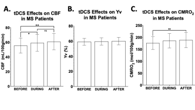 |
Neuronal Response to Transcranial Direct Current Stimulation (tDCS) in Multiple Sclerosis
Marco Muccio1, Lillian Walton Masters2, Giuseppina Pilloni2, Lauren Krupp2, Abhishek Datta3, Marom Bikson4, Leigh Charvet2, and Yulin Ge1
1Radiology, NYU Grossman School of Medicine, New York City, NY, United States, 2Neurology, NYU Grossman School of Medicine, New York City, NY, United States, 3Soterix Medical Inc., New York City, NY, United States, 4Department of Biomedical Engineering, City College of New York, New York City, NY, United States
Transcranial direct current stimulation (tDCS) represents an innovative therapeutic tool for neurological diseases such as multiple sclerosis (MS). In this study, MRI measurements of cerebral blood flow (CBF), venous oxygenation (Yv) and cerebral metabolic rate of oxygen (CMRO2) of MS patients and healthy controls (HCs) were acquired pre-, during- and post-tDCS to investigate its simultaneous effects. Whilst HCs showed an immediate increase (~5%) in CMRO2 (during- versus pre-tDCS, p<0.001), MS patients showed a delayed response to tDCS (7.7% CMRO2 increase pre- versus post-tDCS; p=0.007). We suggest that neuronal response to tDCS represents a potential biomarker of neuronal plasticity.
|
|
| 17:12 | 0135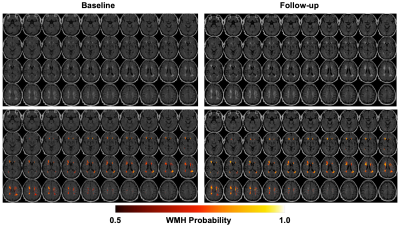 |
Longitudinal Changes of White Matter Hyperintensities Following Hemodialysis Initiation in Old Adults: A Prospective Pilot Study Video Permission Withheld
Xiufeng Li1, Gregory J. Metzger1, and Anne M. Murray2,3,4
1Center for Magnetic Resonance Research, University of Minnesota, Minneapolis, MN, United States, 2Hennepin HealthCare Research Institute, Hennepin Healthcare, Minneapolis, MN, United States, 3Department of Medicine, University of Minnesota, Minneapolis, MN, United States, 4Geriatrics Division, Department of Medicine, Hennepin Healthcare, Minneapolis, MN, United States
Repeated conventional hemodialysis may result in permanent cerebrovascular dysfunction that can initiate and exacerbate other cerebral structural abnormalities. Until today, the longitudinal WMH burden changes in old end-stage renal disease (ESRD) patients following the initiation of hemodialysis treatment have not been specifically studied. We performed a prospective pilot study with a hypothesis that WMH burden increases following the initiation of hemodialysis in old ESRD patient. Our study results suggested that WMH burdens increased significantly after the initiation of hemodialysis treatment.
|
|
17:24 |
0136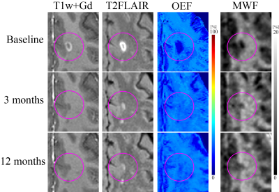 |
Increased oxygen extraction fraction following acute multiple sclerosis (MS) lesion formation is associated with increased myelin repair.
Junghun Cho1, Thanh D Nguyen1, Lily Zexter1, Elizabeth M Sweeney2, Pascal Spincemaille1, Ajay Gupta1, Susan A Gauthier1, and Yi Wang1,3
1Radiology, Weill Cornell Medicine, New York, NY, United States, 2Population Health Sciences, Weill Cornell Medicine, New York, NY, United States, 3Biomedical Engineering, Cornell University, Ithaca, NY, United States
Understanding the cause and progress of remyelination in MS is critical for developing potential therapeutic targets of remyelination to restore neural connectivity and brain functions. In this study, we utilized a novel MRI-based oxygen extraction fraction (OEF) mapping method, namely “QQ”, and found that early lesion oxygen metabolism increase, as measured by QQ-based OEF, is positively associated with lesion myelin recovery, as measured by myelin water fraction. This study suggests that QQ-based OEF mapping may be a useful tool readily and widely available for studying MS lesion oxygen metabolism and its association with MS lesion remyelination.
|
|
| 17:36 |
0137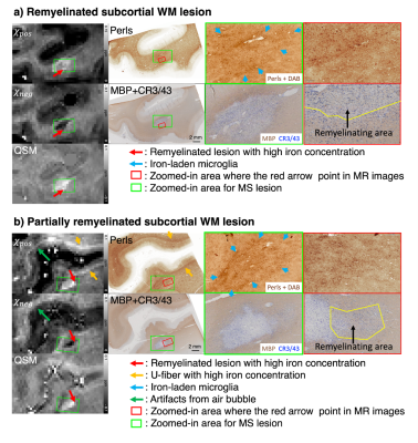 |
Imaging multiple sclerosis histopathology using susceptibility source separation: a postmortem brain study
Hyeong-Geol Shin1, Riccardo Galbusera2,3, Jincheol Seo4, Sooyeon Ji1, Erik Bahn5, Jonas Franz5, Christine Stadelmann-Nessler5, Po-Jui Lu2, Jinhee Jang6, Youngjeon Lee4, Cristina Granziera2, and Jongho Lee1
1Department of Electrical and Computer engineering, Seoul National University, Seoul, Korea, Republic of, 2Department of Biomedical Engineering, Faculty of Medicine, University Hospital Basel and University of Basel, Basel, Switzerland, 3MS Center and Research Center for Clinical Neuroimmunology and Neuroscience Basel (RC2NB), University Hospital Basel and University of Basel, Basel, Switzerland, 4National Primate Research Center, Korea Research Institute of Bioscience and Biotechnology, Cheongju, Korea, Republic of, 5Institute of Neuropathology, University Medical Center, Göttingen, Germany, 6Seoul St Mary’s Hospital, College of Medicine, The Catholic University of Korea, Seoul, Korea, Republic of The pathology of multiple sclerosis (MS) is highly correlated with the dynamics of two susceptibility sources, paramagnetic iron and diamagnetic myelin. Various MRI methods sensitive to the substances have been developed for MS pathology. However, the collective effects of iron and myelin to an MRI signal have hampered monitoring individual changes of the substances. Here, we evaluated the effectiveness of 𝜒-separation, which estimates individual contributions of para-/dia-magnetic susceptibility, for visualizing MS pathology-related iron/myelin changes. The resulting paramagnetic and diamagnetic susceptibility conform to the histopathological features of MS lesion (iron-rim in Perls staining, and re-/de-myelinated lesions in myelin basic protein staining). |
|
| 17:48 | 0138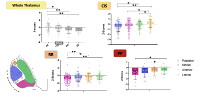 |
Looking for a differential vulnerability of specific thalamic nuclei in multiple sclerosis
Simon Blyau1, Ismail Koubiyr1, Pierrick Coupé2, Manoj Saranathan3, Julie Charré-Morin4, Aurore Saubusse4, Valentin Prevost5, Bei Zhang6, Mathilde Deloire4, Bruno Brochet1, Aurélie Ruet1, and Thomas Tourdias1
1Neurocentre Magendie, Bordeaux, France, 2Laboratoire Bordelais de Recherche en Informatique, TALENCE, France, 3University of Arizona, Tucson, AZ, United States, 4Neurology Department, Hopital Pellegrin, Bordeaux, France, Metropolitan, 5Canon Medical System Corporation, Tochigi, Japan, 6Canon Medical System Europe, Paris, France
Thalamus is one of the first grey matter structure affected in multiple sclerosis (MS). It could undergo a differential vulnerability predominating on nuclei closer to the third ventricle due to pro-inflammatory factors brought by cerebrospinal fluid.First, we validated an atlas-based segmentation method that enables thalamic segmentation over conventional T1 weighted images. Then, using this segmentation method, we brought evidence for a medio-lateral gradient of thalamic atrophy in MS. While less affected, the anterior group was found useful to distinguish cognitively preserved from cognitively impaired patients. This paves the way toward more thalamic nuclei analyses in MS as new biomarkers.
|
|
18:00 |
0139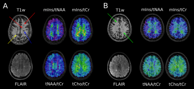 |
Lesion-specific Metabolic Fingerprinting in Relapsing-Remitting Multiple Sclerosis via 7T Magnetic Resonance Spectroscopic Imaging
Alexandra Lipka1, Eva Heckova1, Assunta Dal-Bianco2, Gilbert Hangel1,3, Paulus S. Rommer2, Bernhard Strasser1, Stanislav Motyka1, Lukas Hingerl1, Thomas Berger2, Petra Hnilicová4, Ema Kantorová5, Fritz Leutmezer2, Egon Kurča5, Stephan Gruber1, Siegfried Trattnig1,6, and Wolfgang Bogner1
1High Field MR Centre, Department of Biomedical Imaging and Image-guided Therapy, Medical University of Vienna, Vienna, Austria, 2Department of Neurology, Medical University of Vienna, Vienna, Austria, Vienna, Austria, 3Department of Neurosurgery, Medical University of Vienna, Vienna, Austria, Vienna, Austria, 4Biomedical Center Martin, Jessenius Faculty of Medicine in Martin, Comenius University in Bratislava, Martin, Slovakia, 5Clinic of Neurology, Jessenius Faculty of Medicine in Martin, Comenius University in Bratislava, Martin, Slovakia, 6Karl Landsteiner Institute for Clinical Molecular MRI in Musculoskeletal System, Vienna, Austria Conventional T1/T2-weighted magnetic resonance imaging (cMRI) is the method of choice for diagnosis and treatment monitoring of multiple sclerosis (MS), though not being able to explain underlying pathological processes. In contrast to cMRI-lesions, which only reflect the severity of irreversible tissue damage, MR Spectroscopic Imaging (MRSI) can detect pathologies on a biochemical level. In 51 relapsing-remitting (RRMS) patients, we investigate - enabled through ultra-high resolution FID-MRSI at 7T - the metabolic distribution within lesions and their vicinity as well as their location dependency and correlation to T1-hypointensity.
|
 Back to Meeting Home
 Back to the Program-at-a-Glance
The International Society for Magnetic Resonance in Medicine is accredited by the Accreditation Council for Continuing Medical Education to provide continuing medical education for physicians.
 Back to Meeting Home
 Back to the Program-at-a-Glance
 View the Presentation