Oral Session
Imaging of the Spinal Cord
Joint Annual Meeting ISMRM-ESMRMB & ISMRT 31st Annual Meeting • 07-12 May 2022 • London, UK

09:15 |
0389 |
A 3T/7T multiparametric protocol to characterize amyotrophic lateral sclerosis cervical spinal cord: benefits of ultra-high field MR imaging?
Samira Mchinda1,2,3, Rémi Dintrich1,2, Arnaud Le Troter1,2, Pini Lauriane1,2, Shahram Attarian4, Annie Vershueren4, and Virginie Callot1,2,3
1Aix-Marseille Univ, CNRS, CRMBM, Marseille, France, Marseille, France, 2APHM, Hopital Universitaire Timone, CEMEREM, Marseille, France, Marseille, France, 3iLab-Spine International Associated Laboratory, Marseille-Montreal, France-Canada, Marseille, France, 4APHM, Hôpital Universitaire Timone, Reference Center for Neuromuscular Disorders and ALS, Marseille, France, Marseille, France With comparable acquisition times between 3T and 7T, 7T quantitative protocol offers better resolution and many opportunities to study regional impairments, especially in anterior gray matter subregions. This preliminary study focused on ALS and age-matched healthy controls scanned at both 3T and 7T. With the current postprocessing, abnormalities were mostly detected on the lower part of the cord (GM atrophy, WM/GM T1 impairments). Future work will focus on longitudinal evolution and refined analysis to evaluate the sensitivity for prognostication and to determine individual topological evolution. |
|
| 09:27 | 0390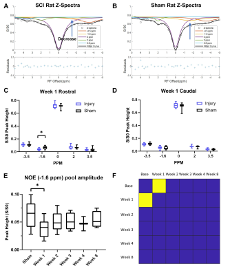 |
Molecular MRI and PET Imaging Detect Biomarkers of Neuroinflammation in Contusion Injured Spinal Cord
Chaoqi Mu1, Jamie L Reed1, Mohammed Noor Tantawy1, Feng Wang1, Li Min Chen1, and John C Gore1
1Vanderbilt University Institute of Imaging Science & Department of Radiology and Radiological Sciences, Vanderbilt University Medical Center, Nashville, TN, United States
Spinal cord injury (SCI) severity/recovery are influenced by complex pathological mechanisms. We used CEST/NOE MR and TSPO PET imaging to evaluate longitudinal molecular changes associated with neuroinflammation in a lumbar contusion SCI rodent model. The NOE(-1.6 ppm) peak amplitude significantly decreased and the CEST(3.5 ppm) peak amplitude increased in SCI rats, 1-week post-injury. Similarly, we detected significant increase in the uptake of a TSPO-targeting PET radiotracer at the SCI epicenter. The CEST/NOE pools can be linked to neuroinflammatory activity associated with glutamate release. The results indicate that CEST/NOE MRI measurements provide complementary information to TSPO PET measurement in SCI.
|
|
| 09:39 | 0391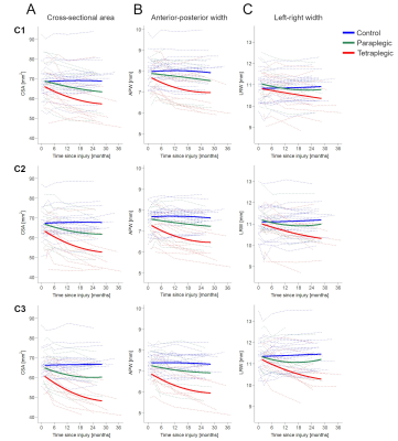 |
Spatiotemporal dynamics of anterograde and retrograde neurodegeneration following spinal cord injury
Simon Schading1, Gergely David1, Tim Max Emmenegger1, Cristian Achim1, Armin Curt1, and Patrick Freund1,2,3
1Spinal Cord Injury Center, Balgrist University Hospital, Zürich, Switzerland, 2Wellcome Trust Centre for Neuroimaging, UCL Institute of Neurology, London, United Kingdom, 3Department of Neurophysics, Max Planck Institute for Human Cognitive and Brain Sciences, Leipzig, Germany We tracked remote neurodegenerative changes in the cervical cord following spinal cord injury over 2 years using macrostructural readouts (cross-sectional area, left-right width, anterior-posterior width) derived from T1-weighted MPRAGE images. Thereby, we evaluated the dependency of the magnitude of atrophy on the distance to the lesion. Over time, differences in atrophy rates along the cervical cord rostral to the lesion gradually formed a lesion gradient. Using anterior-posterior width and left-right width as surrogates for investigating anterograde and retrograde degeneration separately, our results suggest that these two types exhibit different spatiotemporal dynamics. A lesion gradient was only observable for retrograde degeneration. |
|
| 09:51 | 0392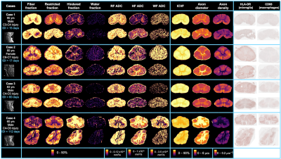 |
Identifying diffusion model biomarkers for inflammation in human traumatic spinal cord injury
Sarah Rosemary Morris1,2,3, Taylor Swift-LaPointe2, Andrew Yung1,3,4, Valentin Prevost1,3,4, Shana George1, Andrew Bauman4, Piotr Kozlowski1,2,3,4, Farah Samadi1,5, Caron Fournier1,5, Lisa Parker6, Kevin Dong1, Femke Streijger1, Veronica Hirsch-Reinshagen1,5,6, G.R. Wayne Wayne Moore1,5,6, Brian K Kwon1,7, and Cornelia Laule1,2,3,5
1International Collaboration on Repair Discoveries (ICORD), Vancouver, BC, Canada, 2Physics & Astronomy, University of British Columbia, Vancouver, BC, Canada, 3Radiology, University of British Columbia, Vancouver, BC, Canada, 4Faculty of Medicine, UBC MRI Research Centre, Vancouver, BC, Canada, 5Pathology & Laboratory Medicine, University of British Columbia, Vancouver, BC, Canada, 6Vancouver General Hospital, Vancouver, BC, Canada, 7Orthopaedics, University of British Columbia, Vancouver, BC, Canada We investigated the relationship between diffusion model biomarkers and inflammatory cells, as measured by HLA-DR and CD68 histological staining for activated microglia and macrophages, after human traumatic spinal cord injury. Fractional anisotropy decreases and radial and mean diffusivity increases were strongly correlated with the presence of activated microglia and macrophages. ActiveAx-derived intra-cellular volume fraction and axon density decreased while axon diameter increased. Fiber fraction from Diffusion Basis Spectrum Imaging decreased and while hindered fraction increased in the presence of inflammation. |
|
| 10:03 | 0393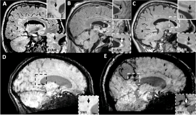 |
Corpus callosum lesions are associated with worse cognitive performance in cerebral amyloid angiopathy
Whitney Freeze1, Maria Clara Zanon Zotin2,3, Ashley Scherlek2, Valentina Perosa2, Andrew Warren2, Louise van der Weerd1, Dorothee Schoemaker Marcotte2, Mitchell Horn2, Edip Gurol2, Elif Gokcal2, Brian Bacskai2, Anand Viswanathan2, Steven Greenberg2, Yael Reijmer4, and Susanne van Veluw1,2
1Leiden University Medical Center, Leiden, Netherlands, 2Massachusetts General Hospital, Boston, MA, United States, 3Ribeirão Preto medical School, São Paulo, Brazil, 4University Medical Center Utrecht, Utrecht, Netherlands
We assessed the contribution of small vessel disease lesions in the corpus callosum (CC) to vascular cognitive impairment in cerebral amyloid angiopathy (CAA). A total number of 21 CC lesions was found in 19/65 (29%) CAA patients. CC lesion presence was associated with reduced microstructural white matter integrity within the CC and in the whole brain white matter. Patients with CC lesions performed significantly worse on multiple cognitive domains compared to those without CC lesions after correcting for relevant covariates. Together, our findings suggest that CC lesions independently contribute to cognitive impairment through strategic microstructural disruption of white matter tracts.
|
|
| 10:15 | 0394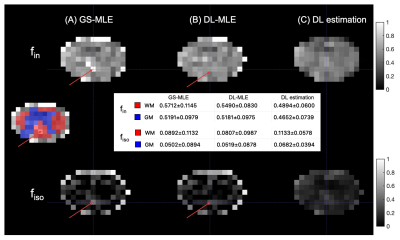 |
Deep-learning-informed parameter estimation improves reliability of spinal cord diffusion MRI
Ting Gong1, Francesco Grussu2,3, Claudia AM Gandini Wheeler-Kingshott4,5,6, Daniel C Alexander1, and Hui Zhang1
1Centre for Medical Image Computing, Department of Computer Science, University College London, London, United Kingdom, 2Radiomics Group, Vall d’Hebron Institute of Oncology, Vall d’Hebron Barcelona Hospital Campus, Barcelona, Spain, 3Queen Square MS Centre, Queen Square Institute of Neurology, Faculty of Brain Sciences, University College London, London, United Kingdom, 4NMR Research Unit, Queen Square Multiple Sclerosis Centre, Department of Neuroinflammation, UCL Queen Square Institute of Neurology, University College London, London, United Kingdom, 5Brain Connectivity Centre, IRCCS Mondino Foundation, Pavia, Italy, 6Department of Brain and Behavioural Sciences, University of Pavia, Pavia, Italy
The very low SNR in spinal cord diffusion MRI can make robust microstructure parameter estimation challenging. To address unreliable parameter estimations under such low SNR, we propose a deep-learning-informed maximum likelihood estimation (MLE) approach, where a deep-learning model is trained to initialise MLE efficiently and optimally. In testing NODDI-derived parameters, simulation and in vivo experiments suggest the DL-informed method can reduce outlier estimates from conventional grid-search MLE, and at the same time, avoid biases from pure DL estimation under the low SNR. The proposed method also speeds up the computation, making it a promising tool for future applications.
|
|
| 10:27 | 0395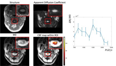 |
Measuring Spinal Cord Blood Flow with Multi-delay pseudo Continuous Arterial Spinal Labeling (pCASL)
Qinyang Shou1, Xingfeng Shao1, and Danny JJ Wang1
1Laboratory of Functional MRI Technology (LOFT), Stevens Neuroimaging and Informatics Institute, University of Southern California, Los Angeles, CA, United States
The purpose of this study was to measure the spinal cord blood flow using high-resolution multi-delay pCASL. An optimized background suppression scheme was applied to suppress the cerebrospinal fluid signal. In-vivo study was performed on 5 healthy subjects. The results show stable cervical spinal cord (C1-C3) blood flow values within the spinal cord ROI, supporting the potential for further development and evaluation studies on noninvasive MRI of spinal cord perfusion.
|
|
| 10:39 | 0396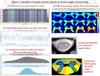 |
Neptune: a toolbox for spinal cord functional MRI data processing and quality assurance Video Not Available
D Rangaprakash1 and Robert L Barry1,2
1Athinoula A. Martinos Center for Biomedical Imaging, Massachusetts General Hospital, Harvard Medical School, Boston, MA, United States, 2Harvard-Massachusetts Institute of Technology Division of Health Sciences & Technology, Cambridge, MA, United States
Spinal cord fMRI data analysis involves niche pre- and post-processing steps due to the cord’s unique anatomy and higher distortions/physiological noise in fMRI data, requiring extensive and careful quality control (QC). However, there is paucity of open-source tools tailored to this need. Building upon 10 years of research and development, we present Neptune, a user-interface-based MATLAB toolbox. It can perform an array of tailored pre- and post-processing steps, including functional connectivity and statistics. It generates extensive QC plots and presents them to view in an elegant webpage format. We demonstrate the utility of Neptune on a 7T dataset.
|
 Back to Meeting Home
 Back to the Program-at-a-Glance
The International Society for Magnetic Resonance in Medicine is accredited by the Accreditation Council for Continuing Medical Education to provide continuing medical education for physicians.
 Back to Meeting Home
 Back to the Program-at-a-Glance
 View the Presentation