Oral Session
Advances in Quantitative Neuroimaging
Joint Annual Meeting ISMRM-ESMRMB & ISMRT 31st Annual Meeting • 07-12 May 2022 • London, UK

Oral Session
Advances in Quantitative Neuroimaging
09:15 |
0591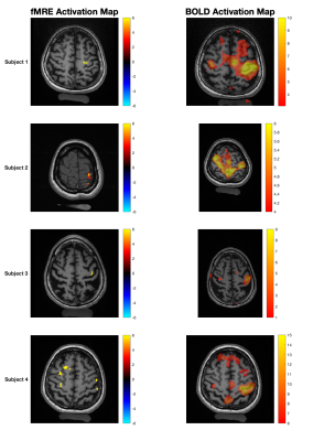 |
Imaging Neuronal Activity at Fast Timescales in Humans using MR Elastography
Shruti Mishra1,2, Bin Deng2,3,4, W. Scott Hoge1,2,3, Yanmei Tie2,5, Giacomo Annio6, Ralph Sinkus6, and Samuel Patz1,2
1Radiology, Brigham & Women's Hospital, Boston, MA, United States, 2Harvard Medical School, Boston, MA, United States, 3Athinoula A. Martinos Center for Biomedical Imaging, Charlestown, MA, United States, 4Radiology, Massachusetts General Hospital, Boston, MA, United States, 5Neurosurgery, Brigham & Women's Hospital, Boston, MA, United States, 6Laboratory for Vascular Translational Science (LVTS), Institut National de la Santé et de la Recherche Médicale (INSERM), Paris, France A stimulus-interleaved magnetic resonance elastography (MRE) sequence was utilized to demonstrate fast functionally mediated localized changes in shear wavelength during a motor task. Four healthy adult subjects underwent a visual-cue mediated right-hand finger-tapping task, switching between tapping and no-tapping blocks every 2 seconds. Areas of greatest significance between the stimulus states were localized to the left primary motor cortex. A decreased stiffness of ~30% was observed during task performance compared to rest. Compared to traditional BOLD fMRI with a 20-second block, functional MRE areas of greatest statistical difference demonstrated greater percentage change and greater spatial localization. |
|
| 09:27 | 0592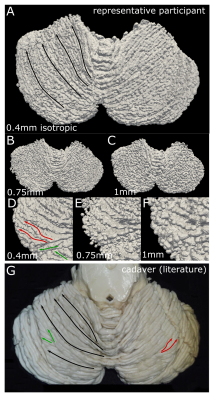 |
Ultra-high field motion-corrected MRI can visualize the mesoscale organization of the human cerebellum.
Nikos Priovoulos1, Mads Andersen2,3, Vincent Oltman Boer4, and Wietske van der Zwaag1
1Spinoza Center for Neuroimaging, Amsterdam, Netherlands, 2Philips Healthcare, Copenhagen, Denmark, 3Lund University Bioimaging Center, Lund University, Lund, Sweden, 4Danish Research Centre for Magnetic Resonance, Centre for Functional and Diagnostic Imaging and Research, Copenhagen University Hospital Amager and Hvidovre, Copenhagen, Denmark
The cerebellum is an important but challenging to image part of the human brain, due to its thin and highly-folded cortex. Here we demonstrate that using motion-corrected high-resolution 7T-MRI data, we can derive segmentations and surfaces of the cerebellar cortex with unprecedented accuracy in vivo. We expect this technique to greatly facilitate cognitive and clinical neuroscience research in the cerebellum.
|
|
| 09:39 | 0593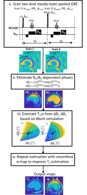 |
TWo Interleaved Steady-states for T2 And Rf Estimation (TWISTARE) - a fast 3D-GRE phase-based method of T2 and B1 mapping in 7T brain imaging
Dana Yacobi1, Amir Seginer2, and Rita Schmidt1
1Department of Brain Sciences, Weizmann Institute of Science, Rehovot, Israel, 2Siemens Healthcare Ltd, Rosh Ha’ayin, Israel Quantitative T2 mapping is an important tool towards standardization of MRI in clinics and research, whose implementation in 7T MRI will gain SNR and resolution. In this work, we demonstrate a phase-based method with fast 3D spoiled GRE acquisition to estimate T2 and B1 maps in 7T brain imaging. We introduce a new approach employing a dual steady-state with interleaving flip angles - denoted as TWISTARE. This approach is suited to address the RF field inhomogeneity in 7T and can shorten the total scan duration. The new approach was examined using a 3D head-shaped phantom and in human imaging. |
|
| 09:51 | 0594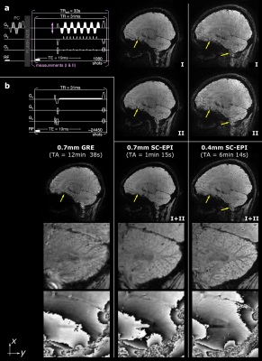 |
T2*-weighted dual-polarity skipped-CAIPI 3D-EPI: 400 microns isotropic whole-brain QSM at 7 Tesla in 6 minutes
Rüdiger Stirnberg1, Andreas Deistung2, and Tony Stöcker1,3
1German Center for Neurodegenerative Diseases (DZNE), Bonn, Germany, 2University Clinic and Outpatient Clinic for Radiology, University Hospital Halle (Saale), Halle, Germany, 3Department of Physics and Astronomy, University of Bonn, Bonn, Germany
We propose a dual-polarity readout technique to eliminate residual segmentation artifacts in interleaved multi-shot EPI despite established echo time shifting. The method only requires retrospective averaging of two or more complex-valued images acquired with alternating EPI readout polarity across measurements. The approach has been integrated into a custom skipped-CAIPI 3D-EPI sequence, combining highly segmented EPI with CAIPIRINHA parallel imaging. We present dual-polarity skipped-CAIPI 3D-EPI for ultrahigh-resolution, rapid, motion-robust whole-brain T2*-weighted imaging and quantitative susceptibility mapping at 7 Tesla. We demonstrate its capability to acquire exquisite imaging contrast at 0.7mm and 0.4mm isotropic resolution in only 1:15 and 6:14 [min:s], respectively.
|
|
| 10:03 |
0595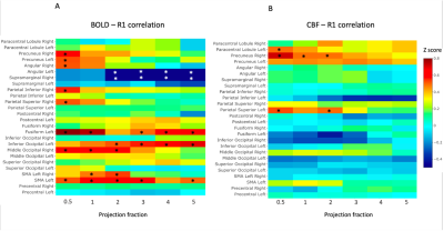 |
Investigating the relationship between cortical myelin and functional brain activity
Eleonora Patitucci1, Michael Germuska1, Valentina Tomassini2,3,4,5,6, and Richard G Wise1,2,3
1School of Psychology, CUBRIC, Cardiff University, Cardiff, United Kingdom, 2Department of Neuroscience, Imaging and Clinical Sciences, University “G. d'Annunzio” of Chieti-Pescara, Chieti, Italy, 3Institute for Advanced Biomedical Technologies, University “G. d'Annunzio” of Chieti-Pescara, Chieti, Italy, 4MS Centre, Neurology Unit, “SS. Annunziata” University Hospital, Chieti, Italy, 5Division of Psychological Medicine and Clinical Neurosciences, School of Medicine, Cardiff University, Cardiff, United Kingdom, 6Helen Durham Centre for Neuroinflammation, University Hospital of Wales, Cardiff, United Kingdom
The role of cortical myelin for cortical function is still under debate. Here we investigate the relationship between cortical myelination (represented by R1) and functional activation (represented by BOLD-fMRI and arterial spin labelling CBF signals) during the execution of a motor task. We demonstrate associations between cortical myelination and functional activity in a subset of task-responding regions. We observe a decrease of the significance of these relationships in the deeper cortical layers.
|
|
| 10:15 | 0596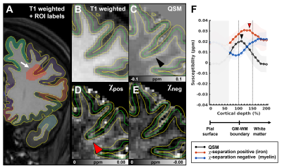 |
Laminar profiling in advanced susceptibility imaging reveals variations in iron and myelin concentrations
Subin Lee1, Hyeong-Geol Shin1, and Jongho Lee1
1Department of Electrical and Computer Engineering, Seoul National University, Seoul, Korea, Republic of The in-vivo imaging of iron and myelin concentrations along cortical laminae in the brain has significance in advancing knowledge about the roles of iron and myelin in brain function and in disease. In this study, we apply χ-separation, a recently developed advanced susceptibility imaging method, to explore changes in iron and myelin content throughout the cortex and into the white matter. Our findings show that iron and myelin peak at different laminar surfaces and that there is regional differences in the intensity profiles. The results suggest regional and laminar variation of iron and myelin throughout the brain. |
|
| 10:27 | 0597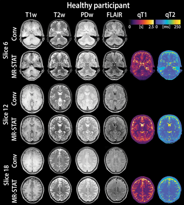 |
Synthetic MRI with MR-STAT: results from a clinical trial
Jordi P.D. Kleinloog1,2, Stefano Mandija1,2, Federico D'Agata3, Oscar van der Heide1,2, Beyza Koktas1, Sarah M. Jacobs4, Cornelis A.T. van den Berg1,2, Jeroen Hendrikse2, Anja G. van der Kolk2, and Alessandro Sbrizzi1,2
1Computational Imaging Group for MR diagnostic and therapy, Center for Image Sciences, University Medical Center Utrecht, Utrecht, Netherlands, 2Department of Radiotherapy, University Medical Centre Utrecht, Utrecht, Netherlands, 3Department of Neurosciences, University of Turin, Turin, Italy, 4Department of Radiology and Nuclear Medicine, University Medical Centre Utrecht, Utrecht, Netherlands Magnetic Resonance Spin TomogrAphy in Time-domain (MR-STAT) reconstructs multiple quantitative MR parameters from a single fast scan. This quantitative information can be leveraged for several purposes, including the synthetization of clinically desired image contrasts. Preliminary results of the first clinical trial using MR-STAT in patients with neurological diseases show that the synthetically generated contrast images (i.e., T1w, T2w, PDw and FLAIR) were acceptable for diagnostic use (5-point Likert scale ≥ 3), although the score was lower compared to conventional contrasts. This highlights the potential of MR-STAT to provide synthetic contrasts on top of quantitative maps, while reducing scan time. |
 Back to Meeting Home
 Back to the Program-at-a-Glance
The International Society for Magnetic Resonance in Medicine is accredited by the Accreditation Council for Continuing Medical Education to provide continuing medical education for physicians.
 Back to Meeting Home
 Back to the Program-at-a-Glance
 View the Presentation