Oral Session
Quantitative Neuroimaging Across the Human Lifespan
Joint Annual Meeting ISMRM-ESMRMB & ISMRT 31st Annual Meeting • 07-12 May 2022 • London, UK

Oral Session
Quantitative Neuroimaging Across the Human Lifespan
| 14:45 | 0065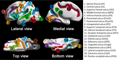 |
Quantification of sulcal emergence in the fetal brain: hemispheric asymmetry and sex difference
Hyuk Jin Yun1, Hyun Ju Lee2, Joo Young Lee2, Tomo Tarui3, Caitlin K. Rollins1, Cynthia M. Ortinau4, Henry A. Feldman1, P. Ellen Grant1, and Kiho Im1
1Boston Children's Hospital, Boston, MA, United States, 2Hanyang University, Seoul, Korea, Republic of, 3Tufts Medical Center, Boston, MA, United States, 4Washington University in St. Louis, St. Louis, MO, United States
Sulcal emergence is important to assess normality of early fetal brain development. Thus, we proposed a quantitative approach to build an accurate timetable of sulcal emergence. Using a large sample of fetuses, we automatically extracted cortical surfaces and detected presence and absences of each sulcus. By fitting a logistic curve to sulcal presence/absence, the timing and inter-subject variability of sulcal emergence were estimated. We also found hemispheric asymmetry and sex difference in the timing and variability of sulcal emergence. Our quantitative timetable concurs with previous reports but provides more precise information and allow statistical comparison.
|
|
| 14:57 | 0066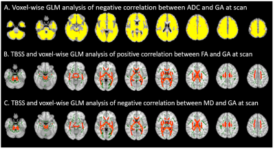 |
Temporal evolution of diffusion metrics in neonatal brains within the first few weeks postpartum Video Not Available
Pratheek Bobba1, Clara F Weber1,2, Adrian Mak1,3, Ajay Malhotra4, Kevin Sheth5, Sarah N Taylor6, Arastoo Vossough7,8, Patricia Ellen Grant9,10, Todd Constable1, Laura R Ment5,6, and Seyedmehdi Payabvash1
1Radiology, Yale School of Medicine, New Haven, CT, United States, 2Psychiatry and Psychotherapy, Social Neuroscience Lab, Department of Psychiatry and Psychotherapy, Lübeck University, Lübeck, Germany, Lübeck, Germany, 3CLAIM - Charité Lab for Artificial Intelligence in Medicine, Charité Universitätsmedizin Berlin, Berlin, Germany, Berlin, Germany, 4Radiology and Biomedical Imaging, Yale School of Medicine, New Haven, CT, United States, 5Neurology, Yale School of Medicine, New Haven, CT, United States, 6Pediatrics, Yale School of Medicine, New Haven, CT, United States, 7Radiology, Children's Hospital of Pennsylvania, Philadelphia, CT, United States, 8Radiology, University of Pennsylvania, Philadelphia, PA, United States, 9Division of Newborn Medicine, Boston Children's Hospital, Boston, MA, United States, 10Radiology, Boston Children's Hospital, Boston, MA, United States
In this retrospective cohort study, we characterized the age-related topography of quantitative diffusion metric evolution in 569 neonates scanned at our institution. We also studied the temporal rate of these metrics across different regions of the brain. and developed online interactive atlases depicting age-specific normative values of ADC and FA/MD in neonatal brains.
|
|
| 15:09 | 0067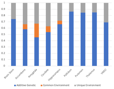 |
Genetic and Environmental Contributions to Subcortical Gray Matter Microstructure and Volume in the Developing Brain
Richard Watts1, BJ Casey1, and Christopher Filippi2
1Department of Psychology, Yale University, New Haven, CT, United States, 2Tufts Medical Center, Boston, MA, United States
Using 9-10 year old monozygotic and dizygotic twins from the Adolescent Brain and Cognitive DevelopmentSM Study, we investigated the genetic and environmental contributions to microstructure (assessed using diffusion MRI and a restriction spectrum imaging model) and volume in nine subcortical gray matter regions. Heritability estimates range from 0.45 to 0.86 for microstructure, and 0.61 to 0.88 for volumes. Only the microstructure of the amygdala showed evidence of sensitivity to the shared environment.
|
|
| 15:21 | 0068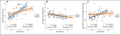 |
Choroid plexus function and structure across the lifespan as evaluated using perfusion-weighted MRI: implications for glymphatic dysfunction
Jarrod J. Eisma1, Colin D. McKnight2, Kilian Hett1, Jason Elenberger1, Daniel O. Claassen1, and Manus J. Donahue1,3
1Department of Neurology, Vanderbilt University Medical Center, Nashville, TN, United States, 2Department of Radiology and Radiological Sciences, Vanderbilt University Medical Center, Nashville, TN, United States, 3Department of Psychiatry and Behavioral Sciences, Vanderbilt University Medical Center, Nashville, TN, United States
The choroid plexus (ChP) has gained recent attention due to its potential functional relevance for cerebral glymphatic circulation. Here, a novel arterial spin labelling sequence and deep learning algorithm were applied to quantify ChP volume, perfusion, and volumetric flow across the healthy lifespan and in neurodegenerative participants with Parkinson’s disease (PD) to test the hypotheses that ChP hypertrophy and hypoperfusion are associated with increasing age and neurodegeneration. ChP volume increased with age, whereas perfusion decreased with age (p<0.01). In PD, ChP volumetric flow increases were more pronounced relative to age-matched healthy controls, consistent with compensatory stimulation of cerebrospinal fluid production.
|
|
15:33 |
0069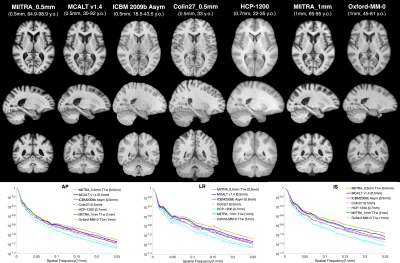 |
MIITRA atlas: T1w and DTI templates of the older adult brain in a common space with 0.5mm resolution
Yingjuan Wu1, Abdur Raquib Ridwan1, Mohammad Rakeen Niaz1, David A. Bennett2, and Konstantinos Arfanakis1,2
1Biomedical Engineering, Illinois Institute of Technology, Chicago, IL, United States, 2Rush Alzheimer's Disease Center, Rush University Medical Center, Chicago, IL, United States
As part of the Multichannel Illinois Institute of Technology & Rush university Aging (MIITRA) atlas project, the present work (A) developed high quality 0.5mm isotropic resolution T1-weighted (T1w) and diffusion tensor imaging (DTI) templates in common space using data from a large, diverse, community cohort of non-demented older adults, and (B) compared the new templates to existing templates of the older and younger adult brain. The new templates exhibited higher image quality, were more representative of the older adult brain, and allowed higher spatial normalization accuracy across subjects and across modalities compared to other templates.
|
 Back to Meeting Home
 Back to the Program-at-a-Glance
The International Society for Magnetic Resonance in Medicine is accredited by the Accreditation Council for Continuing Medical Education to provide continuing medical education for physicians.
 Back to Meeting Home
 Back to the Program-at-a-Glance
 View the Presentation