Oral Session
Tractography & Microstructural Studies in the Brain
Joint Annual Meeting ISMRM-ESMRMB & ISMRT 31st Annual Meeting • 07-12 May 2022 • London, UK

Oral Session
Tractography & Microstructural Studies in the Brain
| 16:45 | 0517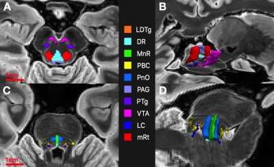 |
Automated Superstructure-based Segmentation of Ascending Arousal Network Nuclei for Diffusion Tractography
Mark D Olchanyi1,2,3, Brian L Edlow1,4, Emery N Brown2,3,5,6, and Juan E Iglesias 4,7,8
1Center for Neurotechnology and Neurorecovery, Department of Neurology, Massachusetts General Hospital, Boston, MA, United States, 2Neurostatistics Research Laboratory, Massachusetts Institute of Technology, Cambridge, MA, United States, 3Institute for Medical Engineering and Science, Massachusetts Institute of Technology, Cambridge, MA, United States, 4Athinoula A. Martinos Center for Biomedical Imaging, Massachusetts General Hospital and Harvard Medical School, Charlestown, MA, United States, 5Picower Institute, Massachusetts Institute of Technology, Cambridge, MA, United States, 6Department of Anesthesia, Critical Care and Pain Medicine, Massachusetts General Hospital and Harvard Medical School, Boston, MA, United States, 7Centre for Medical Image Computing, University College London, London, United Kingdom, 8Computer Science and Artificial Intelligence Laboratory, Massachusetts Institute of Technology, Cambridge, MA, United States
Traumatic brain injury that causes sheering of white matter pathways that connect ascending arousal network (AAN) nuclei in the rostral brainstem to cortical and subcortical targets leads to disorders of consciousness. Connectivity studies involving these nuclei are limited due to their small size, vague boundaries, and lack of reliable annotations. We present a method to automatically segment AAN nuclei from diffusion MR volumes using image registration constrained by brainstem structures with definable contrast boundaries. We test AAN segmentation robustness with probabilistic tractography and provide new insights into connectivity between AAN nuclei and the thalamus.
|
|
| 16:57 | 0518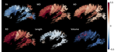 |
Short superficial U-fiber systems and aging: a longitudinal diffusion MRI study
Kurt G Schilling1, Derek Archer2, Fang-Cheng Yeh3, Francois Rheault2, Leon Y Cai2, Timothy Hohman2, Andrea T Shafer4, Susan M Resnick4, Angela Jefferson2, Adam W Anderson2, Hakmook Kang2, and Bennett A Landman2
1Vanderbilt University Medical Center, Nashville, TN, United States, 2Vanderbilt University, Nashville, TN, United States, 3University of Pittsburgh Medical Center, Pittsburg, PA, United States, 4National Institute on Aging, Baltimore, MD, United States
Superficial U-fiber systems are understudied using diffusion tractography, despite making up a majority of the white matter connections. Here, we used a large, longitudinal dataset from the Baltimore Longitudinal Study of Aging, in combination with innovations in U-fiber tractography dissection, to study U-fiber systems in an aging population. We characterize microstructural features, and for the first time, macrostructural features of length and volume, and find significant associations with age. Characterizing what changes occur, and where they occur, in U-fiber systems will complement traditional long-range tractography research to better understand the biological mechanisms of normal aging.
|
|
17:09 |
0519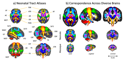 |
Tractography protocols for the neonatal brain, standardised against the adult human and macaque
Shaun of Warrington1, Elinor Thompson1, Jessica Dubois2, Luke Baxter3, Rebeccah Slater3, Rogier B Mars4,5, Saad Jbabdi4, Matteo Bastiani1, and Stamatios N Sotiropoulos1,4,6
1Sir Peter Mansfield Imaging Centre, School of Medicine, University of Nottingham, Nottingham, United Kingdom, 2University of Paris, Inserm, NeuroDiderot Unit; University Paris-Saclay, CEA, NeuroSpin center, Paris, France, 3Department of Paediatrics, University of Oxford, Oxford, United Kingdom, 4Wellcome Centre for Integrative Neuroimaging, FMRIB Centre, Nuffield Department of Clinical Neurosciences, University of Oxford, Oxford, United Kingdom, 5Donders Institute for Brain, Cognition and Behaviour, Radboud University, Nijmegen, Nijmegen, Netherlands, 6National Institute for Health Research (NIHR) Nottingham Biomedical Research Centre, Nottingham, United Kingdom
The neonatal brain undergoes rapid development in the months after birth. Diffusion tractography is a unique method for probing developing white matter connections. We present a novel and comprehensive library of tractography protocols for the neonatal brain, whilst ensuring correspondence with previously developed protocols for the adult and macaque brain. We demonstrate protocol robustness across data quality and show that the resultant tracts capture a-priori known trends in white matter microstructure. We show that these protocols open avenues for quantitative comparisons across the lifespan, but also species, which we exemplify by revealing developmental trends in connectivity patterns.
|
|
| 17:21 | 0520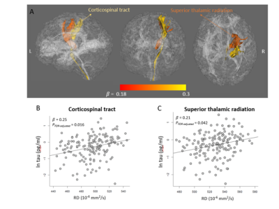 |
Longitudinal associations between blood biomarkers and white-matter MRI in sport-related concussion: A study of the NCAA-DoD CARE Consortium
Yu-Chien Wu1, Qiuting Wen2, Jessica Gill3, Rhea Thukral4, Sujuan Gao5, Kathleen Lane5, Timothy Meier6, Larry Riggen5, Jaroslaw Harezlak7, Christopher Giza8, Joshua Goldman9, Kevin Guskiewicz10, Jason Mihalik 10, Stephen LaConte11, Stefan Duma12, Steven Broglio13, Andrew Saykin2, Thomas McAllister14, and Michael McCrea6
1Department of Radiology and Imaging Sciences, Department of RadioIndiana University School of Medicine, Indianapolis, IN, United States, 2Department of Radiology and Imaging Sciences, Indiana University School of Medicine, Indianapolis, IN, United States, 3National Institute of Health, Bethesda, MD, United States, 4Department of Radiology, Indiana School of Medicine, Indianapolis, IN, United States, 5Department of Biostatistics and Health Data Science, Indiana University School of Medicine, Indianapolis, IN, United States, 6Department of Neurosurgery, Medical College of Wisconsin, Milwaukee, WI, United States, 7Department of Epidemiology and Biostatistics, Indiana University School of Public Health, Bloomington, IN, United States, 8Department of Neurosurgery, David Geffen School of Medicine at University of California Los Angeles, Los Angeles, CA, United States, 9Family Medicine, Ronald Reagan UCLA Medical Center, UCLA Health - Santa Monica Medical Center, Los Angeles, CA, United States, 10Department of Exercise and Sport Science, Matthew Gfeller Sport-Related Traumatic Brain Injury Research Center, Chapel Hill, NC, United States, 11School of Biomedical Engineering and Sciences, Wake-Forest and Virginia Tech University, Roanoke, VA, United States, 12School of Biomedical Engineering and Sciences, Wake-Forest and Virginia Tech University, Blacksburg, VA, United States, 13NeuroTrauma Research Laboratory, Michigan Concussion Center, University of Michigan, Ann Arbor, MI, United States, 14Department of Psychiatry, Indiana University School of Medicine, Indianapolis, IN, United States
White matter structural changes occur when an athlete experiences a concussion. To determine if serum biomarkers are a good concussion diagnosis, it seems prudent to associate white matter structural changes to longitudinal biomarker samples. Longitudinal blood biomarkers and DTI scanning from 77 collegiate athletes were collected across 3 timepoints and tested for associations. Tau was found to have the most significant associations. It was concluded that tau is the most sensitive for white-matter micro-structures.
|
|
| 17:33 |
0521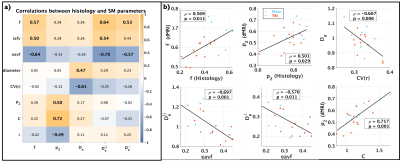 |
Validation of diffusion MRI-derived white matter microstructure metrics using 3D electron microscopy in injured rat brain
Ricardo Coronado-Leija1, Ali Abdollahzadeh2, Hong-Hsi Lee3, Santiago Coelho1, Raimo A Salo2, Jussi Tohka2, Alejandra Sierra2, Dmitry S Novikov1, and Els Fieremans1
1Radiology, New York University School of Medicine, New York, NY, United States, 2University of Eastern Finland, Kuopio, Finland, 3Radiology, Massachusetts General Hospital, Harvard Medical School, Boston, MA, United States
To validate the standard model of diffusion in white matter, we characterized injured rat brain microstructure using 3D EM and ex-vivo MRI. Using histology, we demonstrated that the rotational invariants $$$p_l$$$ of the fiber orientation distribution follow a power-law and that the axon diameter histogram is described well by a generalized extreme value distribution. Comparing 3D EM and dMRI, we found that the SM parameters correlate specifically with their histology counterpart, and revealed the specificity of the intra-axonal diffusivity to axonal beading in traumatic brain injury.
|
|
| 17:45 | 0522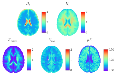 |
First templates of human brain kurtosis sources: relevance of microscopic kurtosis from Correlation Tensor MRI at 3 Tesla
Lisa Novello1, Rafael Neto Henriques2, Andrada Ianuş2, Thorsten Feiweier3, Noam Shemesh2, and Jorge Jovicich1,4
1Center for Mind/Brain Sciences - CIMeC, University of Trento, Rovereto (Trento), Italy, 2Champalimaud Research, Champalimaud Centre for the Unknown, Lisbon, Portugal, 3Siemens Healthcare GmbH, Erlangen, Germany, 4Center for Medical Sciences - CISMeD, University of Trento, Rovereto (Trento), Italy The Correlation Tensor MRI (CTI) framework has been recently formulated to disentangle anisotropic, isotropic, and microscopic kurtosis sources without a priori assumptions from Double-Diffusion-Encoding (DDE) data. In this work, group average maps (templates) are presented for the first time, thereby mapping the contrasts for anisotropic and isotropic kurtosis, and non-vanishing positive microscopic kurtosis in grey and white matter. The relative weight of the microscopic kurtosis within the total kurtosis, and the bias associated with neglecting this component under the Multiple Gaussian Component assumption are investigated and suggest that this component significantly contributes to the total diffusional kurtosis. |
|
| 17:57 | 0523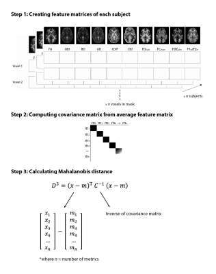 |
Accounting for covariance in studies of multimodal microstructure - a model for multivariate quantification in health and disease
Stefanie A Tremblay1,2, Amir Pirhadi3, Zaki Alasmar4, Felix Carbonell5, Yasser Iturria-Medina6,7,8, Claudine J Gauthier1,2, and Christopher J Steele4,9
1Physics, Concordia University, Montreal, QC, Canada, 2Montreal Heart Institute, Montreal, QC, Canada, 3Electrical and Computer Engineering, Concordia University, Montreal, QC, Canada, 4Psychology, Concordia University, Montreal, QC, Canada, 5Biospective Inc., Montreal, QC, Canada, 6Neurology and Neurosurgery, Montreal Neurological Institute and Hospital, Montreal, QC, Canada, 7McConnell Brain Imaging Centre, Montreal Neurological Institute, Montreal, QC, Canada, 8Ludmer Centre for NeuroInformatics and Mental Health, Montreal, QC, Canada, 9Neurology, Max Planck Institute for Human Cognitive and Brain Sciences, Leipzig, Germany
Multivariate approaches have recently gained in popularity to address the physiological unspecificity of neuroimaging metrics and to better characterize the complexity of biological processes underlying behavior. However, approaches commonly used are biased by the covariance between variables. Here, we propose computing the voxel-wise Mahalanobis distance (MhD), as a measure of deviation from normality that accounts for covariance between metrics. We show that this measure can be linked to behavior and to potential physiological underpinnings by extracting metrics contributing most to the MhD. Integrative multivariate models are crucial to expand our understanding of the multiple factors underlying disease development and progression.
|
|
| 18:09 | 0524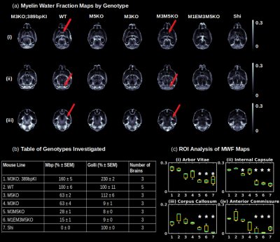 |
Sensitive Quantification of Hypomyelination and Axon g-ratio Using Ultra High Resolution 7T Multi-Echo Gradient Echo MRI with BSS-rPCA
Vladimir Grouza1,2, Hooman Bagheri3, Marius Tuznik1,2, Hanwen Liu1,2, Alan C Peterson2,3,4, and David A Rudko1,2,5
1McConnell Brain Imaging Centre, Montreal Neurological Institute and Hospital, Montreal, QC, Canada, 2Department of Neurology and Neurosurgery, McGill University, Montreal, QC, Canada, 3Department of Human Genetics, McGill University, Montreal, QC, Canada, 4Gerald Bronfman Department of Oncology, McGill University, Montreal, QC, Canada, 5Department of Biomedical Engineering, McGill University, Montreal, QC, Canada
We investigated the application of blind source separation, robust principal component analysis (BSS-rPCA) for myelin water imaging in a panel of mice exhibiting varied myelination profiles. BSS-rPCA exhibited sensitivity to myelin as evidenced by region of interest (ROI) analysis of several white matter tracts as a function of hypomyelinating genotype. By combining BSS-rPCA with estimates of neurite orientation dispersion and density imaging (NODDI)-derived axon water fraction (AWF) maps, we conclude that BSS-rPCA is sensitive to variations in myelin content in the presence of stable axon density.
|
 Back to Meeting Home
 Back to the Program-at-a-Glance
The International Society for Magnetic Resonance in Medicine is accredited by the Accreditation Council for Continuing Medical Education to provide continuing medical education for physicians.
 Back to Meeting Home
 Back to the Program-at-a-Glance
 View the Presentation