Oral Session
High-Resolution fMRI & Functional Connectivity
Joint Annual Meeting ISMRM-ESMRMB & ISMRT 31st Annual Meeting • 07-12 May 2022 • London, UK

Oral Session
High-Resolution fMRI & Functional Connectivity
| 09:15 | 0397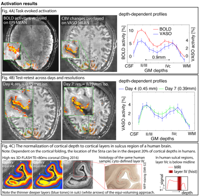 |
Sub-0.1 microliter CBV fMRI on the Next Generation 7T scanner
David A Feinberg1,2,3, Salvatore Torrisi1,2, Alexander JS Beckett1,2, Rüdiger Stirnberg4, Tony Stöcker4, Philipp Ehses4, and Renzo Huber3
1Helen Wills Neuroscience Institute, University of California, Berkeley, Berkeley, CA, United States, 2Advanced MRI Technologies, Sebastopol, CA, United States, 3Faculty of Psychology and Neuroscience, Maastricht University, Maastricht, Netherlands, 4German Center for Neurodegenerative Diseases (DZNE), Bonn, Germany
Laminar-specific fMRI with CBV-sensitive VASO can address neuroscience research questions on directional information processing within and across brain areas. However, conventional 0.8mm resolutions cannot Nyquist sample the individual layer-groups of interest (e.g. Layers II/III from layer IVc in V1). Here we implement, evaluate, and apply a 0.39-0.45mm (iso.) VASO protocol on the NextGen 7T scanner to resolve layer-features that could not be resolved in humans before. We find that 0.45 mm protocols are possible if the sequence parameters of EPI-segmentation and GRAPPA acceleration are differently optimized than one would do for conventional fMRI protocols.
|
|
| 09:27 | 0398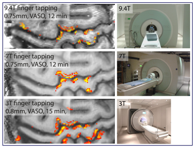 |
Evaluating the capabilities and challenges of layer-fMRI VASO at 3T
Renzo Huber1, Lisa Kronbichler2, Rüdiger Stirnberg3, Luca Vizioli4, Philipp Ehses3, Tony Stöcker3, Sara Fernández-Cabello5, Benedikt A Poser1, and Martin Kronbichler6,7
1FPN, Uni Maastricht, Maastricht, Netherlands, 2Department of Psychiatry, Psychotherapy and Psychosomatics, Christian‐Doppler Medical Centre, PMU, Salzburg, Austria, 3German Center for Neurodegenerative Diseases (DZNE), Bonn, Germany, 4Department of Radiology, University of Minnesota, Center for Magnetic Resonance Research, Minneapolis, MN, United States, 5Norwegian Centre for Mental Disorders Research (NORMENT), Institute of Clinical Medicine, Uni Oslo, Oslo, Norway, 6Neuroscience Institute, Christian Doppler Medical Centre, PMU, Salzburg, Austria, 7Centre for Cognitive Neuroscience & Department of Psychology, University of Salzburg, Salzburg, Austria
Sub-millimeter functional imaging at ultra high field (UHF) strengths can capture cortical layer-specific functional information flow within and across brain systems. However, UHF scanners are only available to appr. 100 privileged imaging centers, globally. In this work, we implement, characterise and apply a CBV-sensitive VASO sequence setup for widely accessible 3T scanners. We find that the longer T2*, and shorter T1 relaxation times at 3T can account for some of the lower z-magnetization in the inversion-recovery VASO sequence compared to 7T and 9.4T. We conclude that layer-fMRI VASO is applicable without too much further ado at 3T.
|
|
| 09:39 | 0399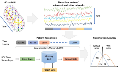 |
Deep Learning Methods for Reversible Cerebral Vasoconstriction Syndrome Classification Based on Resting-state fMRI Images Video Permission Withheld
Tun-Wei Hsu1,2,3, Chia-Hung Wu1,4,5, Hsiu-Mei Wu1,4, Kuan-Lin Lai4,6,7, Shih-Pin Chen4,6,7,8, Shuu-Jiun Wang4,6,7, Jiing-Feng Liring1,4, and Wan-Yuo Guo1,4
1Department of Radiology, Taipei Veterans General Hospital, Taipei, Taiwan, 2Integrated PET/MR Imaging Center, Taipei Veterans General Hospital, Taipei, Taiwan, 3Department of Biomedical Imaging and Radiological Sciences, National Yang-Ming Chiao Tung University, Taipei, Taiwan, 4School of Medicine, College of Medicine, National Yang-Ming Chiao Tung University, Taipei, Taiwan, 5Institute of Clinical Medicine, National Yang-Ming Chiao Tung University, Taipei, Taiwan, 6Department of Neurology, Neurological Institute, Taipei Veterans General Hospital, Taipei, Taiwan, 7Brain Research Center, National Yang-Ming Chiao Tung University, Taipei, Taiwan, 8Division of Translational Research, Taipei Veterans General Hospital, Taipei, Taiwan
Reversible cerebral vasoconstriction syndrome (RCVS) is a reversible segmental and multifocal vasoconstriction of the cerebral arteries and is believed to relate to autonomic network over-activity. We used Long Short-Term Memory networks (LSTMs), a type of deep neural network designed to handle time sequence data, to learn directly from the rs-fMRI time-series for classification of individuals with RCVS and healthy controls based on the regions in autonomic and other functional networks. These results provide methodological implications for rs-fMRI data of RCVS patients involved in the analysis and are a key element in future studies.
|
|
09:51 |
0400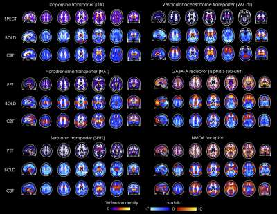 |
Molecular-enriched functional connectivity in the human brain using multi-band multi-echo simultaneous ASL/BOLD fMRI
Ottavia Dipasquale1, Alexander Cohen2, Daniel Martins1, Fernando Zelaya1, Federico Turkheimer1, Mattia Veronese1, Steve Williams1, Baolian Yang3, Suchandrima Banarjee4, and Yang Wang2
1Department of Neuroimaging, Institute of Psychiatry, Psychology and Neuroscience, King's College London, London, United Kingdom, 2Department of Radiology, Medical College of Wisconsin, Milwaukee, WI, United States, 3GE Healthcare, Waukesha, WI, United States, 4GE Healthcare, Menlo Park, CA, United States
This work is a proof of concept for the integration of ASL fMRI and molecular imaging using REACT, a novel analytical strategy that enriches functional data with the molecular information on the neurotransmitter distribution density. We applied REACT to high-resolution, whole-brain simultaneous ASL/BOLD data to estimate BOLD- and perfusion-weighted (PW)-based functional connectivity maps related to specific molecular systems and compared the results from the two fMRI modalities, showing very similar patterns of molecular-enriched functional connectivity. Our findings show that the PW signal is as informative as BOLD in terms of detection of functional circuits associated to specific molecular pathways.
|
|
10:03 |
0401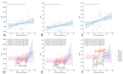 |
Resting state fluctuations in BOLD fMRI might not systematically reflect measures of cerebrovascular physiology between or within subjects
Stefano Moia1, Gang Chen2, Eneko Uruñuela1, Rachael C. Stickland3, Maite Termenon1, César Caballero-Gaudes1, and Molly G. Bright3,4
1Basque Center on Cognition, Brain and Language, Donostia, Spain, 2NIMH/NIH/HHS, Bethesda, MD, United States, 3Physical Therapy and Human Movement Sciences, Feinberg School of Medicine, Northwestern University, Chicago, IL, United States, 4Biomedical Engineering, McCormick School of Engineering, Northwestern University, Evanston, IL, United States
Resting State Fluctuations (RSF) metrics are frequently associated with vascular and physiological factors, to the point of being suggested as alternative estimates of cerebrovascular reactivity (CVR) and as tools for fMRI data calibration. Using a densely sampled fMRI dataset, we demonstrate high individual variability in the inter-session relationships between RSF metrics and CVR. Moreover, while physiological factors such as blood pressure show a significant relationship with CVR, they do not with common RSF metrics. These results indicate that RSF parameters might not be suitable alternatives for CVR, and may not properly account for inter-individual physiological variations in BOLD fMRI data.
|
|
| 10:15 | 0402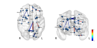 |
Exploring Brain Connectivity in Ageing Using Explainable Deep Learning
Tamar van Asch1, Nathan De Jong1, Walter Backes1, Sebastian Köhler 2, Martin van Boxtel2, Miranda Schram3, and Jacobus Jansen1
1Radiology, Maastricht University Medical Center, Maastricht, Netherlands, 2Psychiatrie & Neuropsychologie, School for Mental Health and Neuroscience, Maastricht, Netherlands, 3Internal Medicine, Maastricht University, Maastricht, Netherlands
There is a growing need for the understanding of the process of ageing and the ability to predict who is at risk of neurodegenerative diseases and mortality. This study aims to develop and train a convolutional neural network on structural brain connectivity data to predict age. dMRI is used to map the structural connectivity of the brain. The dataset comprises 3494 subjects from The Maastricht Study, a cohort study of individuals aged between 40 and 77 years. Brain age prediction on the test set resulted in a Pearson’s correlation coefficient of 0.70 and a mean absolute error of 5.1 years.
|
|
| 10:27 | 0403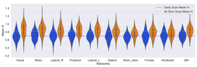 |
Title Fractal Analysis of the BOLD Signal in Preterm Infants Scanned Shortly After Birth and at Term-Equivalent Age
Alexander Mark Weber1,2, Johann Drayne1,2, Olivia Campbell1,2, Cecil Chau2,3, Steven Miller4, and Ruth Grunau2,3
1Brain, Behaviour & Development, BC Children's Hospital Research Institute, Vancouver, BC, Canada, 2University of British Columbia, Vancouver, BC, Canada, 3BC Children's Hospital Research Institute, Vancouver, BC, Canada, 4Division of Neurology and Centre for Brain & Mental Health, SickKids, Toronto, ON, Canada
It has been found that fractal analysis of the BOLD signal in fMRI can be used to measure brain functioning, development, and health. There has to our knowledge, however, not been any fractal analysis fMRI studies done in newborns. By computing the mean Hurst exponent in 9 resting state networks and in a grey matter mask. We found the largest increase in the Hurst exponent between pre-term and term equivalent aged subjects was in the motor and visual network. This motivates the need of further exploration of fractal analysis as a measure of brain dynamics in pre-term infants.
|
|
| 10:39 |
0404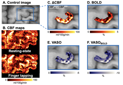 |
Concurrent laminar CBF, CBV, T2-BOLD and CMRO2 fMRI at 7T in human primary motor cortex
Xingfeng Shao1, Jun Hua2, and Danny JJ Wang1
1Laboratory of FMRI Technology (LOFT), Mark & Mary Stevens Neuroimaging and Informatics Institute, Keck School of Medicine, University of Southern California, Los Angeles, CA, United States, 2Department of Radiology, Johns Hopkins University School of Medicine, Baltimore, MD, United States
In this study, we propose a novel pulse sequence to simultaneously acquire laminar ASL, VASO and T2-BOLD signals with high spatial resolution. Experiments were performed on the M1 to characterize concurrent layer-dependent CBF, CBV, T2 BOLD and CMRO2 signal changes to sensory input and motor output. Both positive CBF/CBV/CMRO2 and negative VASO profiles show a clear ‘double-peak’ pattern, which is consistent with the hypothesis that FT engages neural activity of somatosensory input in the superficial layers and motor output in the deep layers. The BOLD response mainly peaked in superficial layers due to dominant contribution of pial veins.
|
 Back to Meeting Home
 Back to the Program-at-a-Glance
The International Society for Magnetic Resonance in Medicine is accredited by the Accreditation Council for Continuing Medical Education to provide continuing medical education for physicians.
 Back to Meeting Home
 Back to the Program-at-a-Glance
 View the Presentation