Oral Session
Tumor Metabolism & Hypoxia
Joint Annual Meeting ISMRM-ESMRMB & ISMRT 31st Annual Meeting • 07-12 May 2022 • London, UK

| 14:30 | 0283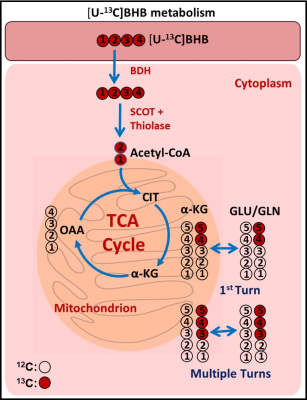 |
Oxidation of ketone body in brain tumor patients
Kumar Pichumani1,2, Omkar B Ijare1, Suzanne Powell3, and David S Baskin1,2
1Kenneth R. Peak Brain and Pituitary Tumor Treatment Center, Department of Neurosurgery, Houston Methodist Research Institute, Houston, TX, United States, 2Weill Cornell Medical College, New York, NY, United States, 3Pathology and Genomic Medicine, Houston Methodist Research Institute, Houston, TX, United States
Normal brain switches to utilization of ketone bodies only during prolonged starvation. It is not known whether brain tumors can utilize ketone body under in vivo conditions. There are ongoing clinical trials on using ketogenic diet as adjuvant therapy for the treatment of brain tumors. Here, we show that brain tumors (meningioma and GBM) can fully oxidize ketone body and therefore ketogenic diet may not be effective in the treatment of these tumor patients. MCT1 is involved in the transport of BHB into tumor cells and therefore it is feasible to target BHB metabolism using MCT1 inhibitor.
|
|
14:42 |
0284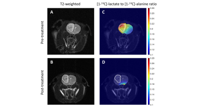 |
Hyperpolarized [1-13C]-alanine monitors tumor immortality and detects response to therapy in patient-derived glioblastoma models in vivo Video Permission Withheld
Georgios Batsios1, Meryssa Tran1, Anne Marie Gillespie1, Celine Taglang1, Sabrina Ronen1, Joseph Costello2, and Pavithra Viswanath1
1Radiology and Biomedical Imaging, University of California San Francisco, San Francisco, CA, United States, 2Neurological Surgery, University of California San Francisco, San Francisco, CA, United States
Telomerase reverse transcriptase (TERT) expression is essential for tumor proliferation. TERT also reprograms metabolism by elevating NADH and upregulating the alanine transporter ASCT2. Here, we exploit these TERT-associated metabolic alterations for non-invasive imaging in live GBM cells and orthotopic tumors using hyperpolarized [1-13C]-alanine. Combined treatment with the TERT inhibitor 6-thio-2’-deoxyguanosine and the ASCT2 inhibitor V-9302 inhibits GBM proliferation, identifying a novel therapeutic opportunity. Importantly, lactate production from hyperpolarized [1-13C]-alanine is an early biomarker of response to treatment in vivo, prior to the onset of anatomical alterations. Our findings pave the way for improved therapy and response assessment for GBM patients.
|
|
14:54 |
0285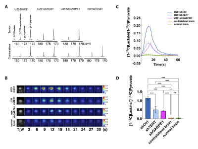 |
MRS-Detectable Metabolic Biomarkers of TERT positive Glioblastoma
Noriaki Minami1, Donghyun Hong1, Nicholas Stevers2, Carter J Barger2, Georgios Batsios1, Anne Marie Gillespie1, Joseph F Costello2, Pavithra Viswanath1, and Sabrina M Ronen1
1Radiology and Biomedical Imaging, UCSF, San Francisco, CA, United States, 2Department of Neurological Surgery, UCSF, San Francisco, CA, United States
Telomerase reverse transcriptase (TERT) expression results from TERT promoter mutations and is a hallmark of cancer. GABPB1, its upstream transcriptional factor, was recently identified as a promising tumor-specific therapeutic target. However, imaging methods that can detect TERT expression and its inhibition with emerging GABPB1-based therapeutics are limited. Here, we demonstrate that the combination of 1H-MRS and hyperpolarized 13C-MRS of [1-13C]pyruvate allow detection of TERT modulation in TERT-expressing GBM cells and in vivo tumors. These findings could be translated to the clinic and thus improve the monitoring and personalized treatment of GBM.
|
|
| 15:06 | 0286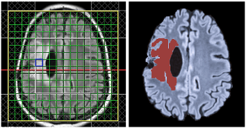 |
Fusion of T2/FLAIR Hyperintensities with Chemical Shift Imaging in Glioma Patients
Wufan Zhao1, Eduardo Coello1, Vicky Liao1, Assaf Tal2, and Alexander Lin1
1Radiology, Brigham and Women's Hospital, Boston, MA, United States, 2Chemical & Biological Physics, Weizmann Institute of Science, Rehovot, Israel
This work fuses manual segmentation of T2/FLAIR hyperintensity regions using 3D Slicer with multivoxel magnetic resonance spectroscopy (MRSI), which profiles neurochemical concentrations in a wide area of the brain. The utility of MRSI is demonstrated in the mapping of metabolites including 2HG, N-acetyl aspartate, myoinositol, glutamate plus glutamine, lactate, and choline in the region of interest. Voxel fractions of hyperintensity were correlated to metabolites, and metabolite-to-creatine ratio intercepts were used to compare hyperintense with healthy tissue.
|
|
| 15:18 | 0287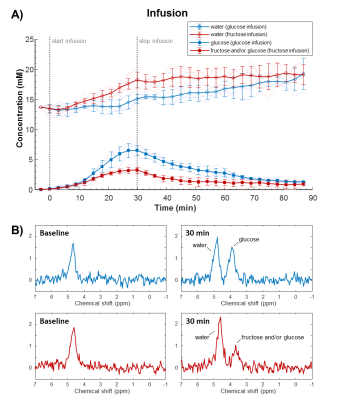 |
Glucose versus fructose metabolism in the liver measured with deuterium metabolic imaging Video Permission Withheld
Arjan D. Hendriks1, Andor Veltien2, Ingmar J. Voogt3, Arend Heerschap2, Tom W.J. Scheenen2, and Jeanine J. Prompers1
1Department of Radiology, University Medical Center Utrecht, Utrecht, Netherlands, 2Department of Medical Imaging (Radiology), Radboud University Medical Center, Nijmegen, Netherlands, 3WaveTronica B.V., Utrecht, Netherlands
The worldwide increase in sugar consumption is a major health concern. The contribution of fructose to the epidemic of metabolic disorders is controversial. This study investigated differences between glucose and fructose metabolism in the liver using dynamic deuterium metabolic imaging (DMI). Deuterium signal time curves after bolus injection and slow infusion were successfully acquired and showed different patterns. It was found that the peak concentration and conversion of deuterated glucose was different from that of fructose, indicating faster turnover of the latter. Overall, these methods could potentially lead to better understanding of healthy liver metabolism and aberrations in metabolic diseases.
|
|
| 15:30 | 0288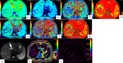 |
Differentiation of hepatocellular carcinoma from intrahepatic cholangiocarcinoma by mDIXON Quant and intravoxel incoherent motion imaging Video Not Available
Tao Lin1, Jiazheng Wang2, Liangjie Lin2, Lihua Chen 1, Qingwei Song1, Renwang Pu1, Ying Zhao1, Xue Ren1, Qihao Xu1, and Ailian Liu1
1The First Affiliated Hospital of Dalian Medical University, Dalian, China, 2Philips Healthcare, Beijing, China
Differentiation between hepatocellular carcinoma (HCC) and intrahepatic cholangiocarcinoma (ICC), the most common types of primary liver cancer, is important for the treatment planning and prognosis, but remains challenging via conventional radiological tools. Results by this study indicated that both mDIXON Quant and IVIM parameters showed significant different between HCC and ICC groups, and their combination can be of great potential value for clinical differential diagnosis between the two lesions.
|
|
| 15:42 | 0289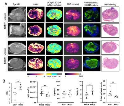 |
Imaging hypoxia in murine oral cavity squamous cell carcinomas with oxygen-enhanced MRI
Upasana Roy1, Elise Y. Lepicard1, Jessica K.R. Boult1, Carol Box1, Kevin J. Harrington1, James P.B. O’Connor1, Yann Jamin1, and Simon P. Robinson1
1Division of Radiotherapy and Imaging, The Institute of Cancer Research, Sutton, United Kingdom
Oxygen-enhanced (OE)-MRI was used to map and quantify hypoxia (pOxyR) in two murine models of oral cavity squamous cell carcinoma, a tumour type in which hypoxia adversely affects patient prognosis. OE-MRI relies on quantifying changes in the longitudinal MRI relaxation rate R1 induced by excess paramagnetic oxygen molecules dissolved in blood plasma and interstitial fluid with inhalation of oxygen. When combined with susceptibility-contrast MRI for determination of fractional blood volume and diffusion-weighted MRI to assess cellularity, OE-MRI enables an assessment of the spatial distribution and extent of tumour hypoxia, patent vasculature and necrosis.
|
|
| 15:54 | 0290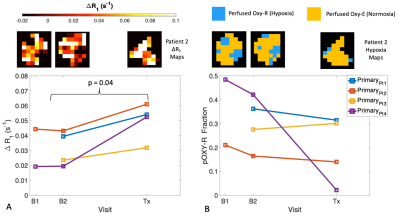 |
First in-human technique translation of OE-MRI for hypoxia imaging onto an MR Linac system in patients with head and neck cancer
Michael J Dubec1,2, Anubhav Datta1,3, Ross A Little1, Abigael Clough4, David L Buckley2,5, Christina Hague6, Damien McHugh1,2, Michael Berks1, Susan Cheung1, Amal Salah7, David Higgins8, Cynthia L Eccles1,4, Robert Bristow1,6, Josephine H Naish9,10, Julian C Matthews11, Peter Hoskin1,6,12, Marcel van Herk1, Geoff JM Parker10,13, Ananya Choudhury1,6, Andrew McPartlin6,
and James PB O'Connor1,3,14
1Division of Cancer Sciences, University of Manchester, Manchester, United Kingdom, 2Christie Medical Physics and Engineering, The Christie NHS Foundation Trust, Manchester, United Kingdom, 3Radiology, The Christie NHS Foundation Trust, Manchester, United Kingdom, 4Radiotherapy, The Christie NHS Foundation Trust, Manchester, United Kingdom, 5Biomedical Imaging, University of Leeds, Leeds, United Kingdom, 6Clinical Oncology, The Christie NHS Foundation Trust, Manchester, United Kingdom, 7Proton Beam Therapy, The Christie NHS Foundation Trust, Manchester, United Kingdom, 8Philips, Manchester, United Kingdom, 9MCMR, University of Manchester NHS Foundation Trust, Manchester, United Kingdom, 10Bioxydyn Ltd, Manchester, United Kingdom, 11Neuroscience and Experimental Psychology, University of Manchester, Manchester, United Kingdom, 12Radiotherapy, Mount Vernon, London, United Kingdom, 13Centre for Medical Image Computing, Unversity College London, London, United Kingdom, 14Radiotherapy and Imaging, Institute of Cancer Research, London, United Kingdom
Tumour hypoxia is associated with poor treatment outcome, tumour progression and treatment resistance. Oxygen-enhanced (OE)-MRI has shown promise as a non-invasive method for mapping and quantifying hypoxia. We demonstrate, for the first time, the application of the OE-MRI technique in patients with head and neck carcinoma to monitor changes in hypoxia due to treatment, and translate the technique onto an MR Linac system, demonstrating feasibility in a first-in-human study.
|
 Back to Meeting Home
 Back to the Program-at-a-Glance
The International Society for Magnetic Resonance in Medicine is accredited by the Accreditation Council for Continuing Medical Education to provide continuing medical education for physicians.
 Back to Meeting Home
 Back to the Program-at-a-Glance
 View the Presentation