Oral Session
Understanding Tumor Biology Through Specific Contrast Mechanisms
Joint Annual Meeting ISMRM-ESMRMB & ISMRT 31st Annual Meeting • 07-12 May 2022 • London, UK

Oral Session
Understanding Tumor Biology Through Specific Contrast Mechanisms
| 16:45 | 0543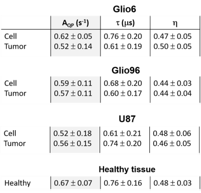 |
1H-14N quadrupolar relaxation of protein backbones: a potential intracellular biomarker of glioma invasion Video Permission Withheld
Hana Lahrech1, Manuel Petit1, Sandra Pierre1, Maxime Leclercq1, Lionel Marc Broche2, and François Berger1
1BrainTech Lab INSERM U1205, Grenoble, France, 2University of Aberdeen, Aberdeen, United Kingdom
In FFC-NMR, R1-dispersion curves of biological systems frequently show quadrupolar peaks (QP) between 1.5 and 3.5 MHz that correspond to the cross relaxation of water 1H and protein 14N. QP of glioma invasion tissues (Glio6 and Glio96 mouse models) and of high glioma proliferation (U87 model) were acquired ex vivo and compared to cell pellets in vitro, showing significant differences. This result suggests that QP signals are intracellular, a result which was confirmed by measurements of cells after trypsin treatment which showed unchanged QP amplitude. This result highlights the potential of the QPs as an intracellular biomarker of glioma invasion.
|
|
| 16:57 | 0544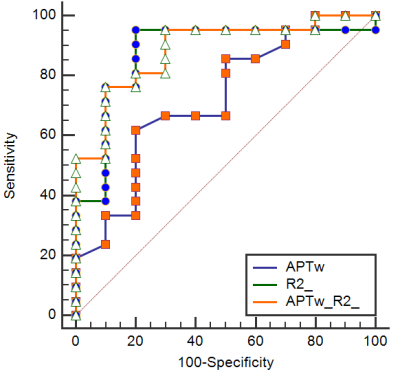 |
Differential diagnosis of primary liver cancer using APT and quantitative DIXON techniques Video Not Available
Tao Lin1, Jiazheng Wang2, Peng Sun2, Lihua Chen1, Qingwei Song1, Renwang Pu1, Ying Zhao1, Xue Ren1, Qihao Xu1, and Ailian Liu1
1The First Affiliated Hospital of Dalian Medical University, Dalian, China, 2Philips Healthcare, Beijing, China
Hepatocellular carcinoma (HCC), intrahepatic cholangiocarcinoma (ICC) are the most common types of primary liver cancer, which differ greatly in terms of pathogenesis, biological behavior, histological morphology, treatment, and prognosis. This retrospective study revealed that APT combined with quantitative DIXON technique could improve the performance for differentiating HCC from ICC.
|
|
| 17:09 | 0545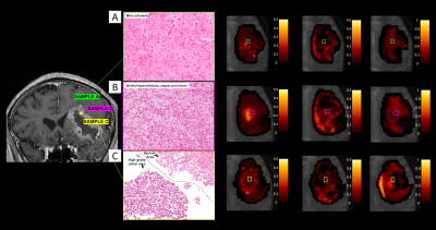 |
VERDICT MRI estimates match histopathology features in brain tumours
Matteo Figini1, Antonella Castellano2, Michele Bailo3, Marcella Callea4, Valentina Pieri2, Marcello Cadioli5, Marco Palombo1,6,7, Pietro Mortini3, Andrea Falini2, Daniel C Alexander1, Mara Cercignani6,8, and Eleftheria Panagiotaki1
1Centre for Medical Image Computing, Computer Science Department, University College London, London, United Kingdom, 2Neuroradiology Unit and CERMAC, Vita-Salute San Raffaele University and IRCCS Ospedale San Raffaele, Milan, Italy, 3Department of Neurosurgery and Gamma Knife Radiosurgery, Vita-Salute San Raffaele University and IRCCS Ospedale San Raffaele, Milan, Italy, 4Pathology Unit, IRCCS Ospedale San Raffaele, Milan, Italy, 5Philips Healthcare, Milan, Italy, 6Cardiff University Brain Research Imaging Centre (CUBRIC), School of Psychology, Cardiff University, Cardiff, United Kingdom, 7School of Computer Science and Informatics, Cardiff University, Cardiff, United Kingdom, 8Clinical Imaging Sciences Centre, Brighton and Sussex Medical School, Brighton, United Kingdom
We recently adapted the VERDICT framework to characterize both the core and peritumoural areas of brain tumours. We report here its first clinical application in the differentiation of brain tumour histotypes. Comparing groups of lesions with increasing aggressiveness (from lower to higher grades to metastases) we observed a significant increase in the intracellular and vascular fraction in the lesion core. VERDICT maps matched the features showed by histopathology in lower grades and in metastases; in the most heterogeneous higher grades, VERDICT maps showed differences between subregions compatible with histopathology results in multiple biopsy samples.
|
|
| 17:21 | 0546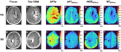 |
APTw imaging outperforms relaxation-compensated CEST-MRI at 3T in assessing glioma progression after radiotherapy - a preliminary analysis
Nikolaus von Knebel Doeberitz1, Florian Kroh2, Johannes Breitling2, Srdjan Maksimovic1, Laila Koenig3, Juergen Debus3,4, Peter Bachert2,5, Heinz-Peter Schlemmer1,6, Mark E. Ladd2,5,6, Steffen Goerke2, and Daniel Paech1,7
1Division of Radiology, German Cancer Research Center (DKFZ), Heidelberg, Germany, 2Division of Medical Physics in Radiology, German Cancer Research Center (DKFZ), Heidelberg, Germany, 3Department of Radiation Oncology, Heidelberg University Hospital, Heidelberg, Germany, 4Clinical Cooperation Unit Radiation Oncology, German Cancer Research Center (DKFZ), Heidelberg, Germany, 5Department of Physics and Astronomy, University of Heidelberg, Heidelberg, Germany, 6Faculty of Medicine, University of Heidelberg, Heidelberg, Germany, 7Department of Neuroradiology, Bonn University Hospital, Bonn, Germany
In this interim analysis we compared in patients with gliomas the ability of asymmetry-based amide proton transfer weighted (APTw) imaging with Lorentzian-fit-based relaxation-compensated CEST-MRI (MTRRex) of the APT, rNOE and MT signal to differentiate disease progression from treatment induced changes six weeks after completion of radiotherapy. In progressive gliomas, APTw displayed significantly higher signal intensities in contrast enhancing tissue compared to non-progressive gliomas with an AUC of 0.77 in receiver operator characteristic analyses (p 0.03). MTMTRrex showed a trend towards higher signal intensities in progressive tumors. All CEST metrics displayed significant differences in white and gray brain matter signal intensities.
|
|
| 17:33 | 0547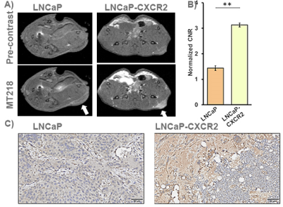 |
MR Molecular Imaging of Extradomain-B Fibronectin for Non-invasive Active Surveillance of Prostate Cancer
Amita Vaidya1, Aman Shankardass1, Megan Buford1, Ryan Hall1, and Zheng-Rong Lu1
1Department of Biomedical Engineering, Case Western Reserve University, Cleveland, OH, United States
C-X-C motif chemokine receptor 2 (CXCR2) expression is associated with high-grade and advanced prostate cancer. LNCaP-CXCR2+ prostate cancer cells with stable expression of CXCR2 exhibited mesenchymal features and became more aggressive with elevated expression of an ECM oncoprotein EDB-FN as compared to the slow-growing parental cell line, LNCaP. MRMI of EDB-FN with a targeted contrast agent MT218 resulted in significant increase of T1-weighted contrast enhancement in LNCaP-CXCR2+ prostate tumors as compared to the LNCaP tumors in mice. The results suggest that MRMI of EDB-FN has the potential to provide non-invasive active surveillance of low-grade prostate cancer.
|
|
| 17:45 | 0548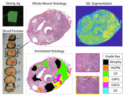 |
Diffusion restriction comparison between Gleason 4 fused and cribriform glands within patients using whole mount prostate pathology
Savannah R Duenweg1, Xi Fang2, Samuel A Bobholz1, Allison K Lowman3, Michael Brehler3, Fitzgerald Kyereme3, Kenneth A Iczkowski4, Kenneth Jacobsohn5, Anjishnu Banerjee2, and Peter S LaViolette3,6
1Biophysics, Medical College of Wisconsin, Wauwatosa, WI, United States, 2Biostatistics, Medical College of Wisconsin, Wauwatosa, WI, United States, 3Radiology, Medical College of Wisconsin, Wauwatosa, WI, United States, 4Pathology, Medical College of Wisconsin, Wauwatosa, WI, United States, 5Urology, Medical College of Wisconsin, Wauwatosa, WI, United States, 6Biomedical Engineering, Medical College of Wisconsin, Wauwatosa, WI, United States
This study used prostate histology and multi-parametric magnetic resonance imaging (MP-MRI) to evaluate diffusion differences in Gleason pattern 4 gland morphology. After surgery, tissue was sliced using slicing jigs, annotated by a board-certified pathologist, and digitally contoured to differentiate lumen and epithelium. Slides were co-registered to the T2-weighted MRI scan. Two linear mixed models were fitted to the MP-MRI data to consider the different hierarchical structures. Model (1) considers patient as random effect, and Model (2) adds a nested effect for slide. We found that cribriform glands were more diffusion restricted than fused glands in Gleason Grade 4 tumors.
|
|
17:57 |
0549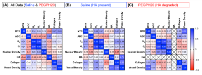 |
Investigating the contribution of hyaluronan (HA) to the breast tumour microenvironment using multiparametric MRI and histology
Emma L Reeves1, Jin Li1, Konstantinos Zormpas-Petridis1, James Sullivan2, Craig Cummings1, Barbara Blouw3, David Kang3, Ralph Sinkus4, Jeffrey C Bamber1, Yann Jamin1, and Simon P Robinson1
1Radiotherapy & Imaging, Institute of Cancer Research, London, United Kingdom, 2Royal Marsden NHS Foundation Trust, London, United Kingdom, 3Halozyme Therapeutics, San Diego, CA, United States, 4Life Sciences and Medicine, King's College London, London, United Kingdom
The impact of HA on tumour characteristics measured by MRI/E is not fully understood. We evaluated relationships between MRI biomarkers and histology in saline (HA present) and PEGPH20 treated (HA absent) breast tumours. ADC had the strongest association with HA, reaffirming that ADC is the most sensitive biomarker of tumour response to PEGPH20. MTR negatively correlated with HA, likely due to a HA-mediated dilution of MT-inducing components including collagen. MRE-derived elasticity (Gd) did not directly correlate with HA, however, HA degradation reduced the contribution of collagen to tumour viscoelasticity and may shift the sensitivity of Gd to the cellular compartment.
|
|
| 18:09 | 0550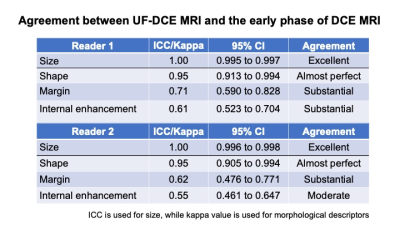 |
Can Ultrafast MRI replace conventional DCE MRI in morphological evaluation and subtype classification in breast mass lesions?
Akane Ohashi1, Masako Kataoka2, Mami Iima2, Maya Honda3, Rie Ota2, Yuta Urushibata 4, Dominic Marcel Nickel5, Masakazu Toi6, Sophia Zackrisson1, and Yuji Nakamoto2
1Translational Medicine, Diagnostic Radiology, Lund University, Skåne University Hospital, Malmö, Sweden, 2Diagnostic Imaging and Nuclear Medicine, Kyoto University Graduate School of Medicine, Kyoto, Japan, 3Diagnostic Radiology, Kansai Electric Power Hospital, Osaka, Japan, 4Siemens Healthcare K.K., Tokyo, Japan, 5Siemens Healthcare GmbH, Erlangen, Germany, 6Breast Surgery, Kyoto University Graduate School of Medicine, Kyoto, Japan
To evaluate the possibility of replacing conventional DCE MRI with UF-DCE MRI, we assessed the concordance of morphological information of the mass lesions between UF-DCE MRI and the early phase of DCE MRI. The size and shape showed excellent concordance between the two phases, while UF-DCE MRI tended to show circumscribed margin and homogeneous enhancement. In a sub-analysis, luminal subtypes demonstrated smaller size with homogeneous enhancement pattern compared with non-luminal subtypes. Predictive model for luminal subtype using the two parameters resulted in the AUC of 0.68, 0.72, for UF-DCE MRI and the early phase of DCE MRI, respectively (p=0.23).
|
 Back to Meeting Home
 Back to the Program-at-a-Glance
The International Society for Magnetic Resonance in Medicine is accredited by the Accreditation Council for Continuing Medical Education to provide continuing medical education for physicians.
 Back to Meeting Home
 Back to the Program-at-a-Glance
 View the Presentation