Oral Session
Antenatal, Neonatal & Pediatric MRI: Structure & Function
Joint Annual Meeting ISMRM-ESMRMB & ISMRT 31st Annual Meeting • 07-12 May 2022 • London, UK

Oral Session
Antenatal, Neonatal & Pediatric MRI: Structure & Function
| 14:30 | 0492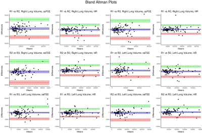 |
Inter-Rater Reliability of Fetal Lung Segmentation in High-Resolution Fetal Body Reconstructions
Kelly Payette1,2, Julia Geiger3, Michael Zellner3, Christian Kellenberger3, Ruth Tuura1, Raimund Kottke3, and Andras Jakab1,2
1Center for MR Research, University Children's Hospital Zurich, Zurich, Switzerland, 2Neuroscience Center Zurich, University of Zurich, Zurich, Switzerland, 3Diagnostic Imaging and Intervention, University Children's Hospital Zurich, Zurich, Switzerland
Reliable measurements of the fetal lung volume are important for the prenatal assessment of various pathologies impacting fetal lung development. Here we compare the inter-rater reliability of fetal lung volume measurements segmented from native ssFSE scans to those from high-resolution fetal body reconstructions to determine if the high-resolution reconstructions provide a clinical benefit. We find that there is increased reliability in the volumes segmented from the high-resolution reconstructions, suggesting that high resolution reconstruction of the fetal lungs may enable more precise assessments of fetal lung volume.
|
|
14:42 |
0493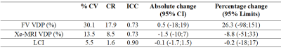 |
Inter- & Intra-visit Reproducibility of Free-Breathing Magnetic Resonance Imaging in Pediatric Cystic Fibrosis Lung Disease
Samal Munidasa1,2, Brandon Zanette1, Marcus Couch1,2,3, Robert Grimm4, Ravi Seethamraju5, Marie-Pier Dumas6, Wallace Wee6, Jacky Au6, Sharon Braganza1, Daniel Li1, Felix Ratjen1,6, and Giles Santyr1,2
1Translational Medicine, The Hospital for Sick Children, Toronto, ON, Canada, 2Medical Biophysics, University of Toronto, Toronto, ON, Canada, 3Siemens Healthcare Limited, Montreal, QC, Canada, 4MR Application Predevelopment, Siemens Healthcare GmbH, Erlangen, Germany, 5MR Collaborations North East, Siemens Healthineers, North East, NY, United States, 6Division of Respiratory Medicine, The Hospital for Sick Children, Toronto, ON, Canada Free-breathing lung MRI has been shown to be a responsive measure to CF pulmonary exacerbations treatments but have not been used to track stable disease progression longitudinally. In this study we determined the intra- and inter-scan reproducibility of free-breathing lung MRI in healthy and stable pediatric CF across 2 visits and compared to hyperpolarized Xenon MRI (Xe-MRI). Xe-MRI and free-breathing lung show a high intra-scan reproducibility in stable CF subjects, but free-breathing lung MRI showed a low inter-scan reproducibility. However, free-breathing lung MRI significantly correlated with Xe-MRI suggesting that it may be an alternative to expensive, and less wide-spread Xe-MRI. |
|
| 14:54 | 0494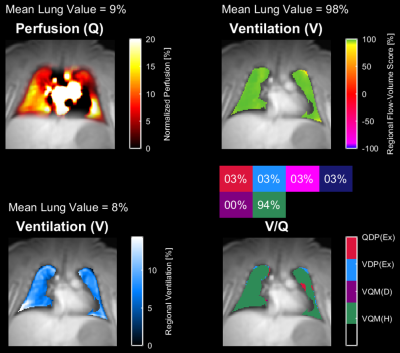 |
Assessment of Lung Ventilation and Perfusion of Premature Infants at 1.5 Tesla Using Phase-Resolved Functional Lung (PREFUL) MRI in the NICU
Jonathan P Dyke1, Andreas Voskrebenzev2,3, Lauren P Blatt4, Jens Vogel-Claussen2, Robert Grimm5, Jeffrey P Perlman4, and Arzu Kovanlikaya1
1Radiology, Weill Cornell Medicine, New York, NY, United States, 2Institute of Diagnostic and Interventional Radiology, Hannover Medical School, Hannover, Germany, 3Biomedical Research in Endstage and Obstructive Lung Disease Hannover (BREATH), German Center for Lung Research (DZL), Hannover, Germany, 4Pediatrics, Weill Cornell Medicine, New York, NY, United States, 5Siemens Healthineers, Erlangen, Germany
The purpose of this study was to evaluate quantitative measures of lung function in premature newborns using (PREFUL) MRI on a 1.5 Tesla scanner located within the neonatal intensive care unit (NICU). MRI can non-invasively assess ventilation and perfusion defects in the infant lung without sedation, ionizing radiation or contrast administration. We performed (PREFUL) MRI in 6 neonates [5 preterm/1 term] without respiratory distress to assess lung function in this normal population. Continuation of this work may allow clinicians to quantitatively assess response to treatment in this vulnerable population with respiratory distress.
|
|
| 15:06 |
0495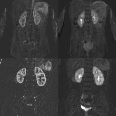 |
4D free-breathing variable density stack-of-stars functional MR urography in young children without sedation: A clinical feasibility study
Jakob Spogis1, Christoph Katemann2, Shuo Zhang2, Ilias Tsiflikas1, Michael Esser1, and Jürgen Schäfer1
1Department of Diagnostic and Interventional Radiology, University Hospital Tuebingen, Tuebingen, Germany, 2Philips GmbH Market DACH, Hamburg, Germany High-quality magnetic resonance urography (MRU) is clinically valuable to provide a comprehensive evaluation of the renal function and urinary tract in children without ionizing radiation. However, respiration-related patient motion remains a major challenge that impairs diagnostic image quality and hampers an accurate quantitative analysis. In this study, we adopt a novel high temporospatial resolution dynamic-contrast-enhanced (DCE) MRI technique based on 3D radial stack-of-stars gradient-echo sequence acquisition in combination with elliptical variable density k-space scheme for morphological and quantitative MRU and demonstrate its motion robustness in small children in free breathing without sedation. |
|
| 15:18 | 0496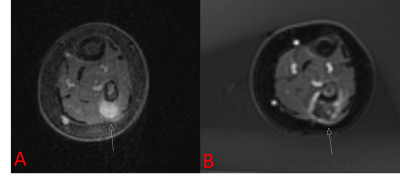 |
Ablative Efficacy of MR-guided Focused Ultrasound in Pediatric Patients: A Retrospective Study
Ali B Syed1, Suzanna Ackroyd1, Daniel Duex1, Victoria Young1, Shreyas Vasanawala1, Pejman Ghanouni1, and Avnesh Thakor1
1Radiology, Stanford University, Palo Alto, CA, United States Performance of MRgFUS in pediatric patients has not been well characterized. Retrospective review of 21 MRgFUS treatments in 9 patients below the age of 18 shows significant reduction in viable tumor volume in a variety of indications including desmoid tumors, vascular malformations, and osteoid osteoma. MRgFUS is an effective ablative therapy in children that warrants further investigation. |
|
| 15:30 | 0497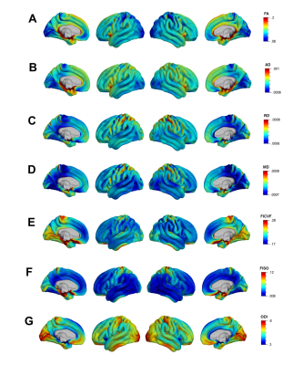 |
The spatiotemporal organization of cortical microstructural development in children and adolescents with diffusion MRI
Kirsten Mary Lynch1, Ryan P Cabeen1, and Arthur W Toga1
1USC Mark and Mary Stevens Institute for Neuroimaging and Informatics, University of Southern California, Los Angeles, CA, United States
Neocortical maturation is a dynamic process that proceeds in a hierarchical manner; however, the spatiotemporal organization of cortical microstructure with diffusion MRI has yet to be fully defined. This study characterized cortical microstructural maturation using fwe-DTI and NODDI in a cohort of 637 children and adolescents between 8 and 21 years of age. We found heterogeneous developmental patterns broadly demarcated into frontal, temporal and occipitoparietal domains where NODDI metrics increased and fwe-DTI metrics decreased with age. Our findings corroborate previous histological and neuroimaging studies that show spatially-varying patterns of cortical maturation that may reflect unique developmental processes of cytoarchitectonically-determined regions.
|
 Back to Meeting Home
 Back to the Program-at-a-Glance
The International Society for Magnetic Resonance in Medicine is accredited by the Accreditation Council for Continuing Medical Education to provide continuing medical education for physicians.
 Back to Meeting Home
 Back to the Program-at-a-Glance
 View the Presentation