Oral Session
Preclinical Brain & Body
Joint Annual Meeting ISMRM-ESMRMB & ISMRT 31st Annual Meeting • 07-12 May 2022 • London, UK

| 17:00 | 0153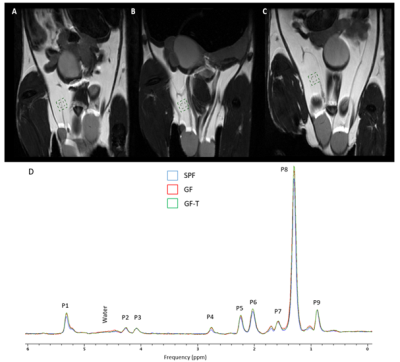 |
Evaluation of the lipid composition in mice transplanted with obese human microbiota using magnetic resonance spectroscopy
Joao Piraquive1,2, Franck Desmoulin2,3, José Manuel Fernandez-Real4, Rémy Burcelin1, Xavier Collet1,2, and Anne Bouloumié1
1Institute of Metabolic and Cardiovascular Diseases (I2MC), Team Dynamix, Toulouse, France, 2Regional center of functional exploration and experimental resources (CREFRE), Noninvasive exploration service (ENI), Toulouse, France, 3Toulouse NeuroImaging Center (ToNIC), INSERM UMR 2314/UPS, Toulouse, France, 4Unitat de Diabetologia, Endocrinologia i Nutricio, University Hospital of Girona Dr. Josep Trueta, Girona, Spain
In this project, mice devoid of microbiota were transplanted with human microbiota from obese patients. Our work focused on evaluating and comparing the lipid composition in the white adipose in mice with different gut microbiota backgrounds using MRS. Our findings showed differences in white adipose tissue storage, lipid composition, and fatty acid composition among the different groups. This suggests that MRS can be used as a noninvasive tool to monitor dysbiotic gut microbiota in obese patients and evaluate a potential therapeutic response for obesity.
|
|
| 17:12 | 0154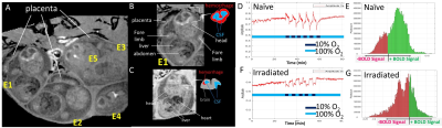 |
Probing Mitochondrial Function in Intact Fetal Brains with Ungated 4D Oxy-wavelet MRI in an Irradiation Injury Mouse Model Video Permission Withheld
Anthony Christodoulou1, Margaret Caroline Stapleton2, Devin Raine Everaldo Cortes3, Eric Goetzman4, Cecilia Lo2, Michael Epperly 5, Joel Greenberger 5, and Yijen L Wu2
1Cedars Sinai Medical Center, Los Angeles, CA, United States, 2Developmental Biology, University of Pittsburgh, Pittsburgh, PA, United States, 3Biomedical Engineering, University of Pittsburgh, Pittsburgh, PA, United States, 4Pediatrics, University of Pittsburgh, Pittsburgh, PA, United States, 5Radiation Oncology, University of Pittsburgh, Pittsburgh, PA, United States
A novel motion-and-time resolved 4D oxy-wavelet MRI (4D-fMRI acquired with oscillating hypoxia challenges, analyzed by a continuous wavelet transform mimicking experimental oscillations) can acquire fetal MRI with high spatiotemporal resolution and can probe mitochondrial functions in live fetal brains. 4D oxy-wavelet MRI outcomes were validated with Oroboros mitochondrial function assays and correlated with mitochondrial targeting drug JP4-039 in a fetal irradiation injury mouse model. The mouse fetuses showed poor 4D oxy-wavelet outcomes had poor mitochondrial functions and vice versa. Furthermore, an automated time-frequency analysis scheme can correctly differentiate normal vs irradiated fetuses, paving the way for future AI-based automatic diagnosis.
|
|
| 17:24 | 0155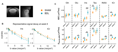 |
Diffusion MRI and MRS probe cerebellar microstructure alterations in the rat developing brain during hepatic encephalopathy
Jessie Mosso1,2,3, Mickaël Rey1,2, Dunja Simicic1,2,3, Katarzyna Pierzchala1,2,3, Ileana Ozana Jelescu4, and Cristina Cudalbu1,2
1CIBM Center for Biomedical Imaging, Lausanne, Switzerland, 2Animal Imaging and Technology (AIT), EPFL, Lausanne, Switzerland, 3LIFMET, EPFL, Lausanne, Switzerland, 4Service de radiodiagnostic et radiologie interventionnelle, Lausanne University Hospital CHUV, Lausanne, Switzerland Children affected by hepatic encephalopathy (HE) suffer from irreversible cognitive damage, and thus exploring the developing brain during HE is of crucial interest. Histology in an adult rat model of HE suggested alterations in the cerebellar microstructure, but there is a need for in vivo probes of these changes. Combining diffusion MRS and diffusion MRI, we measured increased metabolites’ diffusivities, as well as an increased intra-neurite/axon water diffusivity in white and gray matter in the cerebellum of a young rat model of HE compared to control rats, suggesting an alteration of cell density and/or of neurite network complexity. |
|
| 17:36 | 0156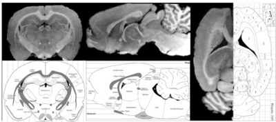 |
Ultra-high-resolution multi-parametric imaging on rats using MULTIPLEX at 9.4T
Yongquan Ye1, Renkuan Zhai2, and Hongxia Lei2
1UIH America, Inc., Houston, TX, United States, 2Wuhan United Imaging Life Science Instrument Co., Ltd., Wuhan, China
In this work, we report the initial experience of a state-of-the-art single-scan, high-resolution multi-parametric method, i.e. MULTIPLEX, on a 9.4T animal system. Ultra-high-resolution multi-parametric images with 0.12mm isotropic voxels were achieved on ex-vivo rat brain with atlas level anatomical details, while high resolution (0.1x0.1x0.4mm3) results with sufficient SNR were achieved with in-vivo rat brain.
|
|
| 17:48 | 0157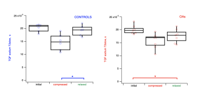 |
Mechanically Induced contrast in OA and control cartilage tissues at histological length scales probed by multi-scale 1H/23Na MRS and MRI at 9.4T.
Galina Pavlovskaya1, Onur Belek2, Thomas Meersmann1, Christopher Philp1, Jane McLaren2, David A Walsh2, and Brigitte E Scammell2
1SPMIC/Medicine, University of Nottingham, Nottingham, United Kingdom, 2Medicine, University of Nottingham, Nottingham, United Kingdom
We reveal intriguing experimental results providing the evidence that triple quantum filtered sodium relaxation time associated with histologically confirmed sodium bound to glycosaminoglycans (GAGs) in the cartilage matrix could be a marker for earlier osteoarthritis. The change appears only when cartilage specimens are subjected to mechanical load corresponding to 200N force applied normal to the cartilage surface. Hence, this mechanically induced change can potentially serve as a health score of cartilage tissues in health and disease in vivo.
|
|
| 18:00 | 0158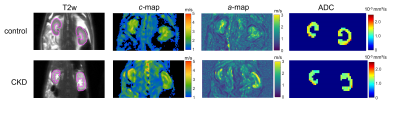 |
Renal fibrosis investigated by in vivo multifrequency MR elastography in a rat model of chronic kidney disease.
Karolina Garczyńska1,2, Julia Hahndorf1, Nicola Stolzenburg1, Matthias Taupitz1, Jürgen Braun3, Ingolf Sack1, Jörg Schnorr1, and Jing Guo1
1Department of Radiology, Charité Universitätsmedizin Berlin, Berlin, Germany, 2Department of Veterinary Pathology, Free University of Berlin, Berlin, Germany, 3Institute of Medical Informatics, Charité Universitätsmedizin Berlin, Berlin, Germany Using in vivo tomoelastography and multiparametric MRI (mp-MRI) we investigated kidneys of 10 rats with adenine-induced chronic kidney disease (CKD) and 8 healthy controls. In CKD rats, increased kidney volume, shear wave speed (SWS, related to stiffness) and wave penetration rate (related to inverse viscosity) were observed, while water diffusivity was reduced. These imaging findings were correlated with histopathologically quantified renal fibrosis. Our results suggested that collagen accumulation during CKD progression transforms soft-compliant renal tissue into a more rigid-solid state with reduced water mobility and shows that tomoelastography is a promising tool for non-invasive monitoring of disease progression. |
|
| 18:12 | 0159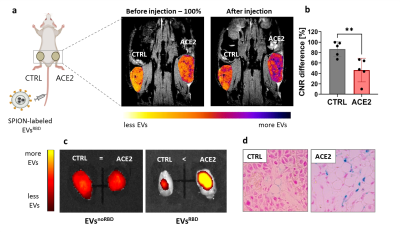 |
MRI tracking of genetically engineered extracellular vesicles as SARS-CoV-2 mimetics for studying viral-induced pathologies
Andrea Galisova1, Jiri Zahradnik1, Hyla Allouche-Arnon 1, Gideon Schreiber1, and Amnon Bar-Shir1
1Weizmann Institute of Science, Rehovot, Israel
Extracellular vesicles (EVs) are cell derived nanoformulations that allow to use genetic tools to engineer their features. Here, we show an MR imaging platform, which is based on genetically engineered magnetically labeled EVs, for mapping the binding of SARS-CoV-2 mimetics to the ACE2 receptor in vivo. The proposed imaging platform can help to better understand the mechanism and development of SARS-CoV-2 infection and in the delivery of proposed therapeutics. The presented principles can be further extended to study other viruses and viral mutations by engineering a tailored peptide on the EVs surface and thus study viral-induced pathologies by MRI.
|
 Back to Meeting Home
 Back to the Program-at-a-Glance
The International Society for Magnetic Resonance in Medicine is accredited by the Accreditation Council for Continuing Medical Education to provide continuing medical education for physicians.
 Back to Meeting Home
 Back to the Program-at-a-Glance
 View the Presentation