Oral Session
Preclinical Cancer
Joint Annual Meeting ISMRM-ESMRMB & ISMRT 31st Annual Meeting • 07-12 May 2022 • London, UK

| 14:45 | 0674 |
Oral delivery of 2H-labeled fumarate for deuterium magnetic resonance spectroscopic imaging of tumor cell death in vivo
Friederike Hesse1, Alan Wright1, Vencel Somai1,2, Flaviu Bulat1,3, and Kevin Brindle1,4
1CRUK CI, University of Cambridge, Cambridge, United Kingdom, 2Department of Radiology, University of Cambridge, Cambridge, United Kingdom, 3Department of Chemistry, University of Cambridge, Cambridge, United Kingdom, 4Department of Biochemistry, University of Cambridge, Cambridge, United Kingdom Cell death is an important imaging target for assessing early tumour treatment response and the effectiveness of therapy. We show here that 2H-labelled fumarate can be administered orally to detect cell death and assess early tumour treatment response in a subcutaneous lymphoma (EL4) model. Following oral gavage, 2H spectra were acquired from tumors with a time resolution of 5 min. Within 48h after chemotherapeutic drug (etoposide) treatment the tumor malate/fumarate signal ratios increased similarly to those measured after intravenous injection. |
|
| 14:57 | 0675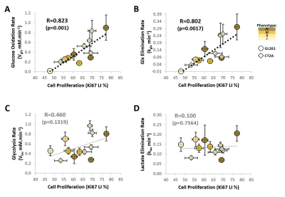 |
Cell proliferation correlates with glucose metabolic fluxes in mouse glioblastoma subtypes as assessed via deuterium MRS
Rui V Simoes1, Rafael N Henriques1, Beatriz M Cardoso1, Francisca F Fernandes1, Tania V Carvalho1, and Noam V Shemesh1
1Champalimaud Research, Champalimaud Centre for the Unknown, Lisbon, Portugal
Dynamic glucose-enhanced deuterium MRS (DGE 2H-MRS) coupled with Marchenko-Pastur PCA denoising has shown potential for in vivo quantification of glucose metabolism through glycolysis and mitochondrial oxidation in a mouse model of glioblastoma, and for assessment of pathway flux modulations according to tumor heterogeneity. Here, we extend this approach to immunocompetent mouse glioblastoma subtypes with marked differences in tumor cell metabolism and histopathologic features, to utterly demonstrate the potential of DGE 2H-MRS for non-invasive metabolic phenotyping of glioma, or other cancers with mitochondrial oxidation dependencies, according to key features of the tumor microenvironment such as cell proliferation.
|
|
| 15:09 | 0676 |
Deuterium magnetic resonance spectroscopy using 2H-pyruvate allows in vivo imaging of tumor burden and response to therapy Video Permission Withheld
Georgios Batsios1, Celine Taglang1, Meryssa Tran1, Anne Marie Gillespie1, Sabrina Ronen1, Joseph Costello2, and Pavithra Viswanath1
1Radiology and Biomedical Imaging, UCSF, San Francisco, CA, United States, 2Neurological Surgery, UCSF, San Francisco, CA, United States
Telomerase reverse transcriptase (TERT) is essential for tumor proliferation and is an attractive therapeutic target. Non-invasive methods of imaging TERT can report on tumor proliferation and response to therapy. Here, we show that 2H-MRS following administration of [U-2H]pyruvate non-invasively monitors TERT expression in clinically relevant patient-derived glioma models. Importantly, imaging TERT using [U-2H]pyruvate provides a readout of response to targeted TERT inhibitors and standard of care chemotherapy in vivo, at early timepoints that precedes MRI-detectable anatomical alterations. Clinical translation of our results can enable non-invasive assessment of TERT expression in vivo and, thereby, aid in imaging response to anti-cancer therapies.
|
|
15:21 |
0677 |
Proton and hyperpolarized 13C MRS based biomarkers response to the BAY-1436032 IDH inhibitor in cell and in vivo glioma models
Donghyun Hong1, Marina Radoul1, Noriaki Minami1, Anne Marie Gillespie1, Russell O. Pieper1, Joseph Costello1, Pavithra Viswanath1, and Sabrina M. Ronen1
1University of California, San Francisco, San Francisco, CA, United States
Mutant IDH1 inhibitor treatment is currently in clinical trials for glioma patients. Therefore, in vivo biomarkers are needed to assess early therapeutic responses. Here we treated mutant IDH1-expressing cells and animals with patient-derived mutant IDH1 brain tumors with the emerging inhibitor BAY-1436032. Using 1H MRS we found a significant decrease in 2HG that was accompanied by an increase in glutamate and phosphocholine, and using 13C MRS we detected a significant decrease in 2HG and a significant increase in glutamate produced from hyperpolarized [1-13C] α-ketoglutarate in cell and animal models.
|
|
| 15:33 | 0678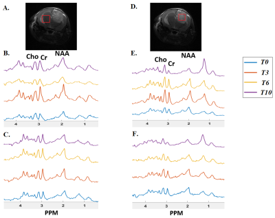 |
MRS to assess changes in total choline as a potential marker of treatment response to choline kinase inhibition in GL261 mouse glioblastomas
Sourav Bhaduri1, Jack Sharkey1, Soham Mukherjee1, Edward J Delikatny2, Sungheon G Kim3, and Harish Poptani1
1Centre for Preclinical Imaging, University of Liverpool, Liverpool, United Kingdom, 2Department of Radiology, University of Pennsylvania, Philadelphia, PA, United States, 3Department of Radiology, Weill Cornell Medical College, New York, NY, United States
This study investigates the effect of JAS239 on GL261 mouse glioblastoma using MRS. Higher reduction in Cho concentration and Cho/Cr ratios were observed in the tumour region than the contralateral normal brain after JAS239, indicating high sensitivity of MRS in as a pharmacodynamic marker of ChoK inhibition in glioblastomas with a potential of being an alternative treatment method.
|
|
| 15:45 | 0679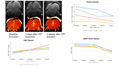 |
Advanced Vessel- and Cell-Size MRI to Assess Chemo-Radiation Treatment Response in Pediatric Ependymoma Models
Natalie J Serkova1, Jane S Manalo2, Jenna L Steiner2, Andrea M Griesinger2, Angela Pierce2, Mark S Brown2, and Nicholas F Foreman3
1Radiology, University of Colorado Anschutz Medical Campus, Aurora, CO, United States, 2University of Colorado Anschutz Medical Campus, Aurora, CO, United States, 3Children's Hospital Colorado, Aurora, CO, United States
We report on an advanced mpMRI protocol for characterizing the cellular and vascular phenotype in an orthotopic mouse model of pediatric ependymoma (EPN). Using diffusion-weighted based cell-size imaging, iron-oxide based vessel-size imaging and quantitative T2-maps, we report on an EPN-specific phenotype, characterized by an increased cell size (S=14 microns), increased vessel density index (Q=0.54), and low ADC values (0.63x10⁻³). Radiation treatment (10 Gy) in combination with 5-fluorouracil chemotherapy resulted in a decrease of gross tumor volumes, tumor necrosis with decreased cell sizes and increased ADC values, as well as a dramatic vascular-inflammatory response (highly decreased Q and DT2 values).
|
|
| 15:57 | 0680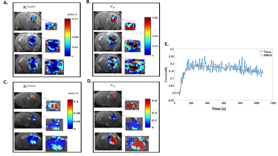 |
Assessing tumour haemodynamic heterogeneity and response to choline kinase inhibition using DCE-MRI in rodent models of glioblastoma
Sourav Bhaduri1, Jack Sharkey1, Clémentine Lesbats2, Claire L. Kelly1, Soham Mukherjee1, Edward J Delikatny3, Sungheon G Kim4, and Harish Poptani1
1Centre for Preclinical Imaging, University of Liverpool, Liverpool, United Kingdom, 2Division of Radiotherapy and Imaging, Institute of Cancer Research, London, United Kingdom, 3Department of Radiology, University of Pennsylvania, Philadelphia, PA, United States, 4Department of Radiology, Weill Cornell Medical College, New York, NY, United States
This study was designed to monitor changes in DCE-MRI based parameters Ktrans, ve, τi and Fp in pre-clinical GBM models in response to choline kinase inhibition using a cluster analysis approach. The study demonstrates that region-based clustered pharmacokinetic parameters obtained using DCE-MRI can be used for detecting and assessing tumour heterogeneities which may be useful in assessing therapeutic response.
|
 Back to Meeting Home
 Back to the Program-at-a-Glance
The International Society for Magnetic Resonance in Medicine is accredited by the Accreditation Council for Continuing Medical Education to provide continuing medical education for physicians.
 Back to Meeting Home
 Back to the Program-at-a-Glance
 View the Presentation