Oral Session
Advances in Treatment of Body Disorders
Joint Annual Meeting ISMRM-ESMRMB & ISMRT 31st Annual Meeting • 07-12 May 2022 • London, UK

Oral Session
Advances in Treatment of Body Disorders
| 09:15 | 0225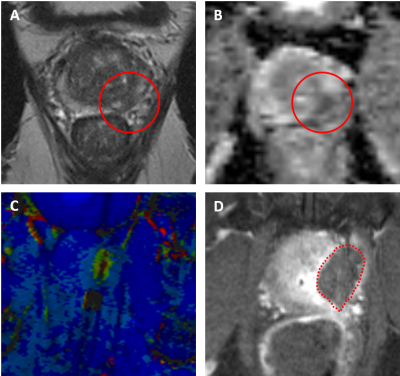 |
Transrectal magnetic resonance imaging and robot- guided focal laser ablation of prostate cancer: preliminary results of a phase 2 study.
Annemarijke Van Luijtelaar1, J.P. Michiel Sedelaar2, Joyce G.R. Bomers1, and Jurgen J. Fütterer1
1Medical Imaging, Radboudumc Nijmegen, Nijmegen, Netherlands, 2Urology, Radboudumc Nijmegen, Nijmegen, Netherlands
Magnetic resonance imaging-guided focal laser ablation is used as alternative to radical treatment while preserving healthy tissue and subsequently reduce treatment-related morbidity in patients with localized prostate cancer. Preliminary results of this prospective, non-randomized, clinical phase 2 study using a remote-controlled manipulator, show promising results in both oncological and functional outcomes in a group of 20 patients with low- to intermediate-risk (Gleason Score ≤ 4+3) prostate cancer. Transrectal magnetic resonance imaging-guided focal laser ablation is a promising outpatient procedure under local anesthesia for localized prostate cancer. It provides a fast and minimally-invasive strategy while reducing treatment related complications and side-effects.
|
|
| 09:27 | 0226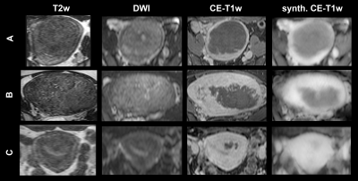 |
Diffusion-weighted MRI with deep learning for visualizing treatment results of MR-guided HIFU ablation of uterine fibroids.
Derk J. Slotman1,2, Lambertus W. Bartels2, Aylene Zijlstra1, Inez M. Verpalen1, Jochen A.C. van Osch1, Ingrid M. Nijholt1, Edwin Heijman3,4, Miranda van 't Veer-ten Kate1, Erwin de Boer1, Rolf D. van den Hoed1, Martijn Froeling2, and Martijn F. Boomsma1
1Radiology, Isala Zwolle, Zwolle, Netherlands, 2Image Sciences Institute, Imaging & Oncology Division, University Medical Center Utrecht, Utrecht, Netherlands, 3Faculty of Medicine and University Hospital of Cologne, Institute of Diagnostic and Interventional Radiology, University of Cologne, Cologne, Germany, 4High Tech Campus, Philips Research Eindhoven, Eindhoven, Netherlands The non-perfused volume (NPV) cannot be assessed repeatedly with contrast-enhanced imaging during MR-HIFU ablations of uterine fibroids, due to contrast agent dose constraints and safety concerns. In this study, synthetic contrast-enhanced (CE)-T1w scans were generated from diffusion weighted imaging (DWI) using deep learning-based image-to-image translation. A significant linear association was found between the NPV-ratios based on synthetic and paired reference CE-T1w scans (r=0.80, p<0.001). Radiologists agreed in 83% on treatment success based on synthetic and reference CE-T1w scans. This indicates that translation of DWI into synthetic CE-T1w scans has potential as method for gadolinium-free imaging of the NPV. |
|
| 09:39 | 0227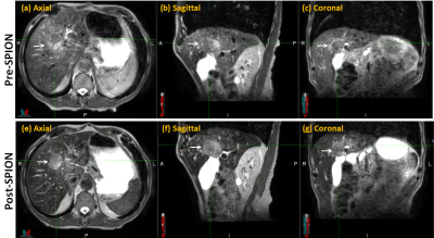 |
Application of Super-Paramagnetic Iron Oxide Nanoparticle to improve tumor visualization for MR-guided liver SBRT using Elekta Unity MR-Linac
Danny Lee1, Seungjong Oh1, Min-sig Hwang1, Mary McCauley1, Daniel Pavord1, Kyung Lim Yun1, Jason Sohn1,2, and Alexander V. Kirichenko1,2
1Radiation Oncology, Allegheny Health Network, Pittsburgh, PA, United States, 2Drexel University, Philadelphia, PA, United States
Ablative SBRT to liver malignancies on MRI-Linac is a novel and rapidly evolving technology allowing visualization of tumor and nearby organs at risk (OAR). Reliable identification of liver tumors has direct impact on radiotherapy planning and outcome. SPION agent Ferumoxytol® (Feraheme, AMAG Pharmaceuticals, Waltham, MA) was applied as MRI contrast during adaptive planning on Elekta Unity MR-Linac. Compared to the non-contrast images, liver tumors after Ferumoxytol® injection, were superiorly visible for accurate delineation during the entire treatment course. This study is the first to report the impact of SPION contrast agent on liver tumor visualization in a 1.5T Elekta MR-Linac.
|
|
| 09:51 | 0228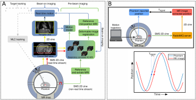 |
A novel 4D-MRI method enabling real-time image streaming for tumor tracking in radiotherapy
Katrinus Keijnemans1, Pim Borman1, Bas Raaymakers1, and Martin Fast1
1Department of Radiotherapy, University Medical Center Utrecht, Utrecht, Netherlands In MR-guided abdominothoracic radiotherapy, fast 2D imaging for MLC tracking and respiratory-correlated 4D imaging for dose accumulation is desirable. We developed a hybrid 2D/4D MRI method for pre-beam and beam-on imaging, which continuously acquires and reconstructs T2w coronal simultaneous multi-slice (SMS) images. Pre-beam 4D-MRIs were used to calculate a 3D mid-position reference image. Beam-on SMS data were used to extract real-time motion using a 4D-based motion model, and continuous 4D-MRIs. Dynamic updating of the 4D-motion model yielded the best real-time motion estimation. The presented hybrid 2D/4D MRI method can potentially facilitate novel MR-guided abdominothoracic radiotherapy workflows. |
|
| 10:03 | 0229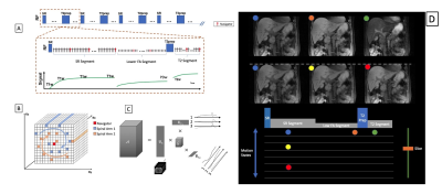 |
Integrated Multi-Task MR For Abdominal Adaptive Radiation Therapy
Junzhou Chen1,2, Pei Han2,3, Ye Tian4, Wensha Yang5, Debiao Li6,7, Krishna Nayak4, Yingli Yang8, Anthony Christodoulou6,9, and Zhaoyang Fan1,5,10
1Radiology, University of Southern California, Los Angeles, CA, United States, 2Bioengineering, University of California, Los Angeles, Los Angeles, CA, United States, 3Biomedical Imaging Research Institute, Cedars-Sinai Medical Center, Los Angeles, CA, United States, 4Electrical Engineering, University of Southern California, Los Angeles, CA, United States, 5Radiation Oncology, University of Southern California, Los Angeles, CA, United States, 6Cedars-Sinai Medical Center, Los Angeles, CA, United States, 7University of California, Los Angeles, Los Angeles, CA, United States, 8Radiation Oncology, University of California, Los Angeles, Los Angeles, CA, United States, 9Bioengineering, Cedars-Sinai Medical Center, Los Angeles, CA, United States, 10Biomedical Engineering, University of Southern California, Los Angeles, CA, United States
MR applications in radiation therapy has shown great promise for highly conformal treatment. However, MRgRT in the abdomen remains challenging due to motion and the sheer number of organs. In this work, we have demonstrated an integrated multi-task MR platform for adaptive radiation planning and treatment monitoring in the abdomen. In our proposed framework, from a single "pre-beam" scan we are able to generate a volumetric, multi-contrast and motion resolved images for adaptive treatment planning as well as enabling multi-contrast 3D real-time display during treatment. We demonstrate this capability in 3T and 0.55T.
|
|
| 10:15 | 0230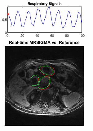 |
Real-time 3D MRI for Low-Latency Volumetric Motion Tracking on a 1.5T MR-Linac System
Can Wu1, Neelam Tyagi1, Marsha Reyngold2, Christopher Crane2, and Ricardo Otazo1,3
1Department of Medical Physics, Memorial Sloan Kettering Cancer Center, New York, NY, United States, 2Department of Radiation Oncology, Memorial Sloan Kettering Cancer Center, New York, NY, United States, 3Department of Radiology, Memorial Sloan Kettering Cancer Center, New York, NY, United States
Real-time 3D MRI for low-latency volumetric motion tracking was successfully implemented on a 1.5T MR-Linac system using the MRSIGMA framework. A first scan was performed for offline learning to obtain a training dictionary of ten 3D motion states. A second scan with the same sequence parameters was performed to generate the motion signature in real-time for online matching and was also used as a reference for retrospective self-validation. The feasibility of the technique was demonstrated on a healthy volunteer and a patient with pancreatic cancer which presented high quantitative concordance between contours of real-time MRSIGMA matching and the reference.
|
|
| 10:27 | 0231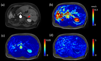 |
Y-90 treatment lung shunting prediction of hepatocellular cancer using DCE MRI with kinetic modeling and quantitative transport mapping
Qihao Zhang1, Kyungmouk Steve Lee2, Thanh Nguyen3, Pascal Spincemaille2, and Yi Wang2
1Cornell University, Ithaca, NY, NY, United States, 2Weill Cornell Medical College, New York, NY, United States, 3Cornell University, New York, NY, United States
Trans-arterial radioembolization with yttrium-90 microspheres (TARE with Y90) is a treatment for patients with liver cancers, but requires the evaluation of lung shunting fraction (LSF). Currently, LSF is estimated using a separate invasive “dry-run” with a transient radioactive Technetium-99m macroaggregated albumin (Tc-99m-MAA) that doubles the cost and risk of TARE. This study proposes to predict LSF from dynamic contrast enhanced MRI (DCE MRI) and perfusion quantification. Our preliminary data demonstrated that it is feasible to estimate LSF as measured by Tc-99m-MAA from noninvasive DCE MRI using quantitative transport mapping (QTM) velocity.
|
 Back to Meeting Home
 Back to the Program-at-a-Glance
The International Society for Magnetic Resonance in Medicine is accredited by the Accreditation Council for Continuing Medical Education to provide continuing medical education for physicians.
 Back to Meeting Home
 Back to the Program-at-a-Glance
 View the Presentation