Power Pitch Session
New Techniques for Processing & Analysis
Joint Annual Meeting ISMRM-ESMRMB & ISMRT 31st Annual Meeting • 07-12 May 2022 • London, UK

Power Pitch Session
New Techniques for Processing & Analysis
Power Pitch Session: How it Works
1st Hour: 2-minute Power Pitches in the Power Pitch Theater.
2nd Hour: 60-minute digital poster presentations at the smaller screens around the perimeter of the Power Pitch Theater.
14:45 |
0686.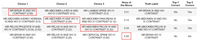 |
Designing a Clinical Decision Support System for MRI Radiology Titles Using Machine Learning Techniques and Electronic Medical Records
Peyman Shokrollahi1, Juan M. Zambrano Chaves1, Jonathan P.H. Lam1, Avishkar Sharma1, Debashish Pal2, Naeim Bahrami2, Akshay S. Chaudhari1, and Andreas M. Loening1
1Radiology, Stanford University, Stanford, CA, United States, 2GE Healthcare, Sunnyvale, CA, United States The use of inappropriate radiology protocols introduces risk of missed and incomplete diagnoses, thus endangering patient health, potentially prolonging treatment, and increasing healthcare costs. A clinical decision support system based on machine learning and electronic medical records of patients undergoing MRI was developed to predict radiology titles and their probabilities for radiologist review. A cumulative F1-score of ~85% was obtained for the top three predicted titles. The proposed system can guide physicians toward selecting appropriate titles and alert radiologists of potentially inappropriate selections, thereby improving imaging utility and increasing diagnostic accuracy, which favors better patient outcomes. |
|
| 14:47 | 0687.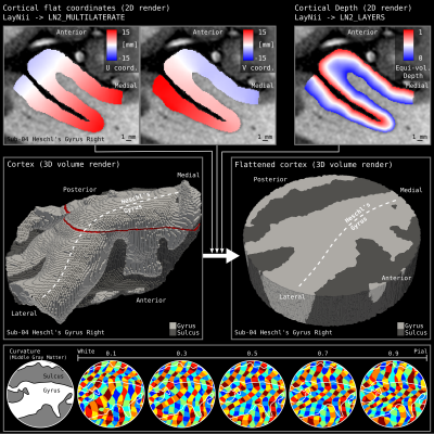 |
Brain in a virtual Petri dish: Flattening high resolution cortex patches in MRI and histology
Omer Faruk Gulban1,2, Renzo Huber3, Konrad Wagstyl4, Saskia Bolmann5, Dimo Ivanov3, Kendrick Kay6, and Rainer Goebel2,3
1Cognitive Neuroscience Department, Maastricht University, Maastricht, Netherlands, 2Brain Innovation, Maastricht, Netherlands, 3Cognitive Neuroscience, Maasticht University, Maastricht, Netherlands, 4UCL, London, United Kingdom, 5Center for Advanced Imaging, The University of Queensland, Queensland, Australia, 6Center for Magnetic Resonance Research, University of Minnesota, Minneapolis, MN, United States
High resolution imaging poses analysis challenges in the daily lives of high-resolution imaging researchers (high res. MRI, histology). One challenge is flattening patches of the cortex to study the cortical topography. We demonstrate a method that works in volume without requiring triangular meshes to represent the cortical surfaces at any stage of the algorithm. This approach for surface mapping enables mesoscopic analysis of cortical architecture. Our method is specifically developed for -but not limited to- flattening patches of the cortex. We implement our patch flattening as a C++ program and make it freely available within LayNii v2.1.0.
|
|
| 14:49 | 0688.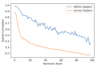 |
On the Reliability of Connectome Harmonic Decompositions of Human Brain Structure and Function
Brian S Winston1, Hoyt Patrick Taylor2, Pew-Thian Yap2, Frederick S Barrett1, and James J Pekar3,4
1Department of Psychiatry and Behavioral Sciences, Johns Hopkins University, Baltimore, MD, United States, 2Department of Radiology and BRIC, University of North Carolina, Chapel Hill, NC, United States, 3F.M. Kirby Research Center, Kennedy Krieger Institute, Baltimore, MD, United States, 4Department of Radiology, Johns Hopkins University, Baltimore, MD, United States
The connectome harmonic decomposition provides a principled approach to dimensionality reduction of human brain functional data using the eigenmodes of the graph Laplacian derived from the anatomical connectome. In the present report, test-retest data from the Human Connectome Project are used to assess the reliability of the structural harmonics, and of decompositions of resting-state functional MRI data using these harmonics. For both structure and function, reliability was higher within than between subjects, and decreased with harmonic rank.
|
|
| 14:51 | 0689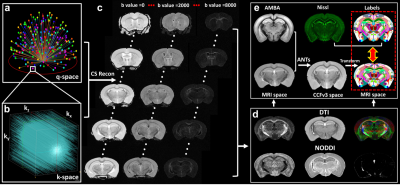 |
Comparing Diffusion MRI Metrics with 3D Nissl Staining of Whole Mouse Brain at Microscopic Resolution Video Permission Withheld
Nian Wang1,2 and Surendra Maharjan1
1Radiology and Imaging Sciences, Indiana University, Indianapolis, IN, United States, 2Stark Neurosciences Research Institute, Indiana University, Indianapolis, IN, United States
Magnetic resonance imaging (MRI) offers a noninvasive method for characterizing brain neuroanatomy and provides insights into various disease processes. Diffusion MRI (dMRI) has been used for assessment of tissue microstructure due to its exquisite sensitivity to finer scales far above imaging resolution. The recently developed Allen Mouse Brain Common Coordinate Framework (CCFv3) has opened the opportunities to integrate multimodal and multiscale datasets of whole mouse brain in 3D, and thus, this approach can be used to compare quantitative MRI parameters with conventional histology features throughout the whole brain.
|
|
| 14:53 | 0690.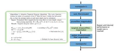 |
Model Pruning for Generalisability Across MRI Imaging Sites
Nicola Katharine Dinsdale1, Mark Jenkinson2,3, and Ana IL Namburete4
1WIN / Oxford Machine Learning in NeuroImaging Lab, University of Oxford, Oxford, United Kingdom, 2WIN, University of Oxford, Oxford, United Kingdom, 3Australian Institute for Machine Learning, University of Adelaide, Adelaide, Australia, 4Computer Science, Oxford Machine Learning in NeuroImaging Lab, University of Oxford, Oxford, United Kingdom
We propose an algorithm to simultaneously prune and train CNNs, leading to networks which have increased generalisability across imaging sites. Through segmentation of data from the ABIDE dataset, we show that through reducing the number of parameters in the network throughout training, we are able to reduce model overfitting, creating a model which is more robust to expect image variations across scanners. We also introduce a novel Targeted Dropout algorithm, which aids the process of model pruning. We demonstrate the approach on a UNet architecture, the basis of nearly all segmentation approaches across medical imaging.
|
|
| 14:55 | 0691.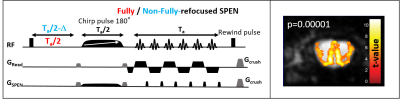 |
Functional MRI of mice olfactory bulbs at 15.2T reveals characteristic activation patterns when stimulated by different odors
Odélia Jacqueline Chitrit1, Qingjia Bao1, Silvia Chuartzman2, Noga Silkha2, Tali Kimchi2, and Lucio Frydman1
1Chemical and biological physics, Weizmann institute of Science, Rehovot, Israel, 2Neurobiology, Weizmann institute of Science, Rehovot, Israel Single-shot fMRI executed at ultrahigh fields can reveal valuable insight about brain function, if it successfully overcomes field inhomogeneity problems. Spatiotemporal Encoding provides a route for achieving this, enabling studies on the olfactory bulbs of mice at 15.2T. Images collected with a 125 µm in-plane resolution yielded remarkably large and well-defined responses to olfactory cues, particularly in males. These were unambiguously linked to olfaction via single-nostril experiments. The experiments highlighted specific activation regions in the external plexiform region and in glomeruli in the lateral part of the bulb, when stimulated by aversive or appetitive odors, respectively. |
|
| 14:57 | 0692.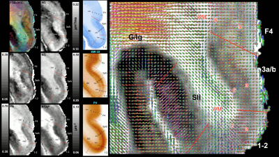 |
High-resolution MAP-MRI reveals cortical area specific laminar patterns observed with histological staining
Alexandru V Avram1,2, Kadharbatcha S Saleem1,2, Michal E Komlosh1,2, and Peter J Basser1
1Eunice Kennedy Shriver National Institute of Child Health and Human Development, National Institutes of Health, Bethesda, MD, United States, 2Center for Neuroscience and Regenerative Medicine, The Henry Jackson Foundation, Bethesda, MD, United States
We investigate the sensitivity of high-resolution MAP-MRI to distinguish cortical architectonic features by directly comparing laminar patterns in whole-brain volumes of MAP-derived parameters to the corresponding histological sections in the same macaque monkey brain. MAP parameters, in particular PA, NG, RTAP, DEC, and fODF maps, show cortical area-specific lamination patterns with excellent conspicuity in the mid-cortical layers, matching, and sometimes complementing, the contrast observed in the immuno- or histochemically stained sections. Delineating cortical areas with distinct cytoarchitectonic features non-invasively based on diffusion propagators could transform our ability to study brain networks and diagnose subtle microstructural changes in many clinical applications.
|
|
| 14:59 | 0693.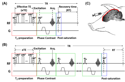 |
Quantitative validation of MRI mapping of cerebral venous oxygenation with direct blood sampling: a graded-O2 study in piglets
Dengrong Jiang1, Raymond C. Koehler2, Xiuyun Liu2, Ewa Kulikowicz2, Jennifer K. Lee2, Hanzhang Lu1,3,4, and Peiying Liu1
1Department of Radiology, Johns Hopkins University School of Medicine, Baltimore, MD, United States, 2Department of Anesthesiology and Critical Care Medicine, Johns Hopkins University School of Medicine, Baltimore, MD, United States, 3Department of Biomedical Engineering, Johns Hopkins University School of Medicine, Baltimore, MD, United States, 4F.M. Kirby Research Center for Functional Brain Imaging, Kennedy Krieger Research Institute, Baltimore, MD, United States
The neonatal brain relies primarily on oxygen metabolism to meet its enormous energy demands. Cerebral venous oxygenation (Yv) is an important parameter of the brain’s oxygen utilization, and has been demonstrated to be a potential biomarker in various neonatal diseases. We previously developed two non-invasive MRI techniques, TRUPC and accelerated-TRUPC, to measure Yv in the neonatal brain. In this work, we validated the accuracy of these two techniques by comparing their Yv measurements with gold-standard blood gas oximetry on a piglet model, and demonstrated that TRUPC and accelerated-TRUPC can provide accurate quantifications of Yv.
|
|
| 15:01 | 0694.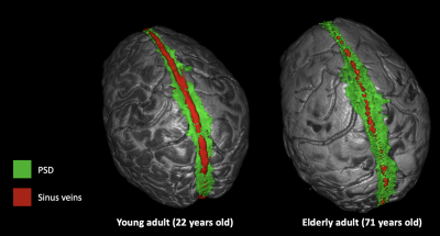 |
Novel non-invasive determination of parasagittal dural volume and cerebrospinal fluid flux: assessment of glymphatic clearance mechanisms
Kilian Hett1, Colin D. McKnight2, Jarrod J. Eisma1, Jason Elenberger1, Ciaran M. Considine1, Daniel O. Claassen1, and Manus J. Donahue1,3
1Neurology, Vanderbilt University Medical Center, Nashville, TN, United States, 2Radiology and Radiological Sciences, Vanderbilt University Medical Center, Nashville, TN, United States, 3Psychiatry and Behavioral Sciences, Vanderbilt University Medical Center, Nashville, TN, United States
Despite prior assumptions that the brain is devoid of lymphatic vessels, emerging evidence indicates that lymphatic vessels are present in the region alongside the superior sagittal sinus, the parasagittal dural (PSD) space, and could play a critical role in waste clearance or glymphatic physiology. However, limited methods are available for interrogating this space non-invasively in vivo. Here, in 62 healthy participants we apply novel deep learning algorithms in sequence with anatomical and phase contrast cerebrospinal fluid (CSF) flow assessments to test fundamental hypotheses regarding how PSD volume evolves across the lifespan and relates to measures of CSF flux and volume.
|
|
| 15:03 | 0695.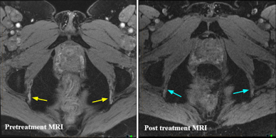 |
Use of Double-Echo Steady-State (DESS) sequence in the evaluation of pudendal nerve- A pilot trial.
Joseph Kus 1, Shakthi Kumaran Ramasamy1, Brett Ploussard 1, Joyce Cara 2, Stacey Bennis3, Collen Fitzgerald 3, Steve Shea 1, Ari D Goldberg 1, Judy James 4, and Anugayathri Jawahar1
1Radiology, Loyola University Medical Center, Maywood, IL, United States, 2Biostatistics, Stritch School of Medicine, Maywood, IL, United States, 3Physical Medicine and Rehabilitation, Loyola University Medical Center, Maywood, IL, United States, 4Radiology and Nuclear Medicine, Loyola University Medical Center, Maywood, IL, United States
Pudendal neuralgia is a common cause of chronic pelvic pain, especially in females. This is caused by pudendal nerve entrapment and can be a severely disabling neuropathic pain syndrome. It is currently a clinical diagnosis and most of the time a diagnosis of exclusion without definitive imaging criteria. We retrospectively compared DESS with the routine T2, T1 and DWI described in literature for the evaluation of pudendal nerves in patients with unresolved pelvic pain. Our study showed that DESS is effective and better than T2 and DWI combined sequences for diagnosing pudendal neuropathy.
|
|
| 15:05 | 0696.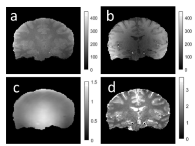 |
Optimization of dual flip angle T1 mapping at 7T with B1 transmit variability
Jyoti Mangal1,2, Sila Ayse Dokumaci1,2, David Leitao1,2, Raphael Tomi-Tricot1,2,3, Amer Ajanovic1,2, Stephen Ogier1,2, Tom Wilkinson1,2, Sharon Giles1,2, Pip Bridgen1,2, Claudia Prieto1, Joseph V Hajnal1,2, Shaihan Malik1,2, and David W Carmichael1,2
1Biomedical Engineering Department, School of Biomedical Engineering and Imaging Sciences, King's College London, London, United Kingdom, 2London Collaborative Ultra high field System (LoCUS), King’s College London, London, United Kingdom, 3MR Research Collaborations, Siemens Healthcare Limited, Frimley, United Kingdom
Quantitative MRI at 7T has advantages of enhanced resolution and uniform tissue contrast however protocol optimisation can be challenging as B1(+) field variability is increased. We maximized the precision of T1 estimation for a dual flip angle SPGR acquisition using Cramer-Rao Lower Bound framework and validated the results using a Monte Carlo simulation and both phantom and in-vivo experiments. For a TR of 20ms, simulations suggested optimal flip angles of 4° and 26° similar to in-vivo results of 4° and 28°. By accounting for other parameters, the CRLB framework can be flexibly extended to optimize more complex mapping protocols.
|
|
| 15:07 | 0697.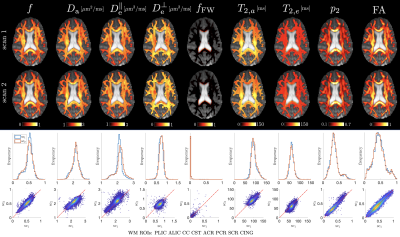 |
Reproducibility of white matter microstructure mapping with diffusion-relaxometry
Santiago Coelho1, Filip Szczepankiewicz2, Jelle Veraart1, Sohae Chung1, Yvonne W. Lui1, Dmitry S. Novikov1, and Els Fieremans1
1Center for Advanced Imaging Innovation and Research (CAI2R), Department of Radiology, New York University, School of Medicine, New York, NY, United States, 2Department of Diagnostic Radiology, Clinical Sciences, Lund University, Lund, Sweden
Joint modeling of diffusion and relaxometry in white matter is attractive due to its potential to provide unique insights into tissue integrity. We designed a diffusion MRI protocol containing varying b-values, b-tensor shapes, and echo times, employed machine learning parameter estimation, and studied the reproducibility of the Standard Model (SM) parameters. We quantified scan-rescan reproducibility in six healthy volunteers for compartmental water fractions, diffusivities, and $$$T_2$$$ relaxation times. We also found good agreement between SM parameters obtained from diffusion-only data at fixed echo time, and those from joint diffusion-relaxometry acquisitions, providing a consistency check for the SM assumptions.
|
|
| 15:09 | 0698.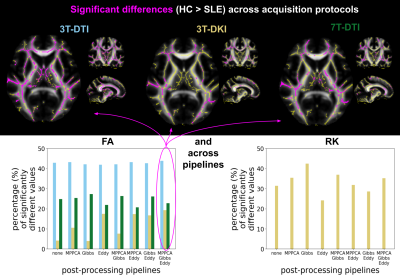 |
Sensitivity of dMRI with TBSS analysis to WM pathology in SLE: influence of field strength, acquisition approach and post-processing strategy
Evgenios N. Kornaropoulos1,2, Stefan Winzeck2,3, Linda Knutsson4,5,6, Marta M. Correia7, Pia C. Sundgren1,8,9, and Markus Nilsson1
1Diagnostic Radiology, Lund University, Lund, Sweden, 2Division of Anaesthesia, University of Cambridge, Cambridge, United Kingdom, 3Computing, Imperial College London, London, United Kingdom, 4Lund University, Lund, Sweden, 5Johns Hopkins University, Baltimore, MD, United States, 6Kennedy Krieger Institute, Baltimore, MD, United States, 7MRC Cognition and Brain Sciences Unit, University of Cambridge, Cambridge, United Kingdom, 8Skane University Hospital, Lund, Sweden, 9BioImaging Center, Lund University, Lund, Sweden
We examined whether more advanced dMRI increase the sensitivity to white matter pathology in systemic lupus erythematosus. Specifically, we examined whether more advanced acquisitions (DKI versus DTI, 7T versus 3T) and more complex processing (e.g. denoising, Gibbs-ringing removal) provide increased sensitivity. Tract-based spatial statistical analysis was applied, and the fraction of significant voxels in the white matter skeleton was used as the sensitivity metric. Results showed that the choice of the acquisition protocol had a greater impact on sensitivity than the choice of the post-processing strategy. 3T-DTI was more sensitive than 3T-DKI and 7T-DTI, regardless of the applied post-processing pipeline.
|
|
| 15:11 | 0699.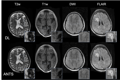 |
Unsupervised Diffeomorphic Registration for Even and Odd Echo Images with Applications to Point-of-Care MRI Reconstruction
Jo Schlemper1, Neel Dey2, Seyed Sadegh Mohseni Salehi1, Kevin Sheth3, W. Taylor Kimberly4, Lorraine Cullen5, and Michal Sofka1
1Hyperfine, Guilford, CT, United States, 2New York University, New York City, NY, United States, 3Yale University, New Haven, CT, United States, 4Massachusetts General Hospital, Boston, MA, United States, 5Gaylord Specialty Healthcare, Wallingford, CT, United States
We present an unsupervised deep image registration framework for MR image reconstruction. Specifically, even and odd echo images from fast spin echo-based sequence are nonlinearly registered using a convolutional network that estimates the deformation field. The registered echo images are then averaged for noise reduction. The proposed framework was evaluated across four imaging contrasts (T1w, T2w, FLAIR, and DWI) from a low-field MR scanner and was found to outperform nonlinear registration from advanced normalization tools, yielding sharper image quality and preserving important pathology features.
|
 Back to Meeting Home
 Back to the Program-at-a-Glance
The International Society for Magnetic Resonance in Medicine is accredited by the Accreditation Council for Continuing Medical Education to provide continuing medical education for physicians.
 Back to Meeting Home
 Back to the Program-at-a-Glance