Power Pitch
Pitch: Motion Correction
Joint Annual Meeting ISMRM-ESMRMB & ISMRT 31st Annual Meeting • 07-12 May 2022 • London, UK

Power Pitch Session: How it Works
1st Hour: 2-minute Power Pitches in the Power Pitch Theater.
2nd Hour: 60-minute digital poster presentations at the smaller screens around the perimeter of the Power Pitch Theater.
16:45 |
0350.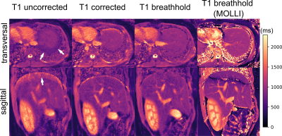 |
Prospective motion correction for 2D T1-Mapping in the liver using an ultra-wideband radar system
Tom Neumann1, Juliane Ludwig1, Kirsten M. Kerkering1, Peter Speier2, Frank Seifert1, and Christoph Kolbitsch1
1Physikalisch-Technische Bundesanstalt, Braunschweig and Berlin, Germany, 2Siemens Healthcare, Erlangen, Germany We present a radar-based approach for prospective motion correction for 2D T1 mapping of the abdomen. An ultra-wideband radar signal is acquired simultaneously but otherwise completely independent of the MR measurement. This allows us to correct for in-plane as well as through-plane motion during MR acquisition. This is especially important for 2D T1 mapping, where motion can cause not only blurring and streaking artifacts, but also inaccurate values when unexcited volumes move through the recorded image plane. It is demonstrated that the method strongly improves T1 maps in phantom and in-vivo scans. |
|
| 16:47 | 0351.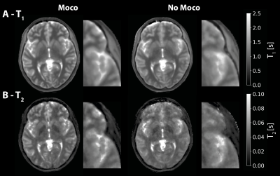 |
Silent Motion-Corrected ZTE for Quantitative T1 and T2 mapping
Emil Ljungberg1,2, Tobias C Wood2, Ana Beatriz Solana3, Gareth J Barker2, and Florian Wiesinger2,3
1Medical Radiation Physics, Lund University, Lund, Sweden, 2Neuroimaging, King's College London, London, United Kingdom, 3GE Healthcare, Munich, Germany
We present a method for retrospective motion-correction of silent ZTE for quantitative T1 and T2 mapping using self-navigation. A spiral phyllotaxis k-space trajectory was used to reconstruct motion navigators, which then were co-registered to obtain motion parameters. The method is demonstrated for neuroimaging, showing improved image quality of quantitative T1 and T2 maps after motion correction. These results further showcase the capacity of ZTE for robust, silent neuroimaging.
|
|
| 16:49 | 0352.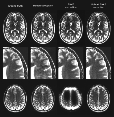 |
Energy dependent z-scores improve parallel imaging motion correction using trimmed autocalibrating k-space estimation (TAKE)
Jeremy Beaumont1, Thomas Troalen2, Swetali Nimje1,3, Stanislas Rapacchi1, and Ludovic de Rochefort1
1Aix Marseille Univ, CNRS, CRMBM, Marseille, France, 2Siemens Healthcare SAS, Saint-Denis, France, 3Aix Marseille Univ, CNRS, LIS, Marseille, France
MRI motion corruption prevents clinical interpretation and image analysis for research purposes. The trimmed autocalibrating k-space estimation based on parallel imaging and structured matrix completion (TAKE) algorithm was previously proposed to retrospectively correct motion corrupted raw k-space data. This study proposes specific modifications of the TAKE algorithm to decrease its computation time and improve the detection of motion-corrupted k-space data. The proposed changes significantly improve the TAKE motion detection sensitivity and specificity along with its motion correction performance, while sufficiently decreasing its computation time to allow for its use in a standard clinical routine workflow.
|
|
| 16:51 | 0353.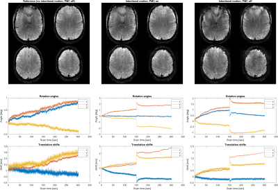 |
Prospective Head Motion Correction Using Orbital K-Space Navigators and a Linear Perturbation Model
Thomas Ulrich1, Malte Riedel1, and Klaas Pruessmann1
1Institute for Biomedical Engineering, ETH Zurich and University of Zurich, Zurich, Switzerland
It has recently been proposed to estimate head motion from orbital k-space navigators based on a linear signal model of the complex-valued navigator signal. In this work, we describe the combination of this algorithm with prospective motion correction, forming a control circuit that tracks the head so that the linear model remains valid also for large motion ranges. This ability is demonstrated by implementation and in-vivo imaging at 7T.
|
|
16:53 |
0354.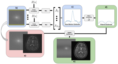 |
Spread-Spectrum Modulated Pilot Tone (SSMPT) for Motion Sensing
Daniel Raz Abraham1, Suma Anand1, and Michael Lustig1
1Electrical Engineering and Computer Sciences, University of California, Berkeley, Berkeley, CA, United States
Pilot Tone (PT) navigators are tones within the MR receiver bandwidth that are used to estimate subject motion. However, the PT must be placed outside the imaging band. This is not always feasible for all types of imaging (e.g. spine imaging) and systems, since the PT may overlap with the image. In this work, we propose Spread-Spectrum Modulated Pilot Tone (SSMPT), which spreads the energy of the pilot tone over the entire imaging band. SSMPT produces comparable motion estimates to PT and respiratory bellows, as demonstrated in a free-breathing scan with a healthy volunteer.
|
|
| 16:55 | 0355. |
Self-supervised Training for Single-Shot Tumor Tracking in the Presence of Respiratory Motion
Marcel Früh1,2, Tobias Hepp1,3, Andreas Schilling4, Sergios Gatidis1,3, and Thomas Küstner1
1Medical Image And Data Analysis (MIDAS.lab), Department of Interventional and Diagnostic Radiology, University Hospital of Tuebingen, Tuebingen, Germany, 2University of Tuebingen, Tuebingen, Germany, 3Max Planck Institute for Intelligent Systems, Tuebingen, Germany, 4Department of Computer Science, Institute for Visual Computing, University of Tuebingen, Tuebingen, Germany
Real-time tumor tracking is a task of growing importance due to the increasing availability of modern linear accelerators paired with MR imaging, called MR-LINAC. Physiological motion can thereby impair focal treatment of moving lesions.Classical tracking approaches often work inadequately because they operate only at the pixel level and thus do not include image-level information.Contrarily, learning based tracking systems typically require a large, fully-annotated dataset which is an arduous task to create. In this work, we propose a framework for example-based single-shot tumor tracking, which is trained without presence of labels and investigated for lesion tracking under respiratory motion.
|
|
| 16:57 | 0356.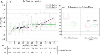 |
Measuring Diffusion of Healthy Human Muscle During Continuous Exercise from Motion-Compensated Intravoxel Incoherent Motion (IVIM) MRI
Anna N Foster1, Jaume Coll-Font1,2,3, Or Perlman2,3, Shi Chen1, Robert A Eder1, Christian T Farrar2,3, and Christopher T Nguyen1,2,3,4
1Cardiovascular Research Center, Massachusetts General Hospital, Charlestown, MA, United States, 2Athinoula A. Martinos Center for Biomedical Imaging, Massachusetts General Hospital, Charlestown, MA, United States, 3Harvard Medical School, Boston, MA, United States, 4Health Science Technology, Harvard-MIT, Cambridge, MA, United States
To date, it is unclear if diffusion increases in skeletal muscles during exercise. We measured diffusion, perfusion, perfusion fraction, and capillary blood flow in the calf during continuous exercise from IVIM fits on both standard (M0) and second order motion-compensated (M2) DWI sequences combined with motion artifact rejection. As diffusion estimates from M2 proved unchanged by artifact rejection at various thresholds, it was used in choosing an outlier threshold. Without artifact rejection, M0 reported diffusion increases. Once artifacts were removed from M0 using this threshold, it measured similar diffusion values to M2 and neither sequence showed diffusion increases during exercise.
|
|
| 16:59 | 0357.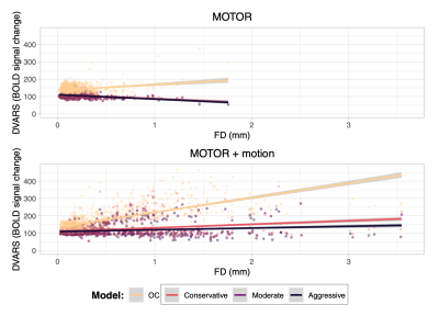 |
Denoising task-correlated head motion in motor-task fMRI data using multi-echo ICA
Neha A. Reddy1,2, Rachael C. Stickland1, Kimberly J. Hemmerling1,2, Kristina M. Zvolanek1,2, César Caballero-Gaudes3, and Molly G. Bright1,2
1Physical Therapy and Human Movement Sciences, Northwestern University, Chicago, IL, United States, 2Biomedical Engineering, Northwestern University, Evanston, IL, United States, 3Basque Center on Cognition, Brain and Language, Donostia, Spain
Multi-echo independent component analysis (ME-ICA) has been shown to differentiate the effects of head motion from desired BOLD signal in fMRI data, but this method has not been tested in motor-task studies with high amounts of task-correlated head motion. We investigated four denoising models on multi-echo motor-task data with limited and amplified task-correlated motion: Aggressive, Moderate, and Conservative ME-ICA nuisance regression models and a conventional optimally combined (OC) model. ME-ICA models were found to better dissociate head motion and BOLD signal variance than the OC model. Among them, the Aggressive model had the most consistent activation results with amplified motion.
|
|
| 17:01 |
0358.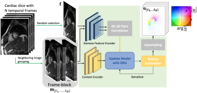 |
A group-wise cardiac motion estimation network leveraging temporal correlation
Jiazhen Pan1, Daniel Rueckert1,2, Thomas Kuestner3, and Kerstin Hammernik1,2
1AI in Medicine, Technical University of Munich, Munich, Germany, 2Department of Computing, Imperial College London, London, United Kingdom, 3Medical Image And Data Analysis (MIDAS.lab), University Hospital of Tübingen, Tübingen, Germany
Cardiac motion estimation is the gold-standard for assessing cardiac function and complementing cardiac image reconstruction. However, previous approaches in this area either suffered from long registration times or low accuracy because the inherent temporal correlation of the cardiac motion is not leveraged. In this work, we propose a method called GRAFT, which takes multiple cardiac frames as inputs to leverage the temporal correlation. Furthermore, temporal coherence is ensured by introducing the temporal smoothness loss during the training. Our experiments indicate that GRAFT can provide competitive deformation estimation results to state-of-the-art methods and outperform them in subsequent motion-compensated MRI reconstruction.
|
|
| 17:03 | 0359.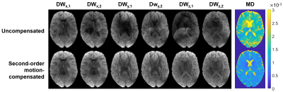 |
Multi-shot DW-EPI using a modified interleaving scheme and second-order motion-compensated diffusion sensitization
Eric Seth Michael1, Franciszek Hennel1, and Klaas Paul Pruessmann1
1Institute for Biomedical Engineering, ETH Zurich and University of Zurich, Zurich, Switzerland
Multi-shot techniques for diffusion MRI are desirable because of the associated improvement potential in image resolution but are inhibited by phase variability across interleaves, which results from subject motion and causes ghosting. Here, an EPI-based acquisition strategy is devised which diminishes the ghosting pattern by employing a modified interleaving scheme and mitigates motion sensitivity by nulling the first- and second-order moments of the diffusion-sensitizing gradient waveforms. This solution yielded unperturbed, high-resolution images in in vivo human brain scans and clear improvements in image quality compared to acquisitions incorporating standard interleaving and uncompensated diffusion sensitization.
|
|
| 17:05 | 0360.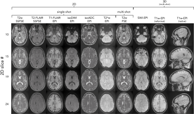 |
NeuroMix - A motion robust single scan brain exam
Tim Sprenger1,2, Adam van Niekerk2, Johan Berglund3, Henric Rydén2,4, Enrico Aventi2,4, and Stefan Skare2,4
1GE Healthcare, Stockholm, Sweden, 2Karolinska Institutet, Stockholm, Sweden, 3Uppsala University Hospital, Uppsala, Sweden, 4Karolinska University Hospital, Stockholm, Sweden
We have presented a new multi-contrast sequence, NeuroMix, which acquires T1w, T2w, T2-FLAIR, T2*w, and DWI contrasts in a single scan, requiring only one prescription and one prescan. NeuroMix includes single-shot EPI and FSE readouts as well as optional multi-shot FSE and 3DEPI acquisitions. The motion robustness of NeuroMix’s single-shot contrasts was generally good even when NeuroMix’s multi-shot sequences were unusable due to motion corruption resulting in a high probability to obtain diagnostic images. In conclusion, NeuroMix enables a fast and motion robust single-scan brain exam allowing higher patient throughput with less discomfort and possibly less need for sedation.
|
|
| 17:07 | 0361.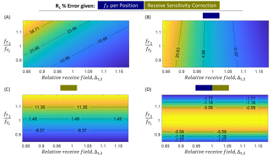 |
The impact and correction of inter-scan motion artefacts in variable flip angle R1 mapping at 3T and 7T
Martina F Callaghan1, Ali Aghaeifar1,2, Yael Balbastre1,3, and Nadège Corbin1,4
1Wellcome Centre for Human Neuroimaging, UCL Queen Square Institute of Neurology, London, United Kingdom, 2MR Research Collaborations, Siemens Healthcare Limited, Frimley, United Kingdom, 3Athinoula A. Martinos Center for Biomedical Imaging, Massachusetts General Hospital and Harvard Medical School, Boston, MA, United States, 4Centre de Résonance Magnétique des Systèmes Biologiques, CNRS/University Bordeaux, Bordeaux, France Images acquired with variable nominal flip angle (VFA) can be combined with a map of the transmit field efficiency and a signal model to estimate the longitudinal relaxation rate, R1. However, if motion occurs between the acquisition of the VFA volumes the inherent assumptions of consistent receive field modulation and transmit field efficiency can be violated causing substantial errors in R1. Through in vivo experiment and simulation we show that the differential receive field modulation introduces the largest amount of error. We further show that the majority of the error can be mitigated with a simple, time efficient correction. |
|
| 17:09 | 0362.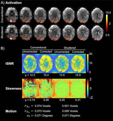 |
Shuttered Echo Planar fMRI with Dynamic Motion and Phase Correction
Saikat Sengupta1,2, Jonathan R Polimeni3,4,5, Kawin Setsompop6, and William A Grissom1,7
1Vanderbilt University Institute of Imaging Science, Nashville, TN, United States, 2Department of Radiology, Vanderbilt University Medical Center, Nashville, TN, United States, 3Athinoula A. Martinos Center for Biomedical Imaging, Charlestown, MA, United States, 4Department of Radiology, Harvard Medical School, Boston, MA, United States, 5Harvard-MIT Division of Health Sciences and Technology, Cambridge, MA, United States, 6Radiological Sciences Laboratory, Stanford University, Standford, CA, United States, 7Department of Biomedical Engineering, Vanderbilt University, Nashville, TN, United States
Achieving submillimeter-resolution BOLD fMRI is a challenge since single-shot EPI suffers from long echo times, blurring, and image distortions, while multishot EPI suffers from motion- and respiration-induced shot-to-shot phase errors. In shuttered multishot EPI, data are acquired in each shot after exciting a set of interleaved “shutters” across the imaged slice. This enables high quality reconstructions from each shot, which in turn enables navigator-free shot-wise phase and motion corrections prior to reconstructing a full-FOV image. Herein we describe a full motion- and phase-correcting image reconstruction for dynamic shuttered EPI and validate it in head motion and BOLD fMRI experiments.
|
|
| 17:11 | 0363.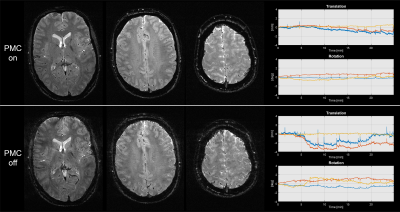 |
Advances in short-wave motion tracking: Jaw-mount markers and prospective motion correction
Christoph Michael Schildknecht1, David Otto Brunner1, Thomas Schmid1, and Klaas Paul Prüssmann1
1Institute for Biomedical Engineering, ETH Zurich and University of Zurich, Zürich, Switzerland
Prospective motion correction has demonstrated significant value for human brain MRI. One issue that all marker-based motion trackers share is the marker fixation to the human head. Short-wave motion tracking can solve this issue since the small wireless marker can directly be attached to the upper jaw with the mouth completely closed. In this work, we present PMC in-vivo imaging with such a sensor. The high temporal and spatial resolution can be observed in the measured physiological motion trajectories, illustrating the high-quality motion of this sensor.
|
|
| 17:13 | 0364.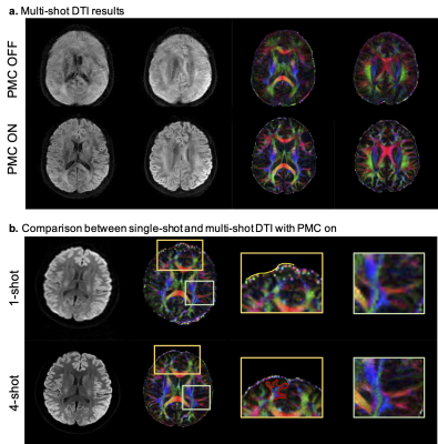 |
High resolution diffusion-weighted MRI combining markerless prospective motion correction and locally low-rank constrained reconstruction
Ke Dai1, Hao Chen1, Sijie Zhong1, Haoran Bai1, Jiaxu Zheng2, Xinyue Zhang2, Shasha Yang2, Tuoyu Cao2, Lucio Frydman3, Chaohong Wang2, and Zhiyong Zhang1
1School of Biomedical Engineering, Shanghai Jiao Tong University, Shanghai, China, 2United Imaging Healthcare, Shanghai, China, 3The Weizmann Institute of Science, Rehovot, Israel
High-resolution diffusion-weighted MRI is susceptible to head movement artifacts and background phase variations. We investigate a crucial combination of prospective motion correction and Spatial-angular local low rank (SPA-LLR) constrained reconstruction to obtain robust multi-shot high-resolution diffusion-weighted MRI under substantial motion. Prospective motion correction with optical markerless motion tracking is used to remove artifacts and blurring due to bulk motion whereas locally low-rank regularization is used to correct remaining artifacts due to the background phase variations from scan to scan. We present the results in healthy adult volunteers, demonstrating high-resolution diffusion-weighted images and FA images under different motion patterns with PMC.
|
 Back to Meeting Home
 Back to the Program-at-a-Glance
The International Society for Magnetic Resonance in Medicine is accredited by the Accreditation Council for Continuing Medical Education to provide continuing medical education for physicians.
 Back to Meeting Home
 Back to the Program-at-a-Glance