Power Pitch
Pitch: Image Acquisition, Reconstruction & Modeling
Joint Annual Meeting ISMRM-ESMRMB & ISMRT 31st Annual Meeting • 07-12 May 2022 • London, UK

Power Pitch
Pitch: Image Acquisition, Reconstruction & Modeling
Power Pitch Session: How it Works
1st Hour: 2-minute Power Pitches in the Power Pitch Theater.
2nd Hour: 60-minute digital poster presentations at the smaller screens around the perimeter of the Power Pitch Theater.
| 14:30 | 0298.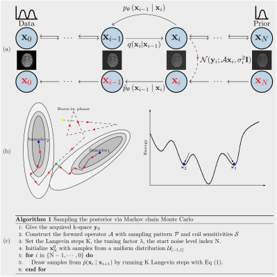 |
Using data-driven Markov chains for MRI reconstruction with Joint Uncertainty Estimation
Guanxiong Luo1, Martin Heide1, and Martin Uecker1,2,3,4
1University Medical Center Göttingen, Göttingen, Germany, 2Institute of Medical Engineering, Graz, Austria, 3German Centre for Cardiovascular Research (DZHK), Partner Site Göttingen, Göttingen, Germany, 4Cluster of Excellence "Multiscale Bioimaging: from Molecular Machines to Networks of Excitable Cells'' (MBExC), University of Göttingen, Göttingen, Germany
This works explores the use of data-driven Markov chains that are constructed from generative models for Bayesian MRI reconstruction, where the generative models utilize prior knowledge learned from an existing image database. Given the measured k-space, samples are then drawn from the posterior using Markov chain Monte Carlo (MCMC) method. In addition to the maximum a posteriori (MAP) estimate for the image which is obtained with conventional methods, also a minimum mean square error (MMSE) estimate and uncertainty maps can be computed from these samples.
|
|
| 14:32 | 0299.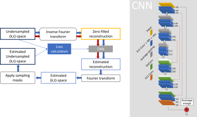 |
The deep SECRET to accelerated first-pass perfusion cardiac MRI
Elena Martín-González1, Ebraham Alskaf2, Amedeo Chiribiri 2, Pablo Casaseca-de-la-Higuera1, Carlos Alberola-López1, Rita G. Nunes3,4, and Teresa Correia2,5
1Laboratorio de Procesado de Imagen, Universidad de Valladolid, Valladolid, Spain, 2School of Biomedical Engineering and Imaging Sciences, King’s College London, London, United Kingdom, 3Institute for Systems and Robotics, Lisbon, Portugal, 4Department of Bioengineering, Instituto Superior Técnico, Universidade de Lisboa, Lisbon, Portugal, 5Centre for Marine Sciences - CCMAR, Faro, Portugal
First-pass perfusion cardiac magnetic resonance (FPP-CMR) is becoming essential to detect blow flow anomalies. However, the need for real-time acquisitions limits the achievable spatial resolution and coverage of the heart. To keep both within a reasonable range, FPP-CMR needs to be accelerated. A SElf-Supervised aCcelerated REconsTruction (SECRET) DL framework is presented to speed-up reconstruction of FPP-CMR images from undersampled (k,t)-space data. The physical reconstruction models are used to train deep neural networks without requiring fully sampled images. SECRET achieves good quality reconstructions at a variety of acceleration rates, with significant speed-ups compared to the state-of-the-art.
|
|
| 14:34 | 0300.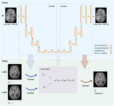 |
Through-plane diffusion MRI super-resolution with autoencoders: validation on outlier replacement scheme for pre-term baby brains
Hamza Kebiri1,2, Erick J. Canales-Rodríguez3, Priscille de Dumast1,2, Athena Taymourtash4, Hélène Lajous1,2, Yasser Alemán-Gómez2, Georg Langs4, and Meritxell Bach Cuadra1,2,3
1CIBM Center for Biomedical Imaging, Lausanne, Switzerland, 2Department of Radiology, Lausanne University Hospital and University of Lausanne, Lausanne, Switzerland, 3Signal Processing Laboratory 5 (LTS5), Ecole Polytechnique Fédérale de Lausanne (EPFL), Lausanne, Switzerland, 4Computational Imaging Research Lab, Department of Biomedical Imaging and Image-guided Therapy, Medical University of Vienna, Vienna, Austria
Although super-resolution diffusion MRI for isotropic volumes has been explored, to date no unsupervised SR techniques have been investigated for anisotropic dMRI. We propose an autoencoder based framework to enhance the through-plane spatial resolution and to replace slice outliers by leveraging existing high-quality datasets. Quantitative evaluation on 31 pre-term subjects show that the proposed framework significantly outperforms conventionally used interpolation methods at the raw data and estimated diffusion tensor maps. This can hence contribute to the depiction of more accurate white matter properties of the developing brain.
|
|
| 14:36 | 0301.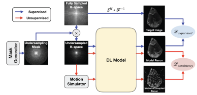 |
Motion2Recon: A Motion-Robust Semi-Supervised Framework for MR Reconstruction
Harris Beg1,2, Beliz Gunel2,3, Batu M Ozturkler2,3, Christopher M Sandino3, John M Pauly3, Shreyas Vasanawala4, Akshay S Chaudhari4,5, and Arjun D Desai3
1Computing and Mathematical Sciences, California Institute of Technology, Pasadena, CA, United States, 2Equal Contribution, Stanford University, Stanford, CA, United States, 3Electrical Engineering, Stanford University, Stanford, CA, United States, 4Radiology, Stanford University, Stanford, CA, United States, 5Biomedical Data Science, Stanford University, Stanford, CA, United States
In this study, we propose Motion2Recon, a semi-supervised consistency-based approach for robust accelerated MR reconstruction of motion-corrupted images. Motion2Recon reduced dependence on fully-sampled (supervised) training data and improves reconstruction performance among motion-corrupted scans. It also maintained superior performance among non-motion, in-distribution scans, which may help eliminate the need for manual motion detection. All code and experimental configurations are openly available in Meddlr (https://github.com/ad12/meddlr).
|
|
| 14:38 | 0302.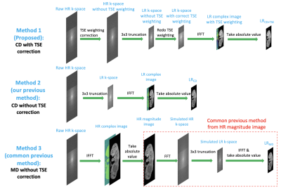 |
T2-deblurred deep learning super-resolution for turbo spin echo MRI
Zihao Chen1,2, Yibin Xie1, Debiao Li1,2, Yijen Wu3, and Anthony Christodoulou1,2
1Biomedical Imaging Research Institute, Cedars-Sinai Medical Center, Los Angeles, CA, United States, 2Department of Bioengineering, University of California, Los Angeles, Los Angeles, CA, United States, 3Department of Developmental Biology, University of Pittsburgh, Pittsburgh, PA, United States
Deep learning MR super-resolution (SR) is a promising approach to reduce scan time without requiring custom sequences and iterative reconstruction. However, previous SR approaches are incompatible with turbo spin echo (TSE) sequence due to differences in T2 blurring effects between high-resolution and low-resolution TSE images. Here we propose a T2-deblurred deep learning SR model for 3x3 in-plane super-resolution of 3D TSE images. Our method accelerated scanning by 9x in both retrospective and prospective testing and provided better image quality than previous SR methods.
|
|
| 14:40 | 0303.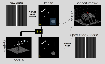 |
Assessment of resolution and noise in MR images reconstructed by data driven approaches
Katja Lauer1,2, Jonas Kleineisel1, Alfio Borzì2, Thorsten Alexander Bley1, Herbert Köstler1, and Tobias Wech1
1Department of Diagnostic and Interventional Radiology, University Hospital Würzburg, Würzburg, Germany, 2Institute of Mathematics, University of Würzburg, Würzburg, Germany
Data-driven reconstruction of undersampled raw data has gained more and more importance in recent years. Due to the non-linear and non-stationary transform characteristics of these imaging methods, objective image quality assessment is difficult. We propose a heuristic approach based on local point spread functions and multiple replica reconstructions, to enable the derivation of resolution- and g-factor-maps for individual images. The method is exemplarily applied in T1- and T2-weighted images of the brain, using a UNet and a Variational Network trained with data from the fastMRI project.
|
|
| 14:42 | 0304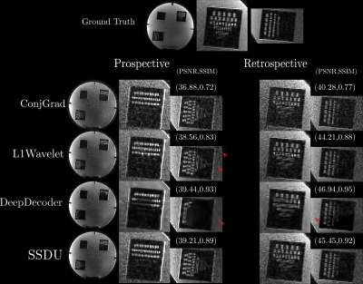 |
Comparison of Self-Supervised Image Reconstruction Methods for Undersampled Image Reconstruction: Validation in a Realistic Setting Video Permission Withheld
Thomas Yu1,2, Tom Hilbert1,3,4, Gian Franco Piredda1,3,4, Erick Canales-Rodrıguez1, Tobias Kober1,3,4, and Jean-Philippe Thiran1,4
1Signal Processing Lab 5 (LTS5), Ecole Polytechnique Fédérale de Lausanne, Lausanne, Switzerland, 2Medical Image Analysis Laboratory, Center for Biomedical Imaging (CIBM), University of Lausanne, Lausanne, Switzerland, 3Advanced Clinical Imaging Technology, Siemens Healthcare, Lausanne, Switzerland, 4Department of Radiology, Lausanne University Hospital and University of Lausanne, Lausanne, Switzerland
Self-supervised reconstruction methods for undersampled acquisitions are becoming increasingly used. We compare different self-supervised reconstruction methods using fully sampled and prospectively/retrospectively accelerated data; we find that prospective and retrospective reconstructions can differ significantly in quantitative metrics and perceptual quality. To test the methods’ generalizability, prospectively accelerated data from multiple field strengths is reconstructed without retraining/retuning. We find that no-reference image quality metrics can distinguish state of the art methods from the baseline, albeit with ambiguity between the state of the art methods.
|
|
| 14:44 | 0305.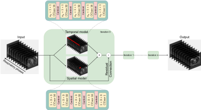 |
Accelerated respiratory-resolved 4D-MRI with separable spatio-temporal neural networks
Maarten Terpstra1,2, Matteo Maspero1,2, Joost JC Verhoeff1, and Cornelis A.T. van den Berg1,2
1Deparment of Radiotherapy, University Medical Center Utrecht, Utrecht, Netherlands, 2Computational Imaging Group for MR Diagnostics & Therapy, University Medical Center Utrecht, Utrecht, Netherlands Four-dimensional (4D) respiratory-resolved imaging is crucial for managing respiratory motion in radiotherapy, enabling irradiation of highly mobile tumours. However, acquiring high-quality 4D-MRI requires long acquisition (typically ≥5 minutes) and long iterative reconstructions, limiting treatment efficiency. Recently, deep learning has been proposed to accelerate undersampled MRI reconstruction. However, it has not been established whether deep learning may reconstruct high-quality 4D-MRI from accelerated acquisitions. This work proposes a small deep learning model that exploits the spatio-temporal information present in 4D-MRI, allowing to split the reconstruction into two separated branches, enabling high-quality retrospectively-accelerated 4D-MRI acquired in ~60 seconds and reconstructed in 16 seconds. |
|
| 14:46 | 0306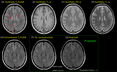 |
Mitigating synthetic T2-FLAIR artifacts in 2D MAGiC using keyhole and deep learning based image reconstruction Video Permission Withheld
Sudhanya Chatterjee1, Naoyuki Takei2, Rohan Patil1, Sugmin Gho3, Suchandrima Banerjee4, Florian Wiesinger5, and Dattesh Dayanand Shanbhag1
1GE Healthcare, Bengaluru, India, 2GE Healthcare, Tokyo, Japan, 3GE Healthcare, Seoul, Korea, Republic of, 4GE Healthcare, Menlo Park, CA, United States, 5GE Healthcare, Munich, Germany
Magnetic resonance image compilation (MAGiC) is a single click scan that provides multiple contrast-weighted images in around 5 mins. This makes it an effective diagnostic option in clinical settings. However, synthetic T2-FLAIR is known to have partial volume artifacts which impacts its diagnostic performance. In this work, we propose a method to mitigate partial-volume artifacts in synthetic T2-FLAIR using a separately acquired fast T2-FLAIR contrast information combined with keyhole and deep learning-based image reconstruction.
|
 Back to Meeting Home
 Back to the Program-at-a-Glance
The International Society for Magnetic Resonance in Medicine is accredited by the Accreditation Council for Continuing Medical Education to provide continuing medical education for physicians.
 Back to Meeting Home
 Back to the Program-at-a-Glance