Power Pitch
Pitch: "Propertography": Contrasts Based on Properties
Joint Annual Meeting ISMRM-ESMRMB & ISMRT 31st Annual Meeting • 07-12 May 2022 • London, UK

Power Pitch
Pitch: "Propertography": Contrasts Based on Properties
Power Pitch Session: How it Works
1st Hour: 2-minute Power Pitches in the Power Pitch Theater.
2nd Hour: 60-minute digital poster presentations at the smaller screens around the perimeter of the Power Pitch Theater.
09:15 |
0439.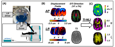 |
Anisotropic Mechanical Properties of White Matter Tracts in Aging via Transversely Isotropic MR Elastography
Diego A Caban-Rivera1, Daniel R Smith1, Matthew DJ McGarry2, L. Tyler Williams1, Grace McIlvain1, Elijah EW Van Houten3, Keith D Paulsen2, Phil V Bayly4, and Curtis L Johnson1
1University of Delaware, Newark, DE, United States, 2Dartmouth College, Hanover, NH, United States, 3Université de Sherbrooke, Sherbrooke, QC, Canada, 4Washington University in St. Louis, St. Louis, MO, United States This study combined multi-excitation MR Elastography (ME-MRE) with a transversely isotropic inversion nonlinear inversion algorithm and diffusion tensor imaging (DTI) fiber directions to investigate anisotropic white matter tract integrity in aging. Participants were 5 older adults and 17 young adults. MRE outcomes include substrate shear modulus, μ, shear anisotropy, φ, tensile anisotropy, ζ, and DTI measures were also examined. Shear and tensile anisotropies were significantly different in the Corona Radiata and Superior Longitudinal Fasciculus. Diffusion measures were significantly different between groups in most tracts. These results suggest sensitivity of anisotropic properties to biological changes along white matter tracts in aging. |
|
| 09:17 | 0440.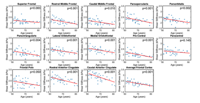 |
MR Elastography of the Frontal Cortex: Impact of Aging and Regional Differences
Kyra E. Twohy1, Peyton L. Delgorio2, Alexa M. Diano2, Mary K. Kramer2, Alexis M. Merritt2, Grace McIlvain2, Lucy V. Hiscox3, and Curtis L. Johnson2
1Mechanical Engineering, University of Delaware, Newark, DE, United States, 2Biomedical Engineering, University of Delaware, Newark, DE, United States, 3Psychology, University of Bath, Bath, United Kingdom
This study tested the utility of high-resolution magnetic resonance elastography (MRE) to analyze frontal cortex degradation due to advancing age. Brain tissue stiffness significantly varied in frontal cortex regions and frontal cortex stiffness also significantly decreased with age. A significant interaction between age and region was found, indicating regions changed differently with age. The stiffness of all frontal cortex regions degraded more per year than reported whole frontal lobe stiffness loss, indicating greater loss in the cortex with age. This approach brings potentially increased sensitivity and specificity to the structure-function relationship found in the frontal cortex with age.
|
|
| 09:19 |
0441.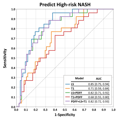 |
Performance of Proton Density Fat Fraction, MRE-based Liver Stiffness, and T1 in Identifying NASH Patients at High Risk of Disease Progression
Jiahui Li1, Xin Lu1, Zheng Zhu1, Kyle J. Kalutkiewicz1, Taofic Mounajjed2, Yi Sui1, Kevin J. Glaser1, Safa Hoodeshenas1, Sudhakar K. Venkatesh1, Armando Manduca1, Vijay H. Shah3, Richard L. Ehman1, Alina M Allen3, and Meng Yin1
1Radiology, Mayo Clinic, Rochester, MN, United States, 2Anatomic Pathology, Mayo Clinic, Rochester, MN, United States, 3Gastroenterology and Hepatology, Mayo Clinic, Rochester, MN, United States Multiparametric MRI/MRE and liver biopsy were performed in 104 patients at risk for NASH. We correlated measurements of proton density fat fraction (PDFF), 3D vector MRE-assessed liver stiffness (LS), and T1 relaxation time across histological features of NASH. We found that PDFF showed superior diagnostic performance in identifying patients with NASH (AUC: 0.84 [0.76, 0.92]), while LS showed superior diagnostic performance in stratifying NASH patients at high risk (0.85 [0.75, 0.94]). The results showed that T1 measurement, either alone or in combination with other biomarkers, did not perform as well as PDFF and LS in diagnosing NASH or stratifying high-risk NASH. |
|
| 09:21 | 0442.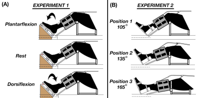 |
Estimation of anisotropic mechanical properties of gastrocnemius during muscle contraction with MR elastography
Daniel R. Smith1, Diego A. Caban-Rivera1, L. Tyler Williams1, Matthew McGarry2, Elijah Van Houten3, Phil V. Bayly4, Keith Paulsen2, and Curtis L. Johnson1
1Biomedical Engineering, University of Delaware, Newark, DE, United States, 2Thayer School of Engineering, Dartmouth College, Hanover, NH, United States, 3Universite de Sherbrooke, Sherbrooke, QC, Canada, 4Mechanical Engineering and Materials Science, Washington University in St. Lous, St. Louis, MO, United States
In this study, we utilized MR elastography (MRE) and transversely isotropic inversion to estimate anisotropic material parameters of muscle during contraction states, including passive muscle lengthening and isometric contraction. We collected MRE and DTI data on six subjects and took averages of the parameters within the medial and lateral heads of the gastrocnemius muscle and compared the results during the contraction states. We found significant differences between the estimates during the various states of contraction. Here we demonstrate that MRE and TI-NLI can generate anisotropic parameter estimates for muscle tissue while capturing changes to functional aspects of the tissue.
|
|
| 09:23 | 0443.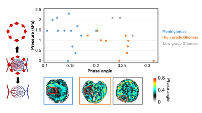 |
Gauging tumour pressure and vasculature organization via Magnetic Resonance Elastography to grade brain tumours.
Giacomo Annio1,2, Robin Bugge3, Siri Fløgstad Svensson3, Omar Darwish4, Giorgio Seano5, Donata Biernat6, Karoline Skogen6, Jon Ramm-Pettersen 7, David Nordsletten8, Einar Vik-Mo 7, Katharina Schregel9, Kyrre Eeg Emblem3, and Ralph Sinkus1
1LVTS - U1148, University Paris, Paris, France, 2School of Biomedical Engineering and Imaging Sciences, King's College London, London, United Kingdom, 3Department of Diagnostic Physics, Division of Radiology and Nuclear Medicine, Oslo University Hospital, Oslo, Norway, 4Department of Biomedical Engineering, King's College London, London, United Kingdom, 5U1021 INSERM, Institut Curie, Paris, France, 6Department of Radiology Ullevål, Division of Radiology and Nuclear Medicine, Oslo University Hospital, Oslo, Norway, 7Department of Neurosurgery, Division of Clinical Neuroscience, Oslo University Hospital, Oslo, Norway, 8Department of Biomedical Engineering and Cardiac Surgery, University of Michigan, Ann Arbor, MI, United States, 9Department of Neuroradiology, Heidelberg University Hospital, Heidelberg, Germany
Distinctive traits of malignant tumours are abnormal angiogenesis and high pressure. Conventional magnetic resonance imaging (MRI) plays a critical role in radiological evaluation of patients and tumour grading, but challenges remain. Pressure and vasculature have a strong impact on the tissue rheology and therefore they can be quantified by Magnetic Resonance Elastography (MRE). We show that MRE allows to quantify non-invasively tumour grade using pressure and tumour vasculature through wave scattering. We believe that MRE could play a central role in tumour grading and diagnosis as well as in therapy planning and dosage, especially in multidrug treatments scenarios.
|
|
| 09:25 | 0444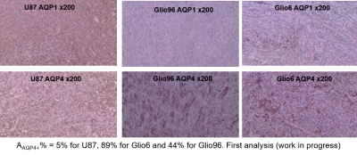 |
Correlation between pathophysiological functions of Glioma invasion and water dynamics by FFC-NMR in vivo Video Permission Withheld
Maria Rosaria Ruggiero1, Hamza Aït Itto2, Simona Baroni1, Jean Boutonnat3, François Berger2, Lionel Marc Broche4, Silvio Aime1, Simonetta Geninatti1, and Hana Lahrech2
1University of Torino, Torino, Italy, 2BrainTech Lab INSERM U1205, Grenoble, France, 3CHU Grenoble, Grenoble, France, 4University of Aberdeen, Aberdeen, United Kingdom
R1-dispersion profiles of in vivo glioma mouse models acquired by Fast-Field-Cycling NMR (FFC-NMR) were found to discriminate invasion from proliferation at fields below 2mT. These differences were correlated to the transcytolemmal water-exchange, demonstrating the water cell influx/outflux role in relaxation mechanisms. Hypoxia and H2O2, two major pathophysiological processes of invasion, were demonstrated to modulate relaxation. Immunohistochemistry of aquaporins AQP1 and AQP4 showed that the water-channel proteins were overexpressed in invasion but not in proliferation, suggesting that relaxations at low-field are modulated by water-exchange under the AQP1 and AQP4 control. The method can be extended to FFC imaging.
|
|
| 09:27 | 0445.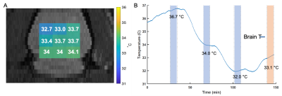 |
In-vivo Absolute Multinuclear Thermometry (AMT) in a Rat Model
Omid Yaghmazadeh1, Mihaly Voroslakos1, Mark Mattingly2, Zakia Ben Youss Gironda3, Youssef Zaim Wadghiri3, Seena Dehkharghani3,4, and Leeor Alon3,4
1Neuroscience, New York University School of Medicine, New York, NY, United States, 2Bruker cooperation, Billerica, MA, United States, 3Department of Radiology, New York University School of Medicine, New York, NY, United States, 4Center for Advanced Imaging Innovation and Research (CAI2R), New York University School of Medicine, New York, NY, United States
We introduce an in-vivo multinuclear MR thermometry technique which utilizes the unique frequency shift between water and sodium resonant frequencies. Reconstruction of the absolute temperature is validated using chemical shift imaging conducted on a rodent model implanted with a thin fiber optic probe for monitoring.
|
|
| 09:29 | 0446. |
Quantitative assessment of cerebral oxygen extraction fraction (OEF) in the medial temporal lobe
Dengrong Jiang1, Peiying Liu1,2, Zixuan Lin1, Abhay Moghekar3, Jay J. Pillai1,4, and Hanzhang Lu1,5,6
1Department of Radiology, Johns Hopkins University School of Medicine, Baltimore, MD, United States, 2Department of Diagnostic Radiology and Nuclear Medicine, University of Maryland School of Medicine, Baltimore, MD, United States, 3Department of Neurology, Johns Hopkins University School of Medicine, Baltimore, MD, United States, 4Department of Neurosurgery, Johns Hopkins University School of Medicine, Baltimore, MD, United States, 5Department of Biomedical Engineering, Johns Hopkins University School of Medicine, Baltimore, MD, United States, 6F.M. Kirby Research Center for Functional Brain Imaging, Kennedy Krieger Research Institute, Baltimore, MD, United States
The medial temporal lobe (MTL), including the hippocampus, is a key area implicated in many brain diseases, such as Alzheimer’s disease. Since neural activity is tightly coupled to the brain’s oxygen consumption, oxygen-extraction-fraction (OEF) in the MTL (MTL-OEF) may serve as a functional biomarker for this critical region. In this work, we developed a novel non-invasive MRI technique, AS-aTRUPC, to specifically measure the MTL-OEF in the human brain. We found that in healthy volunteers, the MTL-OEF is lower than the cortical OEF. The sensitivity of our technique in detecting changes in MTL-OEF was demonstrated in caffeine challenge experiments.
|
|
| 09:31 | 0447.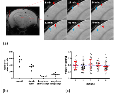 |
Time-lapse MRI: in vivo single-cell tracking of patrolling immune cells
Enrica Wilken1, Ina Fredrich1, Asli Havlas1, Felix Freppon1, Max Masthoff1, and Cornelius Faber1
1TRIC, Clinic of Radiology, University Hospital Muenster, Muenster, Germany
We validate that time-lapse MRI is able to resolve and follow single immune cells patrolling the brain vasculature. For that reason, we simulate motion-dependent contrast based on artificial k-space, imitate migrating cells with a rotating phantom system, and track iron-labeled monocytes in vivo with repeated T2* weighted imaging. Furthermore, we show that time-lapse MRI allows to differentiate between different motion patterns non-invasively and with whole-brain coverage, and to study altered motion behavior upon inflammatory stimulus and the onset of immune responses distant from the site of inflammation.
|
|
| 09:33 | 0448. |
Low-Dose Ferumoxytol Infusion Provides Effective Vascular Suppression for Magnetic Resonance Neurography
Emily Pedrick1, Darryl B Sneag1, Philip G Colucci1, Mylinh Duong1, Alyssa Vanderbeek1, and Ek T Tan1
1Hospital for Special Surgery, New York, NY, United States
This study investigated the effectiveness of a low-dose (25% of a single treatment dose for anemia) ferumoxytol-infusion for vascular suppression in T2-weighted MR neurography of the brachial plexus. 3D STIR-FSE and 2D Dixon-FSE sequences of the brachial plexus were acquired in 12 healthy volunteers pre- and post-ferumoxytol. Quantitative and subjective assessments by two readers demonstrated superior vascular suppression and nerve visualization post-ferumoxytol without negatively affecting soft tissue contrast or fat suppression.
|
|
| 09:35 | 0449.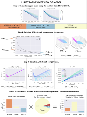 |
Modelling the effect of hyperoxia on the spin-lattice relaxation rate R1 of tissues
Emma Bluemke1, Eleanor Stride1, and Daniel Bulte1
1Institute of Biomedical Engineering, University of Oxford, Oxford, United Kingdom
The paramagnetic relaxivity effect of oxygen on longitudinal relaxation rate (R1) has been explored as a means of inferring partial pressure of oxygen in the tissue or bodily fluids, and examining tissue R1 response to hyperoxic gas breathing is used in research techniques such as oxygen-enhanced MRI. We present a 3-compartment model for estimating the hyperoxia-induced changes in R1 of tissues depending on B0, SO2, blood volume, hematocrit, oxygen extraction fraction, and changes in blood and tissue PO2. It is easy for researchers to tailor this model to their tissue of interest by substituting preferred tissue oxygen diffusion/consumption models.
|
|
| 09:37 | 0450. |
Interplay of Iron and Copper in the Neuromelanin-Related Paramagnetic Relaxation Enhancement
Niklas Wallstein1, Andreas Pöppl2, Andrea Capucciati3, André Pampel 1, Carsten Jäger1, Fabio A. Zucca4, Enrico Monzani3, Luigi Casella3, Luigi Zecca4, and Harald E. Möller1,2
1Max Planck Institute for Human Cognitive and Brain Sciences, Leipzig, Germany, 2Leipzig University, Faculty of Physics and Earth Sciences, Felix Bloch Institute for Solid State Physics, Leipzig, Germany, 3Department of Chemistry, University of Pavia, Pavia, Italy, 4Institute of Biomedical Technologies, National Research Council of Italy, Segrate, Italy Neuromelanin-sensitive MRI receives interest as potential biomarker in neurodegenerative diseases, such as Parkinson’s and Alzheimer’s disease. It is known that human neuromelanin pigments bind large quantities of toxic metal ions, especially iron but also other transition metals including copper. These melanin-iron complexes are a potential source of paramagnetic relaxation enhancement of water proton. In relaxometry investigations, we found deviations from a simple linear concentration-dependent T1 shortening in synthetic neuromelanins containing different amounts of iron and copper. Knowledge of the occupation of distinct metal binding sites seems crucial for contrast optimization or attempts to quantify metal content by MRI. |
 Back to Meeting Home
 Back to the Program-at-a-Glance
The International Society for Magnetic Resonance in Medicine is accredited by the Accreditation Council for Continuing Medical Education to provide continuing medical education for physicians.
 Back to Meeting Home
 Back to the Program-at-a-Glance