Power Pitch
Pitch: Molecular
Joint Annual Meeting ISMRM-ESMRMB & ISMRT 31st Annual Meeting • 07-12 May 2022 • London, UK

Power Pitch Session: How it Works
1st Hour: 2-minute Power Pitches in the Power Pitch Theater.
2nd Hour: 60-minute digital poster presentations at the smaller screens around the perimeter of the Power Pitch Theater.
| 14:45 | 0124.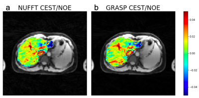 |
GraspCEST: Fast Free-Breathing 3D Steady-State CEST MRI Using Golden-Angle Radial Sparse MRI
Xiang Xu1, Ding Xia1, Kai Tobias Block2, and Li Feng1
1Department of Radiology and BioMedical Engineering and Imaging Institute, Icahn School of Medicine at Mount Sinai, New York, NY, United States, 2Center for Advanced Imaging Innovation and Research (CAI2R) and Department of Radiology, New York University School of Medicine, New York, NY, United States
In this project, we proposed a fast 3D CEST MRI technique called GraspCEST, which is based on the combination of CEST-prepared stack-of-stars golden-angle radial sequence and multicoil compressed sensing reconstruction. Using this technique, we were able to perform lipid suppressed, free-breathing 3D CEST imaging of the liver. We have demonstrated that it is possible to accelerate imaging speed and achieve full Z-spectra acquisition of the liver within 5 min. The GraspCEST technique can be a useful tool in studying glycoNOE in the liver.
|
|
| 14:47 | 0125.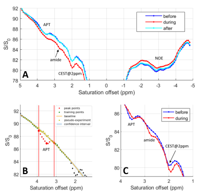 |
Acquisition and data processing considerations in functional metabolic imaging using CEST
Solène Bardin1,2, Fawzi Boumezbeur1,2, and Luisa Ciobanu1,2
1NeuroSpin, CEA, Gif-sur-Yvette, France, 2Paris-Saclay University, Gif-sur-Yvette, France
Accelerated acquisitions with a CEST-linescan sequence coupled with the estimation of the local baseline for the analysis of the Z-spectra allow to simultaneously monitor dynamic changes at several CEST contributions induced by a controlled pH decrease. This approach can be a promising strategy for detecting functional induced metabolic changes devoid of confounding BOLD effects.
|
|
| 14:49 | 0126.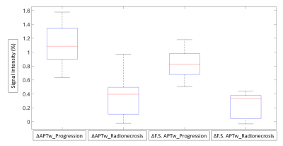 |
Predicting radionecrosis and recurrent disease through Amide Proton Transfer weighted imaging in brain metastases
Lucia Nichelli1,2, Julian Jacob3, Delphine Leclerq1, Farhat Benbelkacem4, Stéphane Lehéricy1,2, and Stefano Casagranda5
1Department of Neuroradiology, Assistance Publique-Hôpitaux de Paris, Groupe Hospitalier Pitié-Salpêtrière-Charles-Foix, Sorbonne Université, Paris, Paris, France, 2Paris Brain Institute – Institute du Cerveau (ICM), Centre de NeuroImagerie de Recherche (CENIR), F-75013, Paris, France, 3Department of Radiation-Oncology, Assistance Publique-Hôpitaux de Paris, Groupe Hospitalier Pitié-Salpêtrière-Charles-Foix, Sorbonne Université, Paris, France, 4Siemens Healthcare SAS, Saint-Denis, Paris, France, 5Department of Research & Innovation, Olea Medical, La Ciotat, France
Distinguishing tumor recurrence from radionecrotic injury of pre-irradiated brain metastases is fundamental to provide optimal patient care. Unfortunately, this distinction is often hard to make even with advanced MRI multimodal protocols. This study aims to evaluate APTw imaging in predicting the differentiation between radio-induced tissue changes from tumor progression at 3T in 20 pre-irradiated metastases. Results show that APTw metrics can significantly separate these two common radiological entities (p<0.0001) and suggest the use of fluid-suppressed APTw to reach higher discriminating values.
|
|
| 14:51 | 0127.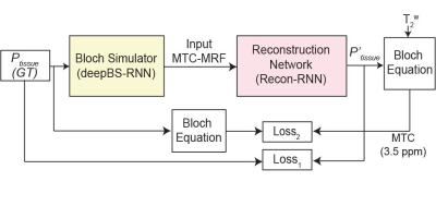 |
Deep-Bloch Simulator Driven Recurrent Neural Network for Reliable Semisolid Magnetization Transfer Contrast MRF and CEST Imaging
Munendra Singh1, Babak Moghadas1, Shanshan Jiang1, Peter van Zijl1, Jinyuan Zhou1, and Hye-Young Heo1
1Johns Hopkins University, Baltimore, MD, United States
High-resolution MRF including a multi-pool saturation transfer model requires a huge dictionary or training dataset simulated with Bloch equations. Furthermore, intensive Bloch simulation tasks are inevitable for MRF schedule optimization. In this study, we developed a deep-learning-based ultrafast Bloch simulator and a recurrent neural network (RNN) for semi-solid macromolecular magnetization transfer contrast (MTC) MRF reconstruction. For the MRF training dataset generation, the deep-learning Bloch simulator required ~200x less time than a conventional Bloch simulation. A test-retest study showed excellent reliability of the tissue parameter quantification using the proposed RNN framework.
|
|
14:53 |
0128.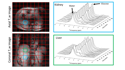 |
Monitoring glucose uptake and metabolism in human kidney with dynamic 3D DMI at 7 T
Ayhan Gursan1, Arjan D. Hendriks1, Dimitri Welting1, Dennis W.J. Klomp1, and Jeanine J. Prompers1
1Department of Radiology, University Medical Center Utrecht, Utrecht, Netherlands
Kidneys play a crucial role in glucose homeostasis via glucose reabsorption and glucose release into and glucose uptake from the circulation. We investigated the feasibility of measuring renal glucose uptake and metabolism with dynamic 3D DMI. A volunteer was scanned twice with two different ways of deuterated glucose administration: in the scanner and before scanning session. Similar glucose uptake was observed in the kidney and liver. We demonstrated the feasibility of measuring renal glucose uptake and metabolism with dynamic 3D DMI. This technique has potential to study the role of renal glucose uptake in glucose homeostasis in diabetes.
|
|
| 14:55 | 0129.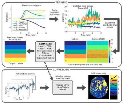 |
Curve shape analysis of dynamic glucose enhanced (DGE) and dynamic contrast enhanced (DCE) MRI in patients with brain tumor
Anina Seidemo1, Ronnie Wirestam1, Gunther Helms1, Karin Markenroth Bloch2, Xiang Xu3,4, Johan Bengzon5,6, Pia C. Sundgren2,7,8, Peter C.M. van Zijl3,9, and Linda Knutsson1,3,9
1Department of Medical Radiation Physics, Lund University, Lund, Sweden, 2Lund University Bioimaging Center, Lund University, Lund, Sweden, 3Russell H. Morgan Department of Radiology and Radiological Science, Johns Hopkins University School of Medicine, Baltimore, MD, United States, 4BioMedical Engineering and Imaging Institute, Icahn School of Medicine at Mount Sinai, New York, NY, United States, 5Lund Stem Cell Center, Department of Clinical Sciences, Lund University, Lund, Sweden, 6Division of Neurosurgery, Department of Clinical Sciences, Lund University and Skåne University Hospital, Lund, Sweden, 7Department of Medical Imaging and Physiology, Skåne University Hospital, Lund and Malmö, Sweden, 8Diagnostic Radiology, Department of Clinical Sciences, Lund University, Lund, Sweden, 9F.M. Kirby Research Center for Functional Brain Imaging, Kennedy Krieger Institute, Baltimore, MD, United States
Dynamic glucose-enhanced (DGE) MRI provides time intensity curves after D-glucose injection and is promising as a less invasive alternative or complement to dynamic contrast-enhanced (DCE) MRI. Here, we calculated non-model-based parameter maps from DGE and DCE MRI of human brain tumors, and created color-coded “curve maps”, a graphic representation of different curve shapes, to further investigate temporospatial enhancement patterns. The results show that DGE MRI can differentiate tumor from healthy brain tissue, that DGE and DCE give similar but not identical information, and that the proposed curve map approach has potential to aid in visual assessment of dynamic images.
|
|
| 14:57 | 0130.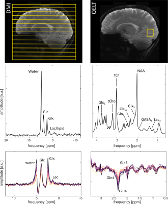 |
Observation of 2H labeled compounds in the human brain with 1H versus 2H magnetic resonance spectroscopy at 9.4T
Loreen Ruhm1,2,3, Theresia Ziegs1,2, Andrew Wright1,2, Nikolai Avdievich1, and Anke Henning1,3
1Max Planck Institute for Biological Cybernetics, Tuebingen, Germany, 2IMPRS for Cognitive and Systems Neuroscience, Eberhard-Karls University of Tuebingen, Tuebingen, Germany, 3Advanced Imaging Research Center, UT Southwestern Medical Center, Dallas, TX, United States
The tracing of glucose metabolism is of interest in many pathologies of the human brain. In this abstract, we compare the information content of 2H MRSI detected deuterium metabolic imaging (DMI) to quantitative exchanged-labeled turnover 1H MRS (QELT), both measured at B0 = 9.4 Tesla. An uptake of 2H labeling was detectable for different metabolites for both methods. Consistent changes in 2H labeled Glx after the oral administration of [6,6’-2H2]-glucose were detected in the brain of healthy human subjects.
|
|
| 14:59 | 0131.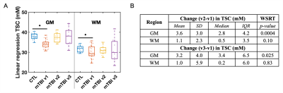 |
Longitudinal 23Na MRI in mild traumatic brain injury
Teresa Gerhalter1, Anna M Chen1, Seena Dehkharghani1,2, Rosemary Peralta1, James S. Babb1, Tamara Bushnik3, Alejandro Zarate3, Jonathan M Silver4, Brian S Im3, Stephen P Wall5, Ryan Brown1,6, Ivan I Kirov1,2,6, and Guillaume Madelin1
1Center for Biomedical Imaging, Department of Radiology, New York University Grossman School of Medicine, New York, NY, United States, 2Department of Neurology, New York University Grossman School of Medicine, New York, NY, United States, 3Department of Rehabilitation Medicine, New York University Grossman School of Medicine, New York, NY, United States, 4Department of Psychiatry, New York University Grossman School of Medicine, New York, NY, United States, 5Ronald O. Perelman Department of Emergency Medicine, New York University Grossman School of Medicine, New York, NY, United States, 6Center for Advanced Imaging Innovation and Research, Department of Radiology, New York University Grossman School of Medicine, New York, NY, United States
In this longitudinal sodium MRI study, mild traumatic brain injury (mTBI) patients and controls were scanned at 3T. Linear regression analysis was used to calculate total sodium concentrations (TSC) in global grey and white matter (GM, WM). Patient GM TSC increased back to control levels at 3-month and 1-year follow-ups. Decreased GM and WM TSC measured at one month were associated with worse cognitive performance, but not worse symptomalogy, at the follow-up visits.
|
|
| 15:01 | 0132.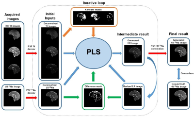 |
A method to increase the resolution of sodium images from simultaneous 1H MRF/23Na MRI
Gonzalo G Rodriguez1, Zidan Yu1,2, Lauren O'Donnell1, Liz Calderon1, Sarah Shaykevich2, Martijn A Cloos3,4, and Guillaume Madelin1,2
1Center for Biomedical Imaging, Department of Radiology, New York University School of Medicine, New York, NY, United States, 2Vilcek Institute of Graduate Biomedical Sciences, NYU Langone Health, New York, NY, United States, 3Centre for Advanced Imaging, The University of Queensland, Brisbane, Australia, 4ARC Training Centre for Innovation in Biomedical Imaging Technology, The University of Queensland, Brisbane, Australia
In this work, we present an algorithm to generate a high-resolution 23Na image from simultaneously-acquired low-resolution 23Na density-weighted MRI (2.85×2.85×5 mm3) and high-resolution 1H density, T1, and T2 maps from MRF (1.5×1.5×5 mm3) in brain at 7 T. As a result, the mean value of the difference between the generated and ground truth high-resolution 23Na images is 0.5% with a standard deviation of 6.2% and multi-scale structural similarity index of 0.97.
|
|
| 15:05 | 0133.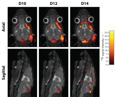 |
High-Resolution Compressed Sensing 19F-MRI Reveals Progression of Neuroinflammation in Autoimmune Encephalomyelitis
Ludger Starke1,2, Paula Ramos Delgado1, Jason M. Millward1, Sabrina Klix1, Thoralf Niendorf1,3, and Sonia Waiczies1
1Berlin Ultrahigh Field Facility, Max Delbrück Center for Molecular Medicine in the Helmholtz Association, Berlin, Germany, 2Digital Health Center, Hasso Plattner Institute, Potsdam, Germany, 3Experimental and Clinical Research Center (ECRC), A Joint Cooperation between the Charité Medical Faculty and the Max-Delbrück Center for Molecular Medicine, Berlin, Germany
Improving sensitivity has been the major challenge towards realizing the promises of 19F-MRI. Here we combine a cryogenic surface RF probe and compressed sensing to achieve high spatially resolved 19F-MRI of neuroinflammation in a challenging longitudinal study. Inflammation in the CNS was observed even in the absence of neurological symptoms and detailed signals along the CNS vasculature are clearly resolved. Together with effective longitudinal registration this allows an in-depth study of disease progression.
|
 Back to Meeting Home
 Back to the Program-at-a-Glance
The International Society for Magnetic Resonance in Medicine is accredited by the Accreditation Council for Continuing Medical Education to provide continuing medical education for physicians.
 Back to Meeting Home
 Back to the Program-at-a-Glance