Power Pitch
Pitch: New Ideas in Coils: Flexibility, Interfaces, Metamaterials & Dielectrics
Joint Annual Meeting ISMRM-ESMRMB & ISMRT 31st Annual Meeting • 07-12 May 2022 • London, UK

Power Pitch
Pitch: New Ideas in Coils: Flexibility, Interfaces, Metamaterials & Dielectrics
Power Pitch Session: How it Works
1st Hour: 2-minute Power Pitches in the Power Pitch Theater.
2nd Hour: 60-minute digital poster presentations at the smaller screens around the perimeter of the Power Pitch Theater.
| 17:00 | 0185.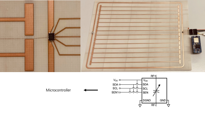 |
Bluetooth-enabled reconfigurable metamaterials for MRI
Dennis Philipp1,2, Endri Stoja3, Simon Konstandin1, Robin Wilke1, Diego Betancourt3, Thomas Bertuch3, Reiner Umathum1,4, Juergen Jenne1,4, and Matthias Guenther1,2
1Fraunhofer MEVIS, Bremen, Germany, 2University of Bremen, Bremen, Germany, 3Fraunhofer FHR, Wachtberg, Germany, 4DKFZ, Heidelberg, Germany
Bluetooth (BLE)-controlled, reconfigurable metamaterials (MTMs) for SNR enhancement in MRI are presented. These metasurfaces allow to be wirelessly interfaced and tuned during an MRI scan by means of a digital capacitor (DCAP), which is connected to a low-power microcontroller with BLE capabilities. Two prototypes are manufactured, one of which is a metasurface with adjustable resonance frequency, and the second one is dynamically tunable at the unit cell scale. It includes multiple DCAPS and, thus, is the first wirelessly reconfigurable MTM for MRI that offers field shaping capabilities, adjustable FoV, focal regions, sequence sync., and active detuning.
|
|
| 17:02 | 0186.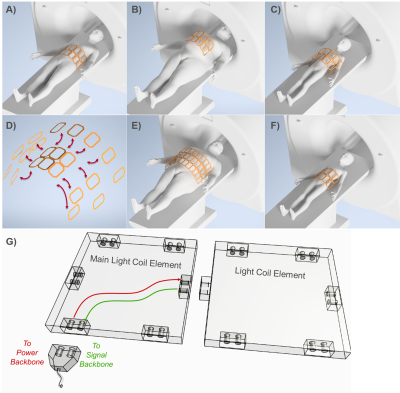 |
Light Coils: MRI with Modular RF Coils Using Optical Power and Data Transmission
Ali Caglar Özen1, Yanis Taege2, Thomas Lottner1, Serhat Ilbey1, Caglar Ataman2, and Michael Bock1
1Dept. Radiology, Medical Physics, Medical Center - University of Freiburg, Freiburg, Germany, 2Gisela and Erwin Sick Laboratory for Micro-Optics, Department of Microsystems Engineering, University of Freiburg, Freiburg, Germany
An optically powered and broadcasted modular receive coil concept (Light Coils) is presented for MRI to simultaneously eliminate the challenges in MRI of pediatric or overweight patients, signal-to-noise ratio losses and potential safety hazards due to electro-magnetic interferences in the transmission cables, and Ohmic losses in the metallic wires. By combining innovative RF antenna architectures, low-noise-low-power front end electronics and state-of-the-art silicon photonics technology, Light Coils might offer a robust and scalable solution for MRI image acquisition. Preliminary experimental results on the power-on-fiber driving of LNAs, and optical active detuning of receive coils are also discussed.
|
|
| 17:04 | 0187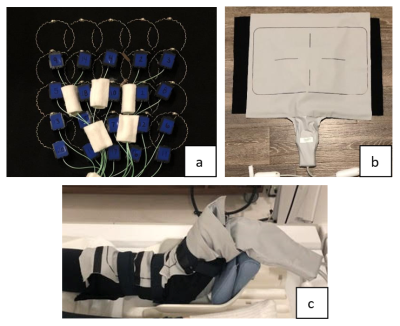 |
Twenty-channel, Highly-stretchable, Overlapped, Receive (THOR) Array Video Permission Withheld
Jana M. Vincent1,2,3,4, Fraser Robb4, Victor Taracila4, and Joseph V. Rispoli1,2
1Weldon School of Biomedical Engineering, Purdue University, West Lafayette, IN, United States, 2School of Electrical and Computer Engineering, Purdue University, West Lafayette, IN, United States, 3Department of Basic Medical Sciences, Purdue University, West Lafayette, IN, United States, 4MR Engineering, GE Healthcare Coils, Aurora, OH, United States
There has been a trend towards lightweight, flexible coils that provide greater SNR and patient comfort. To improve upon these coils and expand on existing stretchable designs, we present an omnidirectionally-stretchable, twenty-channel coil made with conductive thread on athletic fabric for use as a multipurpose coil. This coil was used to obtain in vivo images of anatomies including the wrist and ankle. Greater image quality and sensitivity can be seen with the stretchable coil compared to the commercial flexible coil. The stretchable coil was easier to place, able to be positioned more closely, and was more comfortable for the subjects.
|
|
| 17:06 | 0188.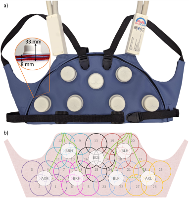 |
BraCoil – a wearable one-size-fits-all breast coil for 3 T MR mammography
Michael Obermann1, Lena Nohava1,2, Sigrun Roat1, Roberta Frass-Kriegl1, Onisim Soanca1, Bernhard Gruber1, Jean-Christophe Ginefri3, Jacques Felblinger2, and Elmar Laistler1
1High Field MR Center, Center for Medical Physics and Biomedical Engineering, Medical University of Vienna, Vienna, Austria, 2IADI, Inserm, Université de Lorraine, Nancy, France, 3Laboratoire d’Imagerie Biomédicale Multimodale Paris Saclay (BioMaps), CEA, CNRS, Inserm, Université Paris-Saclay, Paris, France
In this work, we present a new approach for MR mammography with a wearable light-weight coil array (“BraCoil”) covering both breasts and the sentinel axillary lymph nodes. The coil can be worn like a vest over a T-shirt and used either in prone or supine position. The presented design might be able to overcome numerous shortcomings of the current state-of -the-art in breast imaging by improving sensitivity, comfort and intimacy, shortening measurement time and may thereby eventually enable large-scale breast cancer screening without ionizing radiation.
|
|
| 17:08 | 0189.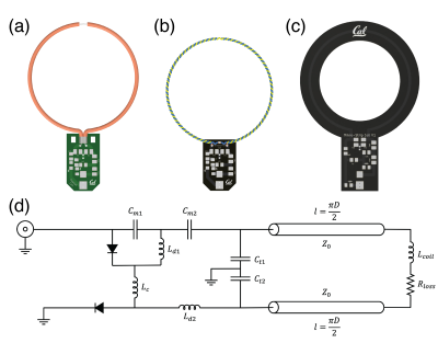 |
Transmission Line Receiver Coils (TLCs) for MRI
Julian Adolfo Maravilla1, Karthik Gopalan1, Ana Claudia Arias1, and Michael Lustig1
1EECS, University of California, Berkeley, Berkeley, CA, United States
Recent advancements in MRI coil design have been made towards designing and efficiently manufacturing tailor-made MRI receive arrays utilizing a multitude of different manufacturing techniques. However, the coils in these arrays must be decoupled geometrically to prevent detuning of the individual channels. This work evaluates Twisted Pair and Microstrip based receiver coils and compares their performance to Coaxial and traditional coils in terms of SNR and decoupling.
|
|
| 17:12 | 0190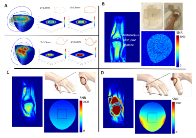 |
Additive manufacturing for the fabrication of subject-specific MRI passive shim and RF coil configurations Video Permission Withheld
Hanne Vanduffel1, Cesar Parra1, Willy Gsell1, Rodrigo de Oliveira Silva1, Uwe Himmelreich1, Wim Vanduffel1, Dimitrios Sakellariou1, and Rob Ameloot1
1KULeuven, Leuven, Belgium
Additive manufacturing methods are cost and time efficient methods, in particular, for the fabrication of subject-specific MRI hardware components. A novel powder-binder jetting method was developed to deposit variable concentrations of magnetic ink at precise pre-calculated positions to sculpt B0 towards a target distribution via the passive response of the 3D printed shims. Discrete spherical harmonic terms were printed as a proof-of-concept towards the manufacturing of subject-specific passive shims. Stereolithography (SLA) and Laser Powder Bed Fusion (L-PBF) manufacturing methods were developed for the fabrication of subject-specific RF coils. Significant gains in image quality, scan time and subject comfort were observed.
|
|
| 17:14 | 0191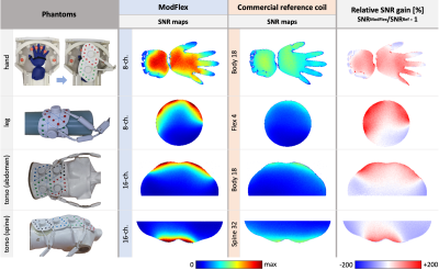 |
ModFlex – a modular system of flexible receive-only coil arrays for 3 T MRI Video Permission Withheld
Lena Nohava1,2, Michael Obermann1, Roberta Frass-Kriegl1, Sigrun Roat1, Onisim Soanca1, Nicolas Weber2, Jean-Christophe Ginefri3, Jacques Felblinger2,4, and Elmar Laistler1
1High Field MR Center, Center for Medical Physics and Biomedical Engineering, Medical University of Vienna, Vienna, Austria, 2IADI, Inserm, Université de Lorraine, Nancy, France, 3Laboratoire d’Imagerie Biomédicale Multimodale Paris Saclay (BioMaps), CEA, CNRS, Inserm, Université Paris-Saclay, Orsay, France, 4CHRU-Nancy, Inserm, Université de Lorraine, CIC, Innovation Technologique, Nancy, France
Flexible form-fitting radiofrequency coils provide high signal-to-noise ratio during MRI, and in array configuration large anatomical areas of interest can be covered. We propose a modular system - “ModFlex”- of flexible lightweight 4-channel coaxial coil arrays for 3T MRI. We investigated the performance difference between commercial reference coils and 8- and 16-channel ModFlex receive-only array systems. The versatility of ModFlex, the robustness of the coil characteristics for different use cases and a signal-to-noise gain compared to reference coils is demonstrated.
|
|
| 17:16 | 0192.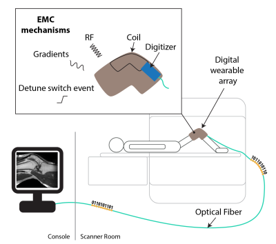 |
On-coil digitization with a 130nm CMOS receiver: Electromagnetic compatibility and use for a wearable knee array
Andreas Port1, Roger Luechinger1, Thomas Burger2, and Klaas Paul Pruessmann1
1Institute for Biomedical Engineering, ETH Zurich and University of Zurich, Zurich, Switzerland, 2Integrated Systems Laboratory, ETH Zurich, Zurich, Switzerland The relevant electromagnetic compatibility mechanisms of an integrated receiver based on 130nm CMOS technology are investigated. Imaging performance is verified with a digital wearable array, which was based on liquid metal and layed out for knee imaging. The study found that gradient switching and transients related to detuning are not critical in terms of EMC. However, coupling of RF to the preamplifier input has been found to exceed the receiver’s acceptable input voltage of 1.7V despite the use of protection diodes. Thus, a B1 limitation was applied and robust imaging performance was demonstrated in phantom and in vivo imaging. |
|
| 17:18 | 0193.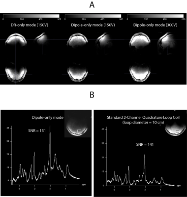 |
8-Channel Dipolectric Antenna Array for MRI at 7T: Proof-of-Principle Study in Human Brain
Daniel Wenz1,2 and Lijing Xin1,2
1CIBM Center for Biomedical Imaging, Lausanne, Switzerland, 2Animal Imaging and Technology, Ecole Polytechnique Federale de Lausanne (EPFL), Lausanne, Switzerland In this work we introduce a new type of antenna for ultrahigh field MRI which combines the advantages of dielectric resonator and dipole antennas: dipolectric antenna. Numerical simulations were performed to benchmark our concept against a loop-dipole antenna, and significant transmit efficiency gains in the peripheral as well as deeper located regions were observed. An 8-channel dipolectric antenna array was developed, and evaluated at the bench as well as in phantom experiments. In vivo MRI/MRS experiments at 7T were conducted and it was demonstrated that dipolectric antennas can be a promising approach for ultrahigh field MRI. |
|
| 17:20 | 0194.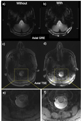 |
Development of a Broadside-Coupled Split-Ring Resonator-Based Metasurface to Improve MRI at Ultra-High Fields
Akbar Alipour1, Alan C Seifert1, Bradley N Delman2, and Priti Balchandani1
1BioMedical Engineering and Imaging Institute (BMEII), Icahn School of Medicine at Mount Sinai, Manhattan, NY, United States, 2Diagnostic, Molecular and Interventional Radiology, Icahn School of Medicine at Mount Sinai, Manhattan, NY, United States
There is a growing interest in ultra-high field magnetic resonance neuroimaging because of the greater signal-to-noise ratio (SNR) compared with conventional field strengths. However, visualization at these higher field strengths and Larmor frequencies is challenged by wavelength effects, compromising anatomical coverage of commercial coils. A novel metasurface based on a hybrid structure consisting of an array of broadside-coupled split-ring resonators and high permittivity materials was designed to augment signal at the 7 Tesla 1H Larmor frequency. This reusable surface device markedly improves SNR in caudal head structures that have historically been limited by poor signal.
|
|
| 17:22 | 0195.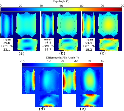 |
Transmit Performance of a Metamaterial Slab as a Passive RF Shimming Element for 3T MRI
Léo Rémillard1, Adam Mitchell Maunder1,2, Ashwin Iyer1, and Nicola De Zanche2,3
1Electrical and Computer Engineering, University of Alberta, Edmonton, AB, Canada, 2Oncology, University of Alberta, Edmonton, AB, Canada, 3Medical Physics, Cross Cancer Institute, Edmonton, AB, Canada
At higher static magnetic field strengths (3T) characteristic regions of low transmit fields occur that degrade the quality and diagnostic efficacy of MRI. Here we present the design, simulation, and measurement of thin (2cm), lightweight metamaterial (MTM) slabs for 3T that manipulate the field produced by a birdcage (BC) volume resonator for improved transmit performance. In a torso-sized phantom, a measured 39% increase in the mean flip angle and 4.9% reduction in the percent coefficient of variation was found, while the specific absorption rate normalized to the mean transmit field was reduced by 32%.
|
|
| 17:24 | 0196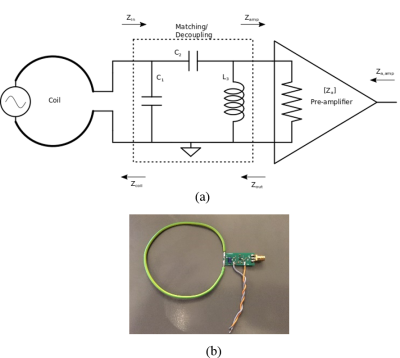 |
Exploiting a trade-off between preamplifier noise figure and bandwidth in the design of multi-nuclear MRI detectors Video Permission Withheld
Anders Nørup Thyrring Simonsen1, Vitaliy Zhurbenko1, Juan Diego Sanchez Heredia1, Wenjun Wang1, and Jan Henrik Ardenkjær-Larsen1
1Techical University of Denmark, Kgs. Lyngby, Denmark
The perspective of extending bandwidth of an MRI detector to ~3.2MHz can enable imaging of 13C, 23Na and 129Xe nuclei with a single coil in 3T scanners. Currently, multinuclear imaging is done using bulky multi-coil setups or triple-tuned matching networks. In this work we propose a different approach to cover several nuclei frequencies by extending the bandwidth of a single receive coil manipulating impedance of the preamplifiers. A trade-off analysis of the achieved bandwidth and SNR is performed. A design example is presented. The approach promises compact and light-weight realization, which is particularly useful for ultra-flexible multinuclear receive arrays.
|
 Back to Meeting Home
 Back to the Program-at-a-Glance
The International Society for Magnetic Resonance in Medicine is accredited by the Accreditation Council for Continuing Medical Education to provide continuing medical education for physicians.
 Back to Meeting Home
 Back to the Program-at-a-Glance