Power Pitch
Pitch: Novel MR Technology
Joint Annual Meeting ISMRM-ESMRMB & ISMRT 31st Annual Meeting • 07-12 May 2022 • London, UK

Power Pitch Session: How it Works
1st Hour: 2-minute Power Pitches in the Power Pitch Theater.
2nd Hour: 60-minute digital poster presentations at the smaller screens around the perimeter of the Power Pitch Theater.
| 09:17 | 0636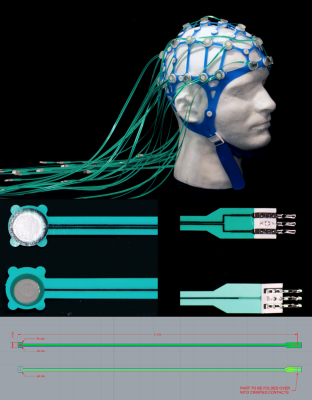 |
The MotoNet: An MRI-Compatible EEG Net with Embedded Motion Sensors Video Permission Withheld
Andre van der Kouwe1, Hongbae Jeong1, Zinong Yang2, Donald Straney1, Robert Frost1, Laura Lewis2, and Giorgio Bonmassar1
1Athinoula A. Martinos Center for Biomedical Imaging, Massachusetts General Hospital, Charlestown, MA, United States, 2Department of Biomedical Engineering, Boston University, Boston, MA, United States
We introduce a new EEG net, which will allow clinicians to monitor EEG while tracking head motion for correction. Motion during MRI limits patient scans, especially of children and patients with seizures. The MotoNet was built using PTF, embedding EEG/motion sensor pairs on opposite sides in one circuit. MRI safety studies at 3T confirmed maximum heating below 1ºC. EEG/motion measurements were made with a standard commercial EEG system. Using a custom MRI sequence with spatial localization gradients only, we showed that the signal on each channel was highly correlated with motion, allowing for electrode positioning and motion tracking.
|
|
| 09:19 | 0637.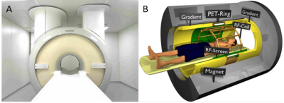 |
A dedicated MRI/PET system for radiotherapy treatment simulation: pathophysiological guidance for small moving target volumes
Casper Beijst1, Woutjan Branderhorst1, Jan Lagendijk1, Bjoern Weissler2,3, David Schug2,3, Florian Mueller2, Harald Radermacher2, Martino Borgo4, Wout Schuth4, Jurgen Mollink5, Govaert Borst5, Marc Verheyen5, Frank van Duin4, Dennis Klomp1, Hugo de Jong1, and Volkmar Schulz2,3
1UMC Utrecht, Utrecht, Netherlands, 2Rheinisch-Westfaelische Technische Hochschule, Aachen, Germany, 3Hyperion Hybrid Imaging Systems, Aachen, Germany, 4Futura Composites, Heerhugowaard, Netherlands, 5Philips Medical Systems, Best, Netherlands
The Unity 1.5T MR-linac has clinically been introduced to provide stereotactic precision for moving targets. We are now developing an integrated 1.5T MRI/PET system with identical MRI performance for planning of MR-linac treatments. The system offers 70 cm bore size and includes PET detectors mounted in the gap of a split gradient coil. Recently, the first ring of PET detectors was installed. We performed extensive interference tests and acquired the first simultaneous images using a brain phantom. Our vision is that the combination of MR-linac with MRI/PET will allow the use of PET information for stereotactic radiation therapy guidance.
|
|
| 09:21 | 0638.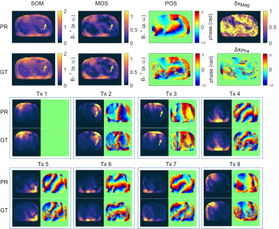 |
Deep learning-based relative B1+-mapping in the human body at 7T
Felix Krüger1, Christoph Stefan Aigner1, Sebastian Dietrich1, Kerstin Hammernik2,3, and Sebastian Schmitter1,4,5
1Physikalisch-Technische Bundesanstalt, Braunschweig and Berlin, Germany, 2Technical University of Munich, Munich, Germany, 3Imperial College London, London, United Kingdom, 4Medical Physics in Radiology, German Cancer Research Center (DKFZ), Heidelberg, Germany, 5Center for Magnetic Resonance Research, University of Minnesota, Minneapolis, MN, United States
In this work, we estimate relative 2D B1+-maps from initial localizer scans using deep learning at 7T. We investigate 7 UNets and MultiResUNets architectures to estimate complex, channel-wise, relative 2D B1+-maps of 8 transmit channels from a single gradient echo localizer obtained with 32 receive channels. The networks are evaluated in 5 unseen volunteers not included in the training library by comparing the prediction with the acquired relative B1+-maps using different evaluation metrics for homogeneous B1+ phase shimming. Our approach saves additional B1+-mapping scans, and, hence, overcomes long calibration times in the human body at 7T.
|
|
| 09:23 | 0639.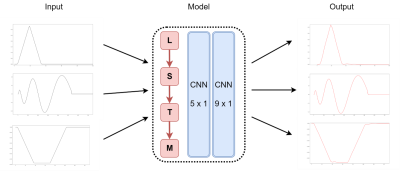 |
Gradient Waveform Prediction Using Deep Neural Network
Qi Liu1, Hoang (Mark) Nguyen1,2, Xucheng Zhu3, Xiaohui Zhai3, Bo Li3, Jian Xu1, and Weiguo Zhang1
1UIH America, Inc., Houston, TX, United States, 2Department of Computer Science and Electrical Engineering, University of Missouri at Kansas City, Kansas City, MO, United States, 3United Imaging Healthcare, Shanghai, China
A versatile, practical, and effective gradient trajectory correction technique that considers gradient system nonlinearity is proposed using deep learning. Its performance was validated on phantoms and human subjects and demonstrated superior quality than the conventional techniques.
|
|
| 09:25 | 0640.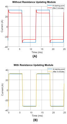 |
Adaptive Feedforward Control of Gradient Currents Using Gradient Heating Prediction
Reza Babaloo1, Ege Aydin1, and Ergin Atalar1,2
1Department of Electrical and Electronics Engineering, Bilkent University, Ankara, Turkey, 2National Magnetic Resonance Research Center (UMRAM), Bilkent University, Ankara, Turkey
The MRI gradient system heating changes its characteristics, and without a feedback system, the gradient waveform distorts resulting image artifacts. The primary effect of temperature rise in the gradient coil winding is the increase in the coil resistance. Feedforward based controllers, preferable in gradient array systems and multi-coil designs, use a linear time-invariant model. However, the gradient heating makes the system time-variant. To address this issue, we propose predicting gradient coil thermal variation using the thermal differential equation and updating feedforward controller parameters continuously.
|
|
| 09:27 | 0641.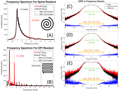 |
MR System Stability and Quality Control using Gradient Impulse Response Functions (GIRF)
Zhe Wu1, Alexander Jaffray2, Johanna Vannesjo3, Kamil Uludag4,5, and Lars Kasper1
1Techna Institute, University Health Network, Toronto, ON, Canada, 2UBC MRI Research Center, University of British Columbia, Vancouver, BC, Canada, 3Department of Physics, Norwegian University of Science and Technology, Trondheim, Norway, 4Techna Institute & Koerner Scientist in MR Imaging, University Health Network, Toronto, ON, Canada, 5Biomedical Engineering, Center for Neuroscience Imaging Research, Institute for Basic Science, Sungkyunkwan University, Suwon, Korea, Republic of
This study investigated the long-term stability of GIRF within an eight-month period, together with the benefits of an optimized GIRF acquisition protocol. These studies provide insight into mechanisms of image quality degrading and potentially lead to improved image quality control, generalizable to arbitrary imaging sequences.
|
|
| 09:29 | 0642.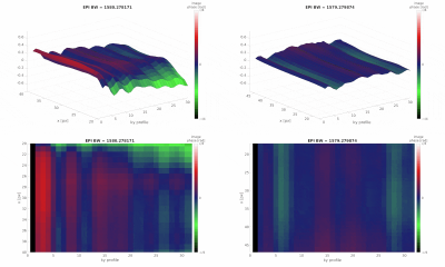 |
Beat Phenomena in MRI - Theoretical and Experimental Description of the Impact of Mechanical Resonances on Fast Readouts
Hannes Dillinger1 and Sebastian Kozerke1
1ETH and University Zurich, Zurich, Switzerland
This work highlights the mechanically resonant behaviour of MRI gradient systems when excited with an oscillating gradient, e.g. echo planar readout. Results demonstrate that a beat phenomenon can be observed in the signal phase with a frequency dependent on readout bandwidth and mechanical resonances of the system. Insights of the beat source are derived theoretically and validated experimentally. Data confirms that the beat phenomenon results in increased blurring in phase encoding direction leading to suggestions for system optimization in order to reduce the impact of the beat phenomenon.
|
|
| 09:31 | 0643. |
A lightweight silent gradient axis design with integrated 32 channel receive array for fast and quiet brain imaging at 3 Tesla
Edwin Versteeg1, Mark van Uden2, Luke van der Hofstad2, Kevin Bax2, Martino Borgo2,3, Wout Schuth3, Jeroen Siero1,4, Mark Gosselink1, and Dennis Klomp1
1Radiology, University Medical Center Utrecht, Utrecht, Netherlands, 2Tesla Dynamic Coils, Zaltbommel, Netherlands, 3Futura Composites, Heerhugowaard, Netherlands, 4Spinoza Centre for Neuroimaging, Amsterdam, Netherlands
The sound level in MRI can be lowered by using a silent gradient axis that adds extra soundless encoding. In this work, we present the field characteristics and first images for a silent gradient designed for a clinical 3 Tesla system. This silent gradient featured a built-in 32-channel receive coil and weighs 21 kg. The gradient field could be oscillated at 23.5 kHz and featured a linear region of 20 cm. The coil had limited interactions with the transmit B1 field and did not affect B1 homogeneity, which was showcased in phantom and in-vivo images.
|
|
| 09:33 | 0644.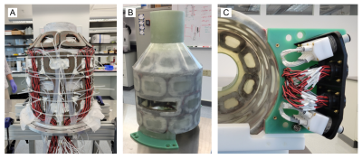 |
Construction and Validation of Multi-Coil Technology for Accessible Head-Only Scanner
Sebastian Theilenberg1, Yun Shang1, Chathura Kumaragamage2, Scott McIntyre2, Terence W. Nixon2, Robin A. de Graaf2, and Christoph Juchem1,3
1Department of Biomedical Engineering, Columbia University, New York, NY, United States, 2Department of Radiology and Biomedical Engineering, Magnetic Resonance Research Center, Yale University School of Medicine, New Haven, CT, United States, 3Department of Radiology, Columbia University Medical Center, New York, NY, United States
As part of a multi-center effort to design and build an accessible 1.5 T head-only MRI scanner (NIH U01EB025153, PI M. Garwood), we designed and constructed a prototype multi-coil array fitting the limited available space and capable of generating linear and non-linear image encoding fields as well as strong concomitant localized B0 shim fields. Here, we describe the construction process, and present system characteristics as well as experimental validation of its field generation capabilities utilizing a conventional 4 T MRI scanner.
|
|
| 09:35 | 0645.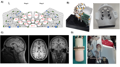 |
The TMSMR 28 channel RF coil: whole head imaging for the 3-axis TMS multichannel system
Lucia Navarro de Lara1,2, Jason Stockmann1,2, Boris Keil3, Azma Mareyam1, Mohammad Daneshzand1,2, Larry Wald1,2, and Aapo Nummenmaa1,2
1Martinos Center - MGH, Charlestown, MA, United States, 2Harvard Medical School, Boston, MA, United States, 3Institute of Medical Physics and Radiation Protection, Technische Hochschule Mittelhessen, Giessen, Germany
Multichannel Transcranial Magnetic Stimulation is an emerging technology for non-invasive stimulation of the human brain. Combining this technique with fMRI offers the unique benefits of studying the causal relationships between the cortical and subcortical nodes of large-scale brain networks. For this reason a whole head 28-channel RF coil array was developed to be integrated with the first 3-axis-TMS multichannel system. We have analyzed how the SNR of the constructed array changes when placing the stimulation units. Additionally, we compared the SNR/g-factor maps with commercial available 32/20-channel RF coils. The results show that the RF coil is performing as expected.
|
|
| 09:37 | 0646. |
Continuous Arterial Spin Labeling with On-Coil Amplification at 7T
Gaël Saïb1, Sherry Huang2, Emily Long1, S. Lalith Talagala3, Hellmut Merkle1, Alan P. Koretsky1, and Natalia Gudino1
1NINDS/LFMI, National Institutes of Health, Bethesda, MD, United States, 2Department of Biomedical Engineering, Case Western Reserve University, Cleveland, OH, United States, 3NINDS/NMRF, National Institutes of Health, Bethesda, MD, United States
Continuous arterial spin labeling using a separate neck labeling coil is an attractive approach for imaging perfusion at 7T. This approach is hindered by specialize hardware including a labeling coil and a separate amplifier located outside the magnet room. On-coil amplifiers have demonstrated higher RF power efficiency with minimal cable loss and patient-coil interaction. This work shows that a separate labeling coil using on-coil amplification can be used for flow driven adiabatic inversion in a flow phantom suggesting that it may provide a suitable approach to simplify the hardware package required as well as to improve CASL studies at 7T.
|
|
| 09:39 | 0647.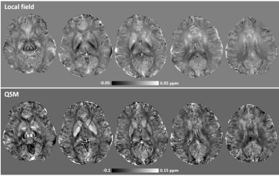 |
Susceptibility-weighted imaging and quantitative susceptibility mapping of the human brain at 10.5 Tesla: an initial experience
Xiaoping Wu1, Andrea Grant1, Xiaodong Ma1, Edward Auerbach1, Jerahmie Ladder1, Alireza Sadeghi-Tarakameh1, Yigitcan Eryaman1, Russell Lagore1, Nader Tavaf2, Pierre‐Francois Van de Moortele1, Gregor Adriany1, and Kamil Ugurbil1
1CMRR, Radiology, University of Minnesota, Minneapolis, MN, United States, 23M, Minneapolis, MN, United States
Susceptibility-weighted imaging (SWI) and quantitative susceptibility mapping (QSM) have been shown to provide unique contrasts that can be used to study pathophysiologic changes of tissue magnetic susceptibility in various brain diseases. As magnetic susceptibility effects increase with the main field strength, there has been a rapidly growing interest in performing SWI and QSM at ultrahigh field (UHF) (7 Tesla and above). The aim of this study was to demonstrate how the use of the UHF of 10.5 Tesla may promote SWI and QSM of the human brain.
|
 Back to Meeting Home
 Back to the Program-at-a-Glance
The International Society for Magnetic Resonance in Medicine is accredited by the Accreditation Council for Continuing Medical Education to provide continuing medical education for physicians.
 Back to Meeting Home
 Back to the Program-at-a-Glance