Power Pitch
Pitch: Flowing Downstream: Kidneys, Bladder, Prostate
Joint Annual Meeting ISMRM-ESMRMB & ISMRT 31st Annual Meeting • 07-12 May 2022 • London, UK

Power Pitch
Pitch: Flowing Downstream: Kidneys, Bladder, Prostate
Power Pitch Session: How it Works
1st Hour: 2-minute Power Pitches in the Power Pitch Theater.
2nd Hour: 60-minute digital poster presentations at the smaller screens around the perimeter of the Power Pitch Theater.
| 14:30 | 0475.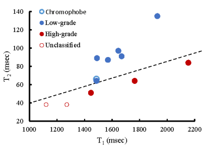 |
Differentiation of tumor grade for renal cell carcinoma using MR Fingerprinting
Sree Harsha Tirumani1,2, Christina J MacAskil1, Michael Markley1, Nicole Pritts1, Jared C Durieux1, Robin Elliott3,4, Adam Calaway5,6, Lee Ponsky5,6, Mark Griswold1, Chris Flask1, and Yong Chen1
1Radiology, Case Western Reserve University, Cleveland, OH, United States, 2Radiology, University Hospitals Cleveland Medical Center, Cleveland, OH, United States, 3Pathology, Case Western Reserve University, Cleveland, OH, United States, 4Pathology, University Hospitals Cleveland Medical Center, Cleveland, OH, United States, 5Urology, Case Western Reserve University, Cleveland, OH, United States, 6Urology, University Hospitals Cleveland Medical Center, Cleveland, OH, United States
In this study, we applied kidney MR Fingerprinting in characterization of renal cell carcinoma (RCC) and correlated the quantitative T1 and T2 values with tumor grade. Our results show significantly lower T2 values between the high-grade/unclassified RCC group and the chromophobe/low-grade RCC group (55±19 vs. 90±24 msec; P<0.05), while no significant difference in T1 values was noticed (P>0.05). More importantly, MRF T1 and T2 measurements provide complementary information in characterization of RCC tumor grades with a sensitivity of 89% and a specificity of 98% in differentiation of the two RCC groups, demonstrating the strength of multi-parametric imaging.
|
|
| 14:32 | 0476.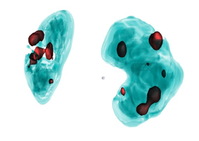 |
Detection of kidney cysts using a Convolutional Neural Network on 11,000 MRI data sets from the German National Cohort
Wilfried Reichardt1, Jan Lipovsek1, Marco Reisert1, Harald Horbach1, Christopher Schlett1, Fabian Bamberg1, Peggy Sekula1, Anna Köttgen1, Elias Kellner1, and Martin Büchert1
1University Medical Center Freiburg, Freiburg, Germany
We implemented a processing pipeline for kidney cyst segmentation using a hierarchical patch-based stack of U-nets and applied it to abdominal MRI images of the German National Cohort (GNC) study. The training data set included 300 cases, and the final net was applied to the dataset of 11,207 MRIs. Kidney cysts could be segmented with 98% sensitivity above a lower detection threshold of 1ml. The relation of first parameters based on the cyst segmentation with age and sex are presented. This result presents an optimal starting point to identify more advanced bimarkaers and their correlates, especially with kidney function parameters.
|
|
| 14:34 | 0477. |
Size Matters: Correct diagnoses of MRI based renal oxygenation assessment by monitoring kidney size
Jason Michael Millward1,2, Kathleen Cantow3, Thomas Gladytz1, Sonia Waiczies1,2, Thoralf Niendorf1,2, and Erdmann Seeliger3
1Berlin Ultrahigh Field Facility, Max Delbrück Center for Molecular Medicine in the Helmholtz Association, Berlin, Germany, 2Experimental and Clinical Research Center, Charité - Universitätsmedizin Berlin, Berlin, Germany, 3Institute of Physiology, Charite´ - Universitätsmedizin Berlin, Berlin, Germany
Tissue hypoxia occurs during acute kidney injury. MRI-based measurement of renal T2* and T2 could become a non-invasive surrogate marker of tissue oxygenation. However, the relationship between renal oxygenation and T2*,T2 is confounded by changes in the blood and tubular volume fractions, which are often accompanied by changes in kidney size (KS). We performed serial MRI scans along with clinically-realistic interventions acutely affecting renal oxygenation, applied directly while rats were in the scanner. We show that changes in KS correlate with changes in renal T2*,T2, underscoring that monitoring KS is necessary for correct interpretation of renal oxygenation derived from MRI.
|
|
| 14:36 | 0478.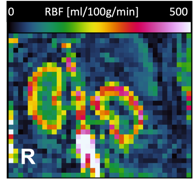 |
Measurement of renal perfusion using ASL-MRI and renal oxygenation using BOLD-MRI in dogs: a pilot study
Amber Hillaert1, Katrien Vanderperren1, and Pim Pullens2
1Department of Morphology, Imaging, Orthopedics, Rehabilitation and Nutrition, Faculty of Veterinary Medicine, Ghent University, Merelbeke, Belgium, 2Department of Radiology and Nuclear Medicine, Ghent University Hospital, Ghent, Belgium ASL- and BOLD-MRI have shown great potential in humans and rodents. To date, however, these techniques have not been used to image the canine kidney. Therefore, renal blood flow (RBF) and relaxation rate (R2*) were assessed using ASL and BOLD sequences on a beagle at 3T to examine the feasibility of both techniques in dogs. Mean RBF was 263.3 ± 39.0 and 277.4 ± 83.3 ml/100g/min in the right and left kidney, respectively. Mean R2* in the right kidney was 22.6 (range 14.8 - 36.8) s-1. This pilot-study demonstrates the feasibility of ASL- and BOLD-MRI for renal assessment in dogs. |
|
14:38 |
0479.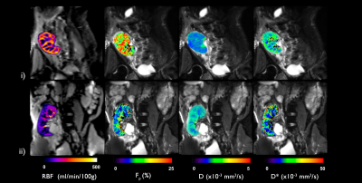 |
Prognostic potential of multiparametric MRI in the assessment of renal allografts early after transplantation
Rebeca Echeverria-Chasco1,2, Paloma L. Martin Moreno3, Nuria Garcia-Fernandez2,3, Marta Vidorreta4, Anne Oyarzun5, Arantxa Villanueva Larre2,5,6, Gorka Bastarrika1,2, and Maria A. Fernandez-Seara1,2
1Radiology, Clinica Universidad de Navarra, Pamplona, Spain, 2IdiSNA, Instituto de Investigación Sanitaria de Navarra, Pamplona, Spain, 3Nephrology, Clinica Universidad de Navarra, Pamplona, Spain, 4Siemens Healthcare, Madrid, Spain, 5Electrical Electronics and Communications Engineering, Public University of Navarre, Pamplona, Spain, 6ISC, Institute of Smart Cities, Pamplona, Spain Purpose: to evaluate the prognostic potential of a multiparametric renal MRI protocol (perfusion, diffusion and T1) for the assessment of the allograft in the very early stages after transplantation. Methods: 18 transplanted patients were imaged 6 days after the transplantation with ASL, IVIM and T1 mapping sequences. 2 groups were made depending on the allograft evolution (group A: no adverse events and group B: any adverse event). Results: eGFR, album-creatinine ratio and cortical and medullary RBF were significantly higher in allografts of group A than in group B. Significant correlations between eGFR and RBF were found. |
|
| 14:40 | 0480.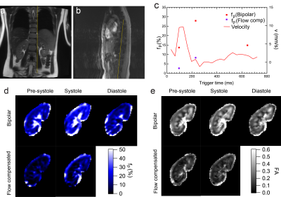 |
Cardiac cycle and flow compensation effects on REnal Flow and Microstructure AnisotroPy (REFMAP) MRI in healthy human kidney
Eric Sigmund1, Artem Mikheev1, Inge Brinkmann2, Thomas Benkert3, and Hersh Chandarana1
1Radiology, NYU Langone Health, New York, NY, United States, 2Siemens Medical Solutions USA, Inc., Malvern, PA, United States, 3Siemens Healthcare GmbH, Erlangen, Germany
Diffusion-weighted imaging in renal tissue is a complex interplay of microstructural and microcirculation effects, which are often quantified with diffusion tensor imaging (DTI) or intravoxel incoherent motion (IVIM), as well as hybrid models such as Renal Flow and Microstructure AnisotroPy (REFMAP). This work measures the modulation of these effects with cardiac phase and with flow compensated (FC) diffusion gradient waveforms. Results show that both cardiac phase and FC affect diffusion metrics signficantly in cortex and medulla, and suggest that these experimental tools may in the future be leveraged to increase biophysical specificity of renal diffusion MRI metrics.
|
|
| 14:42 | 0481.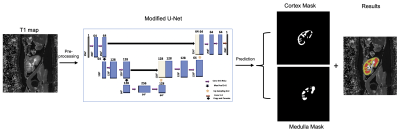 |
Validation of automatically measured ΔT1 values correlated with eGFR and fibrosis assessment of allograft kidneys
Ibtisam Aslam1,2, Fariha Aamir3, Miklos KASSAI1, Lindsey A CROWE1, Sophie de Seigneux4, Solange Moll5, Lena BERCHTOLD4, and Jean-Paul VALLEE1
1Service of Radiology, Geneva University Hospitals and Faculty of Medicine, University Hospital and University of Geneva, Geneva, Switzerland, 2Medical Image Processing Research Group (MIPRG), Deptt. of Electrical & Computer Engineering, COMSATS University Islamabad, ISLAMABAD, Pakistan, 3Medical Image Processing Research Group (MIPRG), Deptt. of Electrical & Computer Engineering, COMSATS University Islamabad (CUI), Pakistan, ISLAMABAD, Pakistan, 4Service and Laboratory of Nephrology, Department for Statistics, Department of Internal Medicine Specialties and of Physiology and Metabolism, University Hospital and University of Geneva, Geneva, Switzerland, 5Department of Clinical Pathology, Institute of Clinical Pathology, University Hospital and University of Geneva, Geneva, Switzerland
Chronic kidney disease (CKD) affects 11% of the population in Switzerland & US per year and is a major public health issue. Also, the CKD is 16th most prominent reason of life lost worldwide. MRI T1-mapping is a non-invasive way to monitor the renal prognosis. Recent studies show that the ∆T1 (cortex-medullary difference) has a strong positive correlation with fibrosis in CKD patients and is an important biomarker for CKD. This work proposes a validation of the T1-map cortico-medullary difference (ΔT1) values measured from automatically segmented cortex and medulla for eGFR and fibrosis assessment of allograft kidneys.
|
|
| 14:44 | 0482.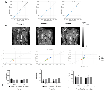 |
A Travelling Kidney study using a harmonised multiparametric renal MRI protocol
Charlotte E Buchanan1, Hao Li2, David M Morris3, Alexander J Daniel1, João Sousa4, Steven Sourbron4, David L Thomas5,6,7, Andrew Nicholas Priest2,8, and Susan T Francis1
1Sir Peter Mansfield Imaging Centre, University of Nottingham, Nottingham, United Kingdom, 2Department of Radiology, University of Cambridge, Cambridge, United Kingdom, 3Centre for Cardiovascular Science, University of Edinburgh, Edinburgh, United Kingdom, 4Department of Infection, Immunity and Cardiovascular Disease, University of Sheffield, Sheffield, United Kingdom, 5Neuroradiological Academic Unit, UCL Queen Square Institute of Neurology, University College London, London, United Kingdom, 6Dementia Research Centre, UCL Queen Square Institute of Neurology, University College London, London, United Kingdom, 7Wellcome Centre for Human Neuroimaging, UCL Queen Square Institute of Neurology, University College London, London, United Kingdom, 8Department of Radiology, Addenbrooke’s Hospital, Cambridge, United Kingdom
Standardisation and multicentre evaluation of renal MRI measures is crucial for clinical translation. Here we present results of a Travelling kidney study on GE, Philips and Siemens using a harmonised multiparametric renal MRI protocol, and show results of B0 and B1 mapping, T1, and T2* mapping. The mode B0 within the kidneys did not vary across vendors, however the FWHM was narrower for Vendor 1. No significant differences were seen in MOLLI T1 values of the renal cortex and medulla after this data was corrected for the offset in T1 seen in the NIST phantom.
|
|
| 14:46 | 0483.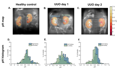 |
Noninvasive assessment of unilateral ureter obstruction using dynamic contrast-enhanced MR-CEST urography
Julia Stabinska1, Aruna Singh1,2, Farzad Sedaghat2, Max Kates3, Yuguo Li1,2, and Michael T. McMahon1,2
1F.M. Kirby Research Center for Functional Brain Imaging, Kennedy Krieger Institute, Baltimore, MD, United States, 2The Russell H. Morgan Department of Radiology and Radiological Science, The Johns Hopkins University School of Medicine, Baltimore, MD, United States, 3The James Buchanan Brady Urological Institute, Department of Urology, The Johns Hopkins University School of Medicine, Baltimore, MD, United States
The extent of recovery of renal function in patients with urinary tract obstructions depends on early diagnosis and prompt intervention. In the present study, we employed dynamic contrast-enhanced MR-CEST urography to noninvasively assess kidney function in mice with unilateral ureter obstruction (UUO) at two time points (day 1 and 2) after UUO surgery. Using a dynamic CEST MRI protocol, we were able to detect a slight increase in pH values (pH 6.74 ± 0.08 and 6.62 ± 0.04 in the UUO and contralateral kidney, respectively) as well as iopamidol retention in the UUO kidney.
|
|
| 14:48 |
0484.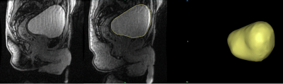 |
MRI Urodynamics: A 3D approach to Bladder Biomechanics
Juan Pablo Gonzalez-Pereira1, Cody Johnson2, Shane A Wells2, Wade Bushman3, and Alejandro Roldan-Alzate1,2
1Mechanical Engineering, University of Wisconsin-Madison, Madison, WI, United States, 2Radiology, University of Wisconsin-Madison, Madison, WI, United States, 3Urology, University of Wisconsin-Madison, Madison, WI, United States
Lower urinary track symptoms (LUTS) affect many older adults. Existing methods to evaluate the lower urinary tract are invasive and provide limited information about changes in bladder anatomy and detrusor muscle function. Use of non-invasive methods for the study of lower urinary tract anatomy and function has been limited. This pilot study demonstrates the feasibility of MRI urodynamics in healthy subjects and in LUTS patients. Future advancements in this study will be aimed at using the data acquired to further deepen the comprehension of the bladder voiding cycle.
|
 Back to Meeting Home
 Back to the Program-at-a-Glance
The International Society for Magnetic Resonance in Medicine is accredited by the Accreditation Council for Continuing Medical Education to provide continuing medical education for physicians.
 Back to Meeting Home
 Back to the Program-at-a-Glance