Power Pitch
Pitch: Hepatobiliary, Pancreas & Bowel Imaging
Joint Annual Meeting ISMRM-ESMRMB & ISMRT 31st Annual Meeting • 07-12 May 2022 • London, UK

Power Pitch
Pitch: Hepatobiliary, Pancreas & Bowel Imaging
Power Pitch Session: How it Works
1st Hour: 2-minute Power Pitches in the Power Pitch Theater.
2nd Hour: 60-minute digital poster presentations at the smaller screens around the perimeter of the Power Pitch Theater.
09:15 |
0598.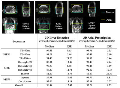 |
AI-based Automated Liver Image Prescription: Evaluation across Patients and Pathologies and Prospective Implementation and Validation
Ruiqi Geng1,2, Collin J. Buelo1,2, Mahalakshmi Sundaresan3, Jitka Starekova1, Nikolaos Panagiotopoulos1,4, Thekla Helene Oechtering1,4, Edward M. Lawrence1, Marcin Ignaciuk1, Scott B. Reeder1,2,5,6,7, and Diego Hernando1,2,3,7
1Radiology, University of Wisconsin-Madison, Madison, WI, United States, 2Medical Physics, University of Wisconsin-Madison, Madison, WI, United States, 3Electrical and Computer Engineering, University of Wisconsin-Madison, Madison, WI, United States, 4Radiology and Nuclear Medicine, Universität zu Lübeck, Lübeck, Germany, 5Medicine, University of Wisconsin-Madison, Madison, WI, United States, 6Emergency Medicine, University of Wisconsin-Madison, Madison, WI, United States, 7Biomedical Engineering, University of Wisconsin-Madison, Madison, WI, United States
An automated AI-based method for liver image prescription from a localizer was recently proposed. In this work, this AI method was further evaluated in a larger retrospective patient cohort (1,039 patients for training/testing), across pathologies, field strengths, and against radiologists’ inter-reader reproducibility performance. AI-based 3D axial prescription achieved a S/I shift of <2.3 cm compared to manual prescription for 99.5% of test dataset. The AI method performed well across all sub-cohorts and better in 3D axial prescription than radiologists’ inter-reader reproducibility performance. The AI method was successfully implemented on a clinical MR system and showed robust performance across localizer sequences.
|
|
| 09:17 | 0599. |
Deep Learning-guided Weighted Averaging for Signal Dropout Compensation in Liver DWI
Fasil Gadjimuradov1,2, Thomas Benkert2, Marcel Dominik Nickel2, and Andreas Maier1
1Pattern Recognition Lab, Department of Computer Science, Friedrich-Alexander-Universität Erlangen-Nürnberg, Erlangen, Germany, 2MR Application Predevelopment, Siemens Healthcare GmbH, Erlangen, Germany
Signal-dropouts caused by cardiac motion are frequently observed artifacts in diffusion-weighted imaging (DWI) of the liver. When several repetitions of a given slice are affected, uniform averaging results in locally reduced liver signal. This work proposes an adaptive weighted averaging of repetitions to locally suppress signal-dropouts. Therefore, weight maps are estimated by an algorithm which computes robust patch statistics under the guidance of a learned classifier which marks corrupted repetitions. In comparison to other methods, the proposed approach enables more homogeneous liver signal and less biased quantitative maps while sacrificing little signal-to-noise ratio (SNR).
|
|
| 09:21 | 0600.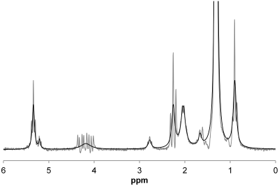 |
Dependence of liver R2* on the fat spectral model.
Gavin Hamilton1, Alexandra N Schlein1, Walter C Henderson1, Danielle N Batakis1, Lael K Ceriani1, Ashley L Louie1, Michael S Middleton1, Yesenia Covarrubias1, Kathryn J Fowler1, and Claude B Sirlin1
1Liver Imaging Group, Department of Radiology, University of California San Diego, San Diego, CA, United States Using MRS, we examined whether the assumed fat spectral model (6-, or 9-peak) altered liver R2* estimation in vivo. We found that liver R2* estimation was model-independent, and liver R2*was not significantly different from water R2* (p = 0.07). However, fat R2* values estimated for these models were different (p < 0.0001), suggesting that fat R2* estimation in high-fat tissues may be model dependent. |
|
| 09:25 |
0601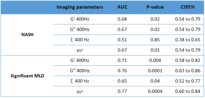 |
Diagnostic performance of magnetic resonance elastography in mice with NASH and significant metabolic liver disease. Video Permission Withheld
Meryem KHALFALLAH1, Sabrina Doblas1, Felicia Julea1, Catherine Postic2, Dominique Valla3, Valérie Paradis3,4, Philippe Garteiser1, and Bernard Van Beers1,5
1Université de Paris, Laboratory of Imaging Biomarkers, Center of Research on Inflammation, UMR 1149, Inserm, F-75018 Paris, France, Paris, France, 2Université de Paris, Institut Cochin, CNRS, INSERM, Paris, France., Paris, France, 3Service d’hépatologie et UMR1149, Hôpital Beaujon, Clichy-la-Garenne ; APHP, Université de Paris et Inserm, Clichy-la-Garenne, France, 4Diderot, CNRS, Centre de Recherche sur l'Inflammation (CRI), Paris, F-75890, France., Paris, France, 5Department of Radiology, AP-HP, Beaujon University Hospital Paris Nord, F-92110 Clichy, France, Clichy, France
Nonalcoholic steatohepatitis (NASH) and significant metabolic liver disease (MLD) are progressive forms of nonalcoholic fatty liver disease (NAFLD). MR elastography parameters have been shown to be useful for assessing NAFLD/NASH and liver inflammation. To assess the role of viscoelastic parameters in diagnosing NASH and significant MLD, MRE hepatic examinations were performed in mice with NAFLD. Our results showed that the diagnostic performance of MR elastography was higher in significant MLD than in NASH. This may be explained by inflammation and fibrosis occurring in indeterminate NASH cases included in MLD, and by poor performance of MR elastography to detect hepatocyte ballooning.
|
|
| 09:27 | 0602.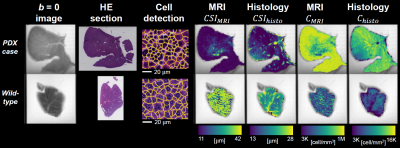 |
Histological correlates of DR-HIGADOS microstructural metrics in the mouse and human liver
Francesco Grussu1, Kinga Bernatowicz1, Ignasi Barba2, Irene Casanova-Salas3, Natalia Castro3, Marco Palombo4, Sara Simonetti3,5, Juan Francisco Corral6,7, Marta Vidorreta8, Xavier Merino6,7, Richard Mast6,7, Núria Roson6,7, Manuel Escobar Amores6,7, Nahúm Calvo‐Malvar6,9, Josep R. Garcia-Bennett9, Rodrigo A. Toledo10, Joaquin Mateo3, Paolo Nuciforo5, Elena Garralda11, and Raquel Perez-Lopez1,7
1Radiomics Group, Vall d'Hebron Institute of Oncology, Vall d'Hebron Barcelona Hospital Campus, Barcelona, Spain, 2NMR Lab, Vall d'Hebron Institute of Oncology, Vall d'Hebron Barcelona Hospital Campus, Barcelona, Spain, 3Prostate Cancer Translational Research Group, Vall d'Hebron Institute of Oncology, Vall d'Hebron Barcelona Hospital Campus, Barcelona, Spain, 4Cardiff University Brain Research Imaging Centre, Cardiff University, Cardiff, United Kingdom, 5Molecular Oncology Group, Vall d'Hebron Institute of Oncology, Vall d'Hebron Barcelona Hospital Campus, Barcelona, Spain, 6Institut de Diagnòstic per la Imatge (IDI), Catalonia, Spain, 7Department of Radiology, Hospital Universitari Vall d'Hebron, Barcelona, Spain, 8Siemens Healthineers, Madrid, Spain, 9Hospital Universitari de Bellvitge, L'Hospitalet de Llobregat, Spain, 10Gastrointestinal and Endocrine Tumors Group, Vall d'Hebron Institute of Oncology, Vall d'Hebron Barcelona Hospital Campus, Barcelona, Spain, 11Early Clinical Drug Development Group, Vall d'Hebron Institute of Oncology, Vall d'Hebron Barcelona Hospital Campus, Barcelona, Spain
“Diffusion-Relaxation Hepatic Imaging via Generalised Assessment of DiffusiOn Simulations” (DR-HIGADOS) is a recent liver diffusion MRI method for intra-cellular fraction, cell size and cellularity measurement. Here we compare DR-HIGADOS metrics to counterparts from histology in two data sets: i) preclinical 9.4T MRI of formalin-fixed patient-derived xenograft mouse livers, with co-localised histological sections; ii) clinical 1.5T in vivo MRI of metastatic and primary liver cancer patients, with co-localised biopsies. Results confirm that DR-HIGADOS cell size and cellularity are histologically meaningful, paving the way to the application of the new technique in clinical studies.
|
|
| 09:29 |
0603.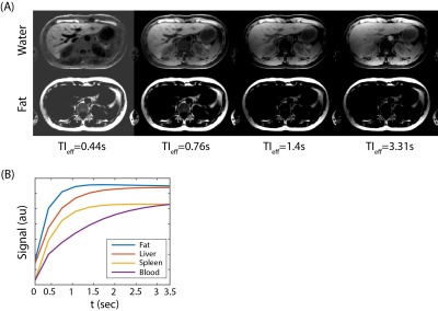 |
Free-Breathing, 3D Phase Sensitive Inversion Recovery T1-Weighted MRI of the Liver
Yavuz Muslu1,2, Ty A. Cashen3, Sagar Mandava4, and Scott B. Reeder1,2,5,6,7
1Department of Biomedical Engineering, University of Wisconsin-Madison, Madison, WI, United States, 2Department of Radiology, University of Wisconsin-Madison, Madison, WI, United States, 3GE Healthcare, Waukesha, WI, United States, 4GE Healthcare, Atlanta, GA, United States, 5Department of Medical Physics, University of Wisconsin-Madison, Madison, WI, United States, 6Department of Medicine, University of Wisconsin-Madison, Madison, WI, United States, 7Department of Emergency Medicine, University of Wisconsin-Madison, Madison, WI, United States
Gadoxetic acid (GA)-enhanced hepatobiliary phase T1-weighted (HBP-T1w)-MRI is widely used in the clinical setting for detection of focal liver lesions (FLLs). However, complete characterization of liver lesions also requires T2w, diffusion weighted (DWI) and dynamic contrast enhanced (DCE) imaging. These methods often have inferior image spatial resolution compared to GA-enhanced HBP-T1w and may be unable to resolve small (<1cm) lesions visible only on HBP-T1w. In this work, we propose a novel phase sensitive inversion recovery (PSIR) T1w-MRI in combination with 3D radial stack-of-stars imaging and model-based water/fat separation for improved detection and characterization of small FLLs.
|
|
| 09:31 | 0604.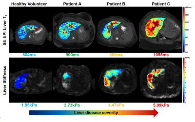 |
The association of Liver Stiffness with Liver tissue T1 and Superior Mesenteric Artery blood flow across disease severity.
Christopher R Bradley1,2, Deirdre Mcgrath1,2, Eleanor F Cox1,2, Naaventhan Palaniyappan2, Robert Scott2, Indra N Guha2, Guruprasad P Aithal2, and Susan T Francis1,2
1Sir Peter Mansfield Imaging Centre, University of Nottingham, Nottingham, United Kingdom, 2NIHR Biomedical Research Centre, Nottingham University Hospitals NHS Trust, Nottingham, United Kingdom We assess liver stiffness measured using magnetic resonance elastography (MRE) and longitudinal relaxation time T1 in three groups: healthy volunteers, non-alcoholic fatty liver disease patients and compensated cirrhosis patients. MRE liver stiffness was measured using the Quantitative Imaging Biomarker Alliance (QIBA) recommendation. T1 longitudinal relaxation time of the liver was measured using a respiratory triggered inversion recovery fat-suppressed spin-echo echo planar imaging scheme. A positive correlation between Liver tissue T1 and Liver stiffness (R=0.70, p<0.0001) across the 3 groups was observed. Superior mesenteric artery flow also correlates with liver stiffness (R=0.62, p<0.002) suggesting worsening hyperdynamic circulation with progressive fibrosis. |
|
| 09:33 | 0605.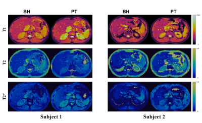 |
Free Breathing Abdominal Tissue Quantification using 2D MR Fingerprinting with quadratic RF phase (qRF-MRF)
Sherry Huang1, Yong Chen2, Reid Bolding3, Leonardo Kayat Bittencourt2,4, Mark A Griswold2, and Rasim Boyacioglu2
1Biomedical Engineering, Case Western Reserve University, Cleveland, OH, United States, 2Radiology, Case Western Reserve University, Cleveland, OH, United States, 3Physics, Case Western Reserve University, Cleveland, OH, United States, 4Radiology, University Hospitals Cleveland Medical Center, Cleveland, OH, United States
This study presents a Pilot Tone (PT) based free-breathing technique for two-dimensional simultaneous quantification of T1, T2, T2*, fat fraction (FF), and water fraction (WF) using quadratic RF phase Magnetic Resonance Fingerprinting (qRF-MRF). We report the quantitative comparison of a cohort of 10 healthy subjects between free-breathing and breath-hold qRF-MRF as well as the comparison to standard clinical protocols. Free-breathing results are comparable to both breath-hold and standard clinical protocols (p > 0.05), indicating the stability and reproducibility of the method.
|
|
| 09:35 | 0606.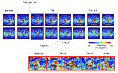 |
Dynamics of insulin secretion and pancreatic blood-flow: a simultaneous ASL perfusion imaging / hyperglycemic clamp study
Manuel Taso1, Donald C Simonson2, Lauren Phung1, Colleen McGrath1, Stephanie Waldman1, Fotini Papadopoulou1, and David C Alsop1
1Division of MRI Research, department of Radiology, Beth Israel Deaconess Medical Center, Harvard Medical School, Boston, MA, United States, 2Division of Endocrinology, Diabetes and Hypertension, Brigham and Women's Hospital, Harvard Medical School, Boston, MA, United States Loss of β-cell function and mass are hallmarks of type-1 and type-2 diabetes. Non-invasive imaging of endocrine pancreatic function would provide valuable insights into the degree and temporal patterns of endocrine dysfunction. For this purpose, Arterial Spin Labeling (ASL) is a uniquely suited non-invasive technique. We therefore conducted a dynamic ASL study during a hyperglycemic clamp to explore the dynamics of insulin secretion and pancreatic blood-flow. This study showed that hyperglycemia and arginine-stimulated insulin secretion causes an increase in pancreatic perfusion and that the rate of insulin concentration change seems to be driving perfusion changes rather than absolute concentration. |
|
| 09:37 | 0607.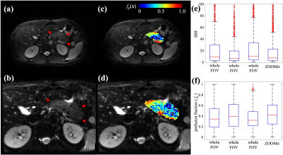 |
Reduced Field-of-View Intravoxel Incoherent Motion of the Human Pancreas Reflects Biphasic Response to Glucose Ingestion
Chengyue Wu1, Thomas E Yankeelov1,2,3,4,5,6, and John Virostko1,2,3,4
1Oden Institute for Computational Engineering and Sciences, The University of Texas at Austin, Austin, TX, United States, 2Livestrong Cancer Institutes, The University of Texas at Austin, Austin, TX, United States, 3Department of Diagnostic Medicine, The University of Texas at Austin, Austin, TX, United States, 4Department of Oncology, The University of Texas at Austin, Austin, TX, United States, 5Department of Biomedical Engineering, The University of Texas at Austin, Austin, TX, United States, 6Department of Imaging Physics, The University of Texas MD Anderson Cancer Center, Houston, TX, United States
Monitoring of the pancreatic islet is needed to assess individuals at risk for developing diabetes and facilitate development of islet-directed therapies. Pancreatic islets may be detectable due to their high vascularity. We imaged pancreas perfusion using both whole field-of-view and reduced field-of-view Intravoxel Incoherent Motion (IVIM) and found that the reduced field-of-view technique achieved similar image quality in half the time. We then performed a reduced field-of-view IVIM acquisition on four healthy controls. We found that pancreas perfusion increased dynamically in response to oral glucose challenge, exhibiting a biphasic increase that parallels the time course of insulin secretion.
|
|
| 09:39 | 0608.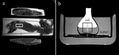 |
Measurement of pancreas graft temperature during cold preservation in MR scanner
Jan Weis1 and Olle Korsgren2
1Department of Medical Physics, Uppsala University Hospital, Uppsala, Sweden, 2Department of Immunology, Genetics and Pathology, Uppsala University, Uppsala, Sweden
Phosphorus (31P) and proton (1H) magnetic resonance spectroscopy (MRS) are the methods of choice in the assessment of the pancreas graft quality before organ or islet of Langerhans transplantation. During the transport and MR scanning hypothermic storage (4±2 oC) has to be maintained. The aim of this study was to investigate if it is possible to measure pancreas graft temperature in a MR scanner using 1H-MRS and temperature constants obtained by the calibration experiments with the water-vegetable oil phantom. The present study has shown that 1H-MRS is able to measure the graft temperature during MR scanning.
|
|
| 09:41 | 0609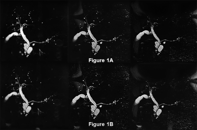 |
Deep Learning Reconstruction: Clinical Utility for 3D MRCPs Obtained by Different k-Space Data Acquisition Methods in IPMN Patients Video Permission Withheld
Takahiro Matsuyama1, Yoshiharu Ohno1,2, Kaori Yamamoto3, Masato Ikedo3, Masao Yui3, Saki Takeda4, Akiyoshi Iwase4, Yuka Oshima1, Nayu Hamabuchi1, Satomu Hanamatsu1, Yuki Obama1, Hiroyuki Nagata1, Takahiro Ueda1, Hirotaka Ikeda1, Kazuhiro Murayama2, and Hiroshi Toyama1
1Radiology, Fujita Health University School of Medicine, Toyoake, Japan, 2Joint Research Laboratory of Advanced Medical Imaging, Fujita Health University School of Medicine, Toyoake, Japan, 3Canon Medical Systems Corporation, Otawara, Japan, 4Radiology, Fujita Health University Hospital, Toyoake, Japan
We hypothesized that deep learning reconstruction (DLR) and multiple k-space data by means of each of the repetition time (TR) techniques (Fast 3D mode multiple: Fast 3Dm) are more useful than parallel imaging (PI) and compressed sensing (CS) for shortening acquisition time and improving image quality and IPMN evaluation capability on 3D MRCP. The purpose of this study was thus to compare the utility of DLR used for PI, Fast 3Dm and CS for improvement of acquisition time, image quality and IPMN evaluation capability on 3D MRCP for patients with IPMN.
|
 Back to Meeting Home
 Back to the Program-at-a-Glance
The International Society for Magnetic Resonance in Medicine is accredited by the Accreditation Council for Continuing Medical Education to provide continuing medical education for physicians.
 Back to Meeting Home
 Back to the Program-at-a-Glance