Power Pitch
Pitch: Quantifying the Heart
Joint Annual Meeting ISMRM-ESMRMB & ISMRT 31st Annual Meeting • 07-12 May 2022 • London, UK

Power Pitch Session: How it Works
1st Hour: 2-minute Power Pitches in the Power Pitch Theater.
2nd Hour: 60-minute digital poster presentations at the smaller screens around the perimeter of the Power Pitch Theater.
09:15 |
0014.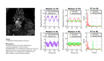 |
Real-time myocardial landmark-tracking for MRI-guided cardiac radio-ablation using Gaussian Processes
Niek R.F. Huttinga1, Osman Akdag1, Martin Fast1, Joost Verhoeff1, Alessandro Sbrizzi1, and Stefano Mandija1
1Department of Radiotherapy, Computational Imaging Group for MR therapy & Diagnostics, University Medical Center Utrecht, Utrecht, Netherlands
Accurate tracking of lesions within or near the heart presents a unique challenge due to cardiorespiratory motion during stereotactic radiotherapy. Mitigating this motion is essential to limit the risk of radiation-induced complications. Here, the feasibility of real-time prospective myocardial landmark-tracking with Gaussian Processes from few k-space readouts is demonstrated on the 1.5T Elekta Unity MR-linac. Retrospective and prospective phantom and in-vivo experiments were conducted in 2D, and preliminary in-silico experiments were done to demonstrate the feasibility in 3D. Results indicate high accuracy, and thereby great potential for real-time 3D myocardial target tracking for cardiac radio-ablation.
|
|
| 09:17 | 0015.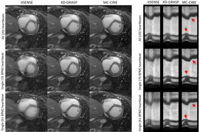 |
Heartbeat-resolved cardiac CINE imaging via motion corrected reconstructions
Gastao da Cruz1, René M. Botnar1, and Claudia Prieto1
1King's College London, London, United Kingdom
Conventional cardiac CINE imaging acquires segmented data over multiple heartbeats to satisfy sampling requirements. Recently, cardiac CINE reconstructions from a single heartbeat have been achieved using motion corrected reconstructions. This approach allows single heartbeat CINE and therefore can image the unique dynamics that occur in each heartbeat. Experiments in healthy subjects demonstrate that single heartbeat CINE is feasible and can detect heartbeat to heartbeat variation; the dynamics of individual heartbeats differ and cannot be detected with conventional CINE performed over multiple heartbeats. Single heartbeat CINE shows promise to characterize arrhythmias and other heart conditions where heartrate variability occurs.
|
|
| 09:19 | 0016.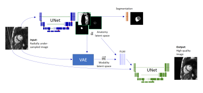 |
A disentangled representation trained for joint reconstruction and segmentation of radially undersampled cardiac MRI
Tobias Wech1, Julius Frederik Heidenreich1, Thorsten Alexander Bley1, and Bettina Baeßler1
1Department of Diagnostic and Interventional Radiology, University Hospital Würzburg, Würzburg, Germany
The network we propose in this work (xSDNet) jointly reconstructs and segments cardiac functional MR images which were sampled below the Nyquist rate. The model is based on disentangled representation learning and factorizes images into spatial factors and a modality vector. The achieved image quality and the fidelity of the delivered segmentation masks promise a considerable acceleration of both acquisition and data processing.
|
|
| 09:21 | 0017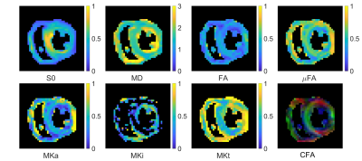 |
Tensor-valued Encoding in the Human Heart In vivo Video Permission Withheld
Irvin Teh1, David Shelley2, Ana-Maria Poenar1, Erica Dall'Armellina1, Sven Plein1, Jürgen E. Schneider1, and Filip Szczepankiewicz3
1Leeds Institute of Cardiovascular and Metabolic Medicine, University of Leeds, Leeds, United Kingdom, 2Leeds Teaching Hospitals Trust, Leeds, United Kingdom, 3Clinical Sciences, Lund University, Lund, Sweden
Tensor-valued encoding holds great promise in enhancing specificity in myocardial characterisation relative to diffusion tensor imaging (DTI). However, its application in the heart is challenging due to motion and limited SNR. In this work, we employed optimized diffusion encoding waveforms with up to 2nd order motion compensation and compensation for concomitant gradient effects to demonstrate the first in vivo application of q-space trajectory imaging (QTI) in the human heart. Baseline myocardial measurements of microscopic fractional anisotropy, as well as isotropic and anisotropic mean kurtosis are reported for the first time in the heart.
|
|
| 09:23 | 0018.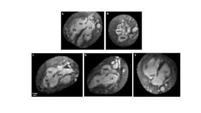 |
Fetal cardiac 4D imaging from multi-planar balanced SSFP MRI
Sharon Portnoy1, Joshua F.P. van Amerom2, Amanda Hope3, Datta Singh Goolaub3, Liqun Sun2, Mike Seed2,4, and Christopher K. Macgowan1,5
1Translational Medicine, Hospital for Sick Children, Toronto, ON, Canada, 2Cardiology, Hospital for Sick Children, Toronto, ON, Canada, 3Hospital for Sick Children, Toronto, ON, Canada, 4Diagnostic Imaging, Hospital for Sick Children, Toronto, ON, Canada, 5Medical Biophysics, University of Toronto, Toronto, ON, Canada
This work explores the feasibility of 4D fetal whole-heart image reconstruction from high resolution 2D multiplanar radial bSSFP acquisitions. High quality 4D reconstructions (1 mm isotropic spatial resolution, ~20 ms temporal resolution) are demonstrated in 3 congenital heart disease cases with distinct cardiac abnormalities. These dynamic, highly detailed visualizations of the fetal heart facilitate prenatal CHD diagnosis and planning for delivery and surgical intervention.
|
|
| 09:25 | 0019.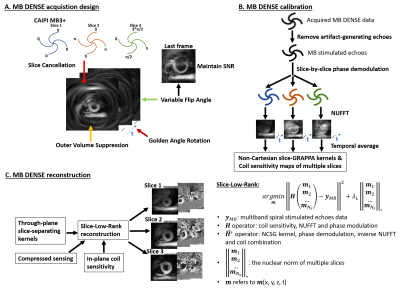 |
Single-breathhold multiband cine DENSE MRI using novel phase modulation and a self-calibrated slice-low-rank reconstruction
Changyu Sun1, Yu Wang1, and Frederick H. Epstein1,2
1Biomedical Engineering, University of Virginia, Charlottesville, VA, United States, 2Radiology, University of Virginia, Charlottesville, VA, United States
Multiband (MB) excitation of spiral cine DENSE MRI has the potential to provide three-slice coverage in single breathhold with 2D displacement encoding. To acquire three slices in one breathhold of 14 heartbeats, we develop MB3+ phase modulation, and use golden-angle rotation, outer volume suppression and variable flip angle. We extend our recent Cartesian slice-L+S method to develop a non-Cartesian slice-low-rank reconstruction model. DENSE data with MB=3 were acquired in 4 volunteers. The MB3+ DENSE method shows fewer artifacts than CAIPIRINHA and Hadamard encoding phase modulations. The slice-low-rank method with MB+ significantly improves slice separation providing fewer artifacts and signal decay.
|
|
| 09:27 | 0020. |
Free-running isotropic whole-heart T2 mapping with ECG-free Pilot Tone navigation
Simone Rumac1, Christopher W. Roy1, Jérôme Yerly1, Mariana B. L. Falcao1, Aurélien Bustin1,2,3, Mario Bacher1,4, Peter Speier4, Matthias Stuber1,5, and Ruud B. van Heeswijk1
1Department of Radiology, Lausanne University Hospital (CHUV) and University of Lausanne (UNIL), Lausanne, Switzerland, 2IHU LIRYC, Electrophysiology and Heart Modeling Institute, INSERM U1045, Centre de recherche Cardio-Thoracique de Bordeaux, Université de Bordeaux, Bordeaux, France, 3Cardiovascular Imaging, Hôpital Cardiologique du Haut-Lévêque, CHU de Bordeaux, Bordeaux, France, 4Siemens Healthcare GmbH, Erlangen, Germany, 5CIBM Center for BioMedical Imaging, Lausanne, Switzerland, Lausanne, Switzerland
The most commonly employed T2 mapping techniques are 2D and make use of ECG-triggering. This may be a limitation in patients with variable heart rate and complex three-dimensional conditions. To address these limitations, we here propose an isotropic free-running 3D T2 mapping technique that avoids ECG triggering by using Pilot Tone navigation. In three healthy volunteers, our technique produced accurate isotropic T2 maps when compared to 2D T2 prepared bSSFP (T2=41.1±4.8ms vs. 44.9±3.3ms, respectively, p=0.1), and cardiac motion was successfully resolved.
|
|
| 09:29 | 0021.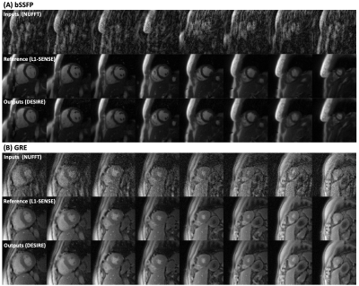 |
Free-breathing High-resolution Spiral Real-time Cardiac Cine Imaging at 1.5 T with DEep learning-based Spiral Image REconstruction (DESIRE)
Junyu Wang1, Ruixi Zhou1,2, Xitong Wang1, Marina Awad1, and Michael Salerno1,3,4,5
1Biomedical Engineering, University of Virginia, Charlottesville, VA, United States, 2School of Artificial Intelligence, Beijing University of Posts and Telecommunications, Beijing, China, 3Medicine, University of Virginia, Charlottesville, VA, United States, 4Radiology, University of Virginia, Charlottesville, VA, United States, 5Medicine, Stanford University, Stanford, CA, United States
Cardiac magnetic resonance (CMR) real-time cine, which does not require breath-holding or ECG gating, is clinically useful particularly for patients with impaired breath-hold capacity and/or arrhythmias. Spiral acquisitions, which provide high acquisition efficiency and insensitivity to motion artifacts, can require a long reconstruction time particularly for compressed-sensing or other iterative reconstruction techniques. As such they cannot provide immediate feedback to the imager. Here, we sought to develop high-resolution real-time cine imaging at 1.5 T using fast spiral acquisitions and deep learning-based rapid imaging reconstruction for both bSSFP and GRE imaging, to make high-quality and online reconstruction for cine imaging feasible.
|
|
| 09:31 | 0022.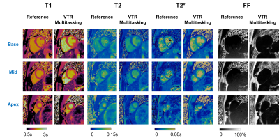 |
Non-ECG-triggered, free-breathing simultaneous T1, T2, T2*, and fat-fraction quantification in the myocardium with MR Multitasking
Tianle Cao1,2, Nan Wang3, Alan C. Kwan4, Hsu-Lei Lee1, Xianglun Mao1, Yibin Xie1, Pei Han1,2, Hui Han1, Anthony G. Christodoulou1,2, and Debiao Li1,2
1Cedars Sinai Medical Center, Los Angeles, CA, United States, 2University of California, Los Angeles, Los Angeles, CA, United States, 3Radiology Department, Stanford University, Stanford, CA, United States, 4Department of Imaging and Cardiology, Cedars Sinai Medical Center, Los Angeles, CA, United States
Myocardium tissue characterization with multi-parametric mapping has gained increased clinical interest. Conventional methods require multiple breath-holding, ECG-triggered scans, which may result in potential image misregistration and patient fatigue. In this work, a free-breathing, non-ECG-triggered approach for simultaneous myocardial T1/T2/T2*/FF (fat-fraction) mapping is presented. Quantitative measurements from the proposed technique agreed with those from single-parameter references.
|
|
| 09:33 |
0023. |
Metabolite-Cycled Echo-Planar Spectroscopic Imaging of the Human Heart
Sophie M. Peereboom1 and Sebastian Kozerke1
1Institute for Biomedical Engineering, University and ETH Zurich, Zurich, Switzerland
Spectroscopic imaging could provide insights into regional cardiac triglyceride variations, but scan times are relatively long. In this work metabolite-cycled echo-planar spectroscopic imaging with motion-adapted gating and weighted acquisition is proposed to assess triglyceride-to-water ratios in different regions of the human heart. Results are compared to metabolite-cycled EPSI with conventional acquisition and to single voxel measurements in the interventricular septum. It is shown that scan time can be reduced by more than half to less than 10 minutes as compared to conventional acquisition, while keeping the quality of triglyceride fitting constant.
|
|
| 09:35 | 0024.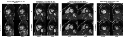 |
Free-Breathing Cardiac Cine MRI with Compressed Sensing Real-Time Imaging and Respiratory Motion Correction: Initial Pediatric Experience
Jianing Pang1, Pedro Itriago Leon2, Xiaoming Bi3, Gary McNeal1, Christoph Forman4, Christianne Leidecker1, Maria M Pereyra5, and Prakash M Masand5
1Siemens Medical Solutions USA Inc., Chicago, IL, United States, 2Siemens Medical Solutions USA Inc., Houston, TX, United States, 3Siemens Medical Solutions USA Inc., Los Angeles, CA, United States, 4Siemens Healthcare, Erlangen, Germany, 5Texas Children's Hospital, Houston, TX, United States
Cardiac cine MRI plays an important role in evaluating cardiac function. The conventional technique, 2D segmented acquisition with retrospective electrocardiogram gating, requires breath-holding and therefore is challenging for many types of patients such as young children. In this work, we report our initial experience of a recently developed free-breathing cardiac cine MRI technique on pediatric patients undergoing routine CMR. Our results showed that the proposed technique, based on compressed sensing real-time imaging and respiratory motion correction, produced comparable quantitative LV measurements and image quality to the reference breath-hold technique in a wide range of patient sizes and ages.
|
|
| 09:37 | 0025.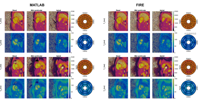 |
Online FIRE Reconstruction of Cardiac MRF T1, T2 and ECV maps with Neural Network Dictionary Generation and Low-Rank Subspace Reconstruction
Alexander Fyrdahl1, Nicole Seiberlich1, and Jesse Hamilton1
1Radiology, University of Michigan, Ann Arbor, MI, United States
The ability to reconstruct research images online may facilitate clinical translation and dissemination of new methods. Online implementations of cardiac MRF have been described previously, although they required long reconstruction times due to the need to perform online Bloch equation simulations. In this work, an efficient online 2D cardiac MRF reconstruction pipeline is introduced, capable of displaying myocardial tissue property maps in less than a minute.
|
 Back to Meeting Home
 Back to the Program-at-a-Glance
The International Society for Magnetic Resonance in Medicine is accredited by the Accreditation Council for Continuing Medical Education to provide continuing medical education for physicians.
 Back to Meeting Home
 Back to the Program-at-a-Glance