Power Pitch
Pitch: MRA & Flow
Joint Annual Meeting ISMRM-ESMRMB & ISMRT 31st Annual Meeting • 07-12 May 2022 • London, UK

Power Pitch Session: How it Works
1st Hour: 2-minute Power Pitches in the Power Pitch Theater.
2nd Hour: 60-minute digital poster presentations at the smaller screens around the perimeter of the Power Pitch Theater.
| 14:45 | 0078.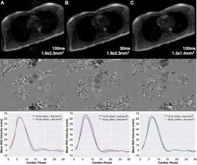 |
Accelerated 2D PC-MRI using a Deep Learning-Based Reconstruction with Complex Difference Estimation: A Prospective Feasibility Study
Matthew J. Middione1, Julio A. Oscanoa1,2, Ali B. Syed1, Shreyas S. Vasanawala1, and Daniel B. Ennis1,3
1Department of Radiology, Stanford University, Stanford, CA, United States, 2Department of Bioengineering, Stanford University, Stanford, CA, United States, 3Cardiovascular Institute, Stanford University, Stanford, CA, United States We have previously demonstrated a Deep Learning (DL) reconstruction of highly accelerated 2D PC-MRI datasets. We trained a DL reconstruction by retrospectively undersampling fully-sampled 2D PC-MRI datasets to enable up to 9x accelerated images with <5% error in accuracy and precision. Herein, we compare the accuracy and precision of 2D PC-MRI measurements for our prospectively deployed sequence and DL reconstruction. In this initial feasibility study, we show that our DL reconstruction provides <5% error in the accuracy of peak velocity and total flow relative to 2x parallel imaging and better accuracy and precision compared to 8x compressed sensing. |
|
| 14:47 | 0079.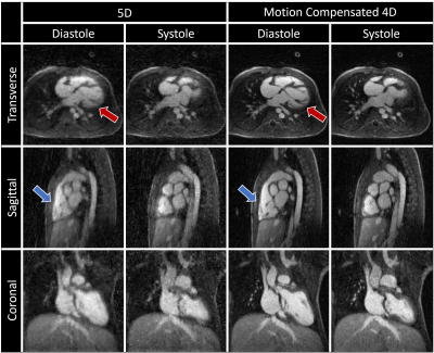 |
Automated compensation of respiratory motion and bulk patient movement in free-running whole-heart 4D MRI
Christopher W Roy1, Jérôme Yerly1,2, Milan Prša3, Estelle Tenisch1, Tobias Rutz4, Davide Piccini1,5, and Matthias Stuber1,2
1Radiology, Lausanne University Hospital (CHUV) and University of Lausanne (UNIL), Lausanne, Switzerland, 2CIBM Center for Biomedical Imaging, Lausanne, Switzerland, 3Woman-Mother-Child, Pediatric Cardiology, Lausanne University Hospital (CHUV) and University of Lausanne (UNIL), Lausanne, Switzerland, 4Cardiology, Lausanne University Hospital (CHUV) and University of Lausanne (UNIL), Lausanne, Switzerland, 5Advanced Clinical Imaging Technology (ACIT), Siemens Healthcare AG, Lausanne, Switzerland
A novel methodology for whole-heart 4D MRI with retrospective compensation of both respiratory motion and bulk patient movement is developed and its initial feasibility demonstrated. This approach enables imaging with high isotropic resolution and allows for retrospective evaluation of the dynamic cardiac anatomy creating a new way to significantly improve image quality in un-cooperative patients and potentially decreasing the need for sedation.
|
|
| 14:49 | 0080.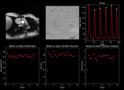 |
FlowSeg: Joint deep artifact suppression and segmentation network for low latency beat-to-beat assessment of cardiac aortic flow.
Olivier Jaubert1,2, Javier Montalt-Tordera1, Simon Arridge2, Jennifer Steeden1, and Vivek Muthurangu1
1Institute of Cardiovascular Sciences, University College London, London, United Kingdom, 2Department of Computer Science, University College London, London, United Kingdom
Real-time cardiac output monitoring is desirable but requires low latency reconstruction and segmentation. A framework based on a spiral real-time flow acquisition and joint deep artifact suppression and segmentation was developed to provide flow volumes with low latency at the scanner. The proposed network was characterized in simulations and the full framework tested at the scanner providing real-time monitoring of flow during exercise. Future work will aim to validate the method in a larger cohort.
|
|
| 14:51 | 0081.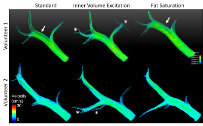 |
Fat mitigation strategies to improve image quality of radial 4D flow MRI in obese subjects
Thekla Helene Oechtering1,2, A M K Muntasir Shamim3, Alma Spahic4, Nikolaos Panagiotopoulos1,2, Oliver Wieben1,4, Alejandro Roldán-Alzate1,5,6, Scott B Reeder1,4,5,6,7, and Kevin M Johnson1,4
1Department of Radiology, University of Wisconsin-Madison, Madison, WI, United States, 2Department of Radiology and Nuclear Medicine, Universität zu Lübeck, Lübeck, Germany, 3Department of Electrical and Computer Engineering, University of Wisconsin-Madison, Madison, WI, United States, 4Department of Medical Physics, University of Wisconsin-Madison, Madison, WI, United States, 5Department of Mechanical Engineering, University of Wisconsin-Madison, Madison, WI, United States, 6Department of Biomedical Engineering, University of Wisconsin-Madison, Madison, WI, United States, 7Department of Emergency Medicine, University of Wisconsin-Madison, Madison, WI, United States 4D flow MRI of the abdomen is challenging in obese patients due to confounding streak artifacts from fat and large imaging volume. This ongoing study aims to test different fat mitigation strategies to improve image quality. We acquired three 10-min contrast-enhanced, radial 4D flow sequences: 1) with clinically used standard parameters, 2) with inner volume excitation, and 3) with fat saturation. Preliminary results confirm best signal-to-noise ratio, longest streamlines and fewest streak-artifacts using inner volume excitation. Flow rate and repeatability did not differ between sequences. |
|
| 14:53 | 0082.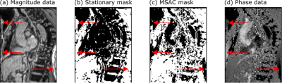 |
Automatic background phase correction on 4D Flow using M-estimate SAmple Consensus (MSAC)
Carola Fischer1,2, Jens Wetzl2, Tobias Schäffter1,3,4, and Daniel Giese2,5
1Department of Medical Imaging, Technical University of Berlin, Berlin, Germany, 2Magnetic Resonance, Siemens Healthcare GmbH, Erlangen, Germany, 3Physikalisch-Technische-Bundesanstalt (PTB), Braunschweig and Berlin, Germany, 4School of Imaging Sciences and Biomedical Engineering, King's College London, London, SE1 7EH, United Kingdom, 5Institute of Radiology, University Hospital Erlangen, Friedrich-Alexander-Universität Erlangen-Nürnberg (FAU), Erlangen, Germany
4D Flow data requires background phase removal to correctly quantify flow velocities. Typical methods are semi-automatic, requiring manual parameter input and remain time-consuming. Furthermore, correction methods based on stationary tissue segmentation fail when in the presence of wrap-around artifacts. In contrast, the proposed M-Estimate SAmple Consensus (MSAC) algorithm robustly and fully automatically corrects background phases by rejecting pixels that show large deviations from the most probable phase offset fit. A parameter sensitivity analysis is proposed following in vivo application in 13 datasets.
|
|
| 14:55 | 0083.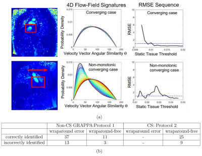 |
Novel Self-calibrating 4D Flow-Field Signature Technique to Simultaneously Optimize Eddy Current Corrections and Detect Wraparound Errors
Thara Nallamothu1,2, Haben Berhane1,2, Liliana Ma1, Justin Baraboo1,2, Daniel C. Lee3, Daniel Kim1, Phillip Greenland4, Michael Markl1,2, Rod Passman3, and Mohammed S.M. Elbaz1
1Radiology, Northwestern University, Chicago, IL, United States, 2Biomedical Engineering, Northwestern University, Chicago, IL, United States, 3Medicine (Cardiology), Northwestern University, Chicago, IL, United States, 4Preventative Medicine, Northwestern University, Chicago, IL, United States
Eddy current corrections are essential in preprocessing 4D flow data due to differing offset errors in each flow component leading to spatiotemporal inaccuracies in the velocity vector-field. Current automated optimization methods may require training data, have limited generalizability to different scan protocols, are sensitive to wraparound errors, and do not account for complex spatiotemporal errors. We introduce a self-calibrating method to optimize eddy current corrections and detect wraparound errors by using our recent 4D flow-field disparity signature technique that stochastically encodes the entire spatiotemporal profile of paired vector disparities and show feasibility and generalizability to two different 4D flow protocols.
|
|
| 14:57 | 0084.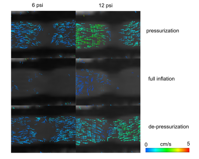 |
Evaluation of operation pressure of a soft robotic extra-aortic cardiac assist device using MRI
Seraina Anne Dual1,2, Mattia Arduini1, Brigitte Schmittlein 3, Judith Zimmermann1,4, Ellen Roche5, and Daniel Ennis2,6,7
1Department of Radiology, Stanford University, Palo Alto, CA, United States, 2Cardiovascular Institute, Stanford University, Palo Alto, CA, United States, 3Department of Mechanical Engineering, Stanford University, Palo Alto, CA, United States, 4Department of Computer Science, Technical University of Munich, Garching, Germany, 5Institute for Medical Engineering & Science, Massachusetts Institute of Technology, Cambridge, MA, United States, 6Department of Radiology, Stanfor University, Palo Alto, CA, United States, 7Division of Radiology, Veterans Affairs Health Care System, Palo Alto, CA, United States
MRI can be used to evaluate the interaction of soft robotic devices with biological tissues. The aim of this study was to use MRI to optimize the actuation pressure of an extra-aortic soft robotic cardiac assist device. We evaluated wall deformations, displaced fluid volumes and qualitative flows induced by the actuator on a fluid filled elastic vessel. Our results show that the displaced volume increases with the actuator pressure and decreases with increasing the vessel pressure. The maximum actuator pressures do not lead to buckling of the vessel wall given the current three-bladder design.
|
|
| 14:59 | 0085. |
4D flow MRI derived global intracranial pulse wave velocity and T1 white matter hypointensities among a population-based group of older people
Cecilia Björnfot1, Anders Eklund1,2, Jenny Larsson3, William Hansson3, Sara Qvarlander1, Lars-Owe Koskinen3, Jan Malm3, and Anders Wåhlin1,2,4
1Department of Radiation Sciences, Umeå University, Umeå, Sweden, 2Umeå Center for Functional Brain Imaging (UFBI), Umeå University, Umeå, Sweden, 3Department of Clinical Science, Neurosciences, Umeå University, Umeå, Sweden, 4Department of Applied Physics and Electronics, Umeå University, Umeå, Sweden
Arterial stiffening occurs with age and could be detrimental to the brain through several pathways. This potentially aggravates factors causing cerebral small vessel disease. We assessed arterial stiffness of the intracranial arteries through a newly developed global intracranial pulse wave velocity (PWV) measurement using 4D flow MRI data in a population-based cohort of older people and compared it to white matter hypointensity volume (WMH). Results revealed a weak but significant correlation between WMH and PWV, suggesting that such PWV measurement could be a useful tool to assess the brain’s vascular health.
|
|
| 15:01 | 0086.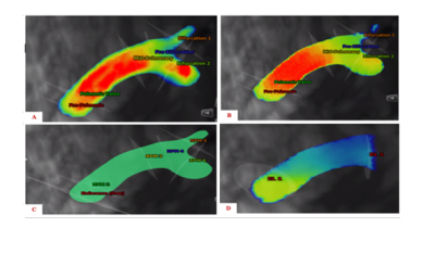 |
Characterization of Pulmonary Flow Hemodynamic Patterns in Patients with repaired Tetralogy of Fallot using 4D Flow MRI
Ashifa Hudani1, David Patton 2, James A White3, Steven Greenway2, and Julio Garcia3
1University of Calgary, Calgary, AB, Canada, 2Alberta Children's Hospital Research Institute, Calgary, AB, Canada, 3Libin Cardiovascular Institute, Calgary, AB, Canada
Tetralogy of Fallot (TOF) occurs in 1 in 3,500 births and is the most common cyanotic congenital heart defect. Patients with repaired TOF (rTOF) require constant monitoring to prevent life-threatening adverse effects. Hence, this study used 4D Flow MRI to assess pulmonary flow hemodynamics in patients with rTOF. All hemodynamic parameters calculated along the main pulmonary were shown to be higher in patients compared to controls.
|
|
| 15:03 | 0087.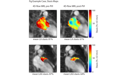 |
Evidence for Improved Left Atrial Blood Flow Dynamics after Pulmonary Vein Isolation in Patients with Atrial Fibrillation
Maurice Pradella1,2, Justin J Baraboo1, Suvai Gunasekaran1, Mitchell A Collins1, Anthony Maroun1, Amanda L DiCarlo1, Rishi Arora3, Phil Greenland4, Rod Passman3, and Michael Markl1
1Department of Radiology, Northwestern University, Chicago, IL, United States, 2Department of Radiology, University Hospital Basel, University of Basel, Basel, Switzerland, 3Department of Cardiology, Northwestern University, Chicago, IL, United States, 4Department of Preventive Medicine, Northwestern University, Chicago, IL, United States
Atrial fibrillation (AF) significantly increases stroke risk which is attributed to thrombus formation in the left atrium (LA) and to a greater extent the LA appendage (LAA). In this study, we investigated 4D-flow MRI-derived blood flow metrics from pre- and post-ablation exams in 26 AF patients undergoing pulmonary vein isolation (PVI). Blood stasis in both the LA and LAA significantly decreased in the post-PVI 4D-flow MRI. Furthermore, LA and LAA volumes also significantly decreased. Our results suggest that 4D-flow MRI was sensitive to detect treatment-related changes of LA and LAA flow metrics implicated on thromboembolic risk in AF patients.
|
|
| 15:05 | 0088.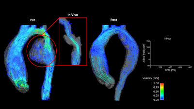 |
In Vitro 4D Flow MRI for the Analysis of Aortic Coarctation
James Rice1,2, Labib Shahid1,2, Haben Berhane3,4, Cynthia K Rigsby5, Joshua D Robinson5, Lindsay M Griffin5, Michael Markl3,4, and Alejandro Roldan-Alzate1,2
1Mechanical Engineering, University of Wisconsin-Madison, Madison, WI, United States, 2Radiology, University of Wisconsin-Madison, Madison, WI, United States, 3Radiology, Northwestern University Feinberg School of Medicine, Chicago, IL, United States, 4Biomedical Engineering, Northwestern University, Evanston, IL, United States, 5Medical Imaging, Lurie Children's Hospital of Chicago, Chicago, IL, United States
4D flow MRI can be used to assess aortic coarctation (COA) repair hemodynamics in-vivo, however, challenges arise depending on hemodynamics and clinical presentation. This study presents a modeling pipeline for in-vitro COA hemodynamic assessment. Three patient-specific in-vitro models were created representing pre- and post-repair COA geometries, connected to a pulsatile flow loop, and imaged with 4D flow MRI to assess model hemodynamics. In all cases, aortic flow profiles are shown to improve after intervention. This agrees with clinical expectation, suggesting in-vitro 4D flow MRI can be used to analyze COA hemodynamics.
|
|
| 15:07 | 0089.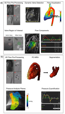 |
Evaluation of BAV Disease in Preserved Ejection Fraction: New Insight from Intra-Cardiac Pressure Drop
Shirin Aliabadi1, Michael S. Bristow1, Carmen Lydell1, Paul WM Fedak1, James White1, and Julio Garcia1
1University of Calgary, Calgary, AB, Canada We aimed to detect the reliability of flow component analysis and intra-cardiac pressure drop analysis in the bicuspid aortic valve (BAV)-induced regurgitation cases compared to age-matched healthy control in the preserved ejection fraction (EF). Flow component analysis did not show differences while intra-cardiac pressure drop differentiated groups. |
|
15:09 |
0090.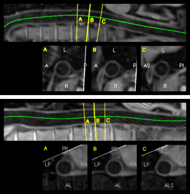 |
Efficient 3D dark-blood thoracic and abdominal aortic imaging with novel motion-corrected water/fat DANTE sequence
Camila Munoz1, Alina Hua1, Karl P Kunze2, Radhouene Neji1,2, Tevfik F Ismail1, René M Botnar1, and Claudia Prieto1
1School of Biomedical Engineering and Imaging Sciences, King's College London, London, United Kingdom, 2MR Research Collaborations, Siemens Healthcare Limited, Frimley, United Kingdom
Dark-blood imaging is a promising tool for vessel wall assessment in cardiovascular disease. Here we introduce a novel water/fat DANTE-prepared sequence for 3D dark-blood imaging of the thoracic and abdominal aorta. The sequence integrates image navigation to enable respiratory motion correction resulting in a predictable scan time. Results from healthy subjects show the feasibility of the sequence for depicting the aortic wall with high-resolution with an overall short scan of ~6 minutes.
|
|
| 15:11 | 0091.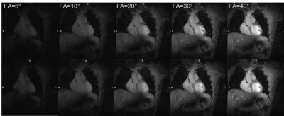 |
High resolution motion compensated contrast-free free-breathing 3D CMRA at 7T using single transmit coil
Shubhajit Paul1, Raphael Tomi-Tricot1,2, Camila Munoz1, Karl Philipp Kunze1,2, Shaihan Malik1,3, Joseph V. Hajnal1,3, Philippa Bridgen1,3, Thomas Wilkinson1,3, Sharon Giles1,3, Radhouene Neji1,2, Reza Razavi1, Claudia Prieto1,4, and René Michael Botnar1,4
1School of Biomedical Engineering and Imaging Sciences, King’s College London, London, United Kingdom, 2MR Research Collaborations, Siemens Healthcare Limited, Frimley, United Kingdom, 3The London Collaborative Ultra high field System (LoCUS), London, United Kingdom, 4Escuela de Ingeniería, Pontificia Universidad Católica de Chile, Santiago, Chile
3D cardiovascular MR imaging at ultra-high field (7T) is very challenging due to magnetic-field inhomogeneity (B0 and B1), SAR constraints and also the coil-depth issues of surface coils. To our best knowledge this is the first report demonstrating contrast-free ultra-high resolution (0.7mm3 isotropic) free-breathing 3D CMRA images with translational respiratory motion compensation. To avoid SAR constraints imposed by the use of T2-preparation pulses, high excitation flip-angles are exploited to obtain high contrast between the coronary arteries and myocardium. This technique may be adopted for clinical applications to produce high-resolution cardiac MR images at 7T scanners.
|
|
| 15:13 | 0092.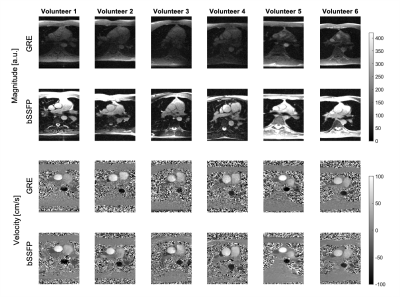 |
Cine Flow Measurements using Phase-Contrast bSSFP at 0.75 Tesla
Eva Peper1, Hannes Dillinger1, Charles McGrath1, Christian Guenthner1, and Sebastian Kozerke1
1Institute for Biomedical Engineering, ETH Zurich, Zurich, Switzerland
The use of balanced steady-state free precession sequences (bSSFP) is an approach to compensate for the signal-to-noise ratio (SNR) loss at lower-field systems (<1T). Low field MRI systems are cheaper, lighter, and less prone to system imperfections than regular 1.5-3T systems, used in clinical practice. In this work, the feasibility and accuracy of velocity encoding using phase-contrast balanced SSFP (PC-bSSFP) is demonstrated and tested on a clinical 3T MRI system ramped down to 0.75T. Phantom and in-vivo results are compared to a standard 2D spoiled gradient echo phase-contrast sequence (PC-GRE).
|
 Back to Meeting Home
 Back to the Program-at-a-Glance
The International Society for Magnetic Resonance in Medicine is accredited by the Accreditation Council for Continuing Medical Education to provide continuing medical education for physicians.
 Back to Meeting Home
 Back to the Program-at-a-Glance