Power Pitch
Pitch: MSK Power Pitch
Joint Annual Meeting ISMRM-ESMRMB & ISMRT 31st Annual Meeting • 07-12 May 2022 • London, UK

Power Pitch Session: How it Works
1st Hour: 2-minute Power Pitches in the Power Pitch Theater.
2nd Hour: 60-minute digital poster presentations at the smaller screens around the perimeter of the Power Pitch Theater.
| 16:45 | 0531.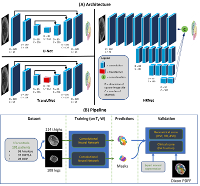 |
Geometric and clinical evaluation of deep learning-based segmentation of individual lower limb muscles from patients with neuropathies
Marc-Adrien Hostin1,2, Augustin C. Ogier2, Constance P. Michel1, Yann Le Fur1, Maxime Guye1,3, Shahram Attarian4, Marc-Emmanuel Bellemare2, and David Bendahan1
1Aix Marseille Univ, CNRS, CRMBM, UMR 7339, Marseille, France, 2Aix Marseille Univ, Université de Toulon, CNRS, LIS, Marseille, France, 3APHM, Hopital Universitaire Timone, CEMEREM, Marseille, France, 4Centre de référence des maladies neuromusculaires et de la SLA, Marseille, France
Quantification of Fat Fraction (FF) in individual lower limb muscles of patients with neuromuscular disorders relies on segmentation. Few studies have indicated that Fully Convolutional Networks (FCNs) can provide reliable automatic segmentations to replace manual tasks. However, their sensitivity to fat infiltration has never been accurately assessed. Four FCN were benchmarked for the segmentation of 114 thigh and 108 calf images (1788 muscles) with FF up to 60%. HRNet was the only network that didn't show any segmentation failures. DSC obtained was comparable to other networks. The FF values calculated from the automatic (FFa) and manual (FFm) segmentations were consistent.
|
|
| 16:47 | 0532.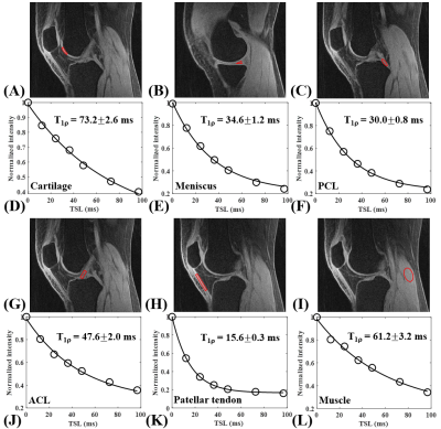 |
Quantitative Assessment of Whole Knee Joint Using a New Phase Modulated Ultrashort Echo Time Adiabatic T1rho (PM-UTE-AdiabT1rho) Sequence
Yajun Ma1, Michael Carl2, Alan Bao1, Hyungseok Jang 1, Saeed Jerban1, Alecio F Lombardi 1, Christine B Chung 1, Eric Y Chang1,3, and Jiang Du1
1Radiology, University of California, San Diego, San Diego, CA, United States, 2GE Healthcare, San Diego, CA, United States, 3Radiology Service, Veterans Affairs San Diego Healthcare System, San Diego, CA, United States
Adiabatic T1rho (AdiabT1rho) is much less sensitive to the magic angle effect compared to the regular continuous wave T1rho (CW-T1rho). In this study we developed a novel phase modulated ultrashort echo time adiabatic T1rho (PM-UTE-AdiabT1rho) sequence for quantitative assessment of both short and long T2 tissue components in the knee joint, including cartilage, meniscus, ligaments, tendons, and muscle, on a clinical 3T scanner. Our results showed excellent single exponential fitting for all the major tissue components in both ex vivo and in vivo normal knee joints.
|
|
| 16:49 | 0533.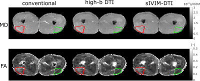 |
Accelerating IVIM and DTI for assessing microstructural changes after acute hamstring injury
Susanne Rauh1, Jithsa Monte2, Melissa Hooijmans2, Joep Suskens3, Oliver Gurney-Champion2, Johannes Tol3, Mario Maas2, Aart Nederveen2, and Gustav Strijkers1
1Department of Biomedical Engineering and Physics, Amsterdam UMC, Amsterdam, Netherlands, 2Department of Radiology and Nuclear Medicine, Amsterdam UMC, Amsterdam, Netherlands, 3Department of Orthopedic Surgery, Amsterdam UMC, Amsterdam, Netherlands DTI and IVIM are sensitive to hamstring injuries, but suffer from long scan times. Using b-values above a certain threshold only (high-b DTI) or a simplified IVIM approach (sIVIM-DTI), which estimates the perfusion fraction and diffusion tensor, can reduce the scan time while inherently correcting DTI-indices for IVIM effects. We showed in this work that those methods provide similar sensitivity to hamstring injuries in comparison to a full IVIM-DTI fit while reducing acquisition time up to 42%. Since no difference in perfusion fraction was found between injury states, we suggest high-b DTI is the method of choice in this application. |
|
| 16:51 | 0534.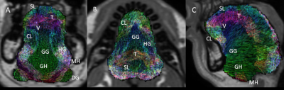 |
Using machine learning to build a population model of tongue muscle architecture based on mDIXON and diffusion tensor imaging
Robert Lloyd1,2, Iain Ball3, and Lynne Bilston1,2
1Neuroscience Research Australia, Sydney, Australia, 2University of New South Wales, Sydney, Australia, 3Philips Australia & New Zealand, Sydney, Australia
Simulations of obstructive sleep apnoea (OSA) require detailed models of the muscle fibres within the tongue, to correctly capture the motion of the tongue. Diffusion weighted images (DWI) of the oral cavity were collected for 5 healthy controls. Fibre-orientation distributions (FOD) estimated from subject DWI, can resolve intersecting muscles in the tongue. The group averaged FOD reduced the influence of spurious peaks, which gave clearer boundaries between the intrinsic muscles of the tongue. These results may help fully automate the segmentation of the muscles within the tongue.
|
|
| 16:53 | 0535.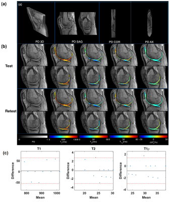 |
3D-MR-Fingerprinting for Rapid Simultaneous T1, T2, and T1ρ Volumetric Mapping of the Human Articular Cartilage at 3T
Azadeh Sharafi1, Marcelo V. W. Zibetti1, Gregory Chang1, Martijn Cloos 2, and Ravinder Regatte1
1Radiology, NYU Langone Health, New York, NY, United States, 2University of Queensland, Brisbane, Brisbane, Australia
Conventional quantitative cartilage MRI (e.g., T1, T2, and T1ρ) approaches measure one single parameter at a time. Magnetic resonance fingerprinting (MRF) is a flexible, non-invasive measurement technique that simultaneously quantifies multiple MR parameters. In this work, we proposed a 3D-MRF sequence for the simultaneous volumetric acquisition of submillimetric proton density (PD) image and T1, T2, T1ρ, and B1+ maps of the knee cartilage in clinically feasible scan time (~11 minutes).
|
|
| 16:55 | 0536.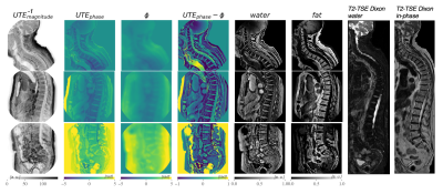 |
On the robustness of single UTE-Dixon for simultaneous short T2*, water and fat imaging across skeletal anatomies
Sophia Kronthaler1, Georg C. Feuerriegel1, Christof Boehm1, Alexandra S. Gersing1, Benedikt J. Schwaiger2, Marcus R. Makowski1, Kilian Weiss3, and Dimitrios C. Karampinos1
1Department of Diagnostic and Interventional Radiology, School of Medicine, Technical University of Munich, Munich, Germany, 2Department of Neuroradiology, Technical University of Munich, Munich, Germany, 3Philips GmbH Market DACH, Hamburg, Germany
In patients with vertebral fractures or degenerative changes often CT and MR imaging are performed, with CT aiming at the characterization of osseous changes and the MR focusing on soft tissue components. Previous work presented a single UTE-Dixon (sUTE-Dixon) approach which enabled the simultaneous assessment of vertebral fractures and edema of the thoracolumbar spine from a single ultrashort-echo time (UTE) image. This work investigates the robustness of the sUTE-Dixon methodology in various skeletal anatomies.
|
|
| 16:59 | 0537.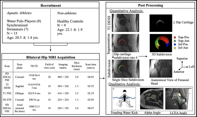 |
FAI Bone Morphology Correlates with Increased T2 Relaxations Times in the Hip Cartilage of Female Water-Treading Athletes
Elka Rubin1, Joanna L Langner2, Marianne S Black2, Arjun D Desai2, James MacKay3, Carly Jones4, Kimberly E Hall2, Marc R Safran2, Feliks Kogan2, and Garry E Gold2
1Radiology, Stanford University, Stanford, CA, United States, 2Stanford University, Stanford, CA, United States, 3University of Easy Anglia, Norwich, United Kingdom, 4University of British Columbia, Vancouver, BC, Canada
Water polo players and synchronized swimmers have previously been found to have an increased prevalence of femoroacetabular impingement morphology compared to the general population. In this study, we used MRI to identify regional patterns in the microstructure of the hip cartilage and joint of high-level female water polo players and synchronized swimmers. Compared to healthy controls, the water-treading athletes had significantly higher T2 relaxation times in the right hip and a significantly higher prevalence of cam morphology in both hips. This study suggests that microstructural and morphological differences are present in these athletes’ hip cartilage compared to controls.
|
|
| 17:01 | 0538.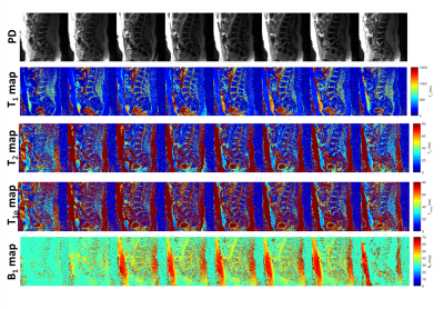 |
Three dimensional multi-parameter quantification of inter-vertebral discs using magnetic resonance fingerprinting
Rajiv G. Menon1, Azadeh Sharafi1, and Ravinder R Regatte1
1Center for Biomedical Imaging, Grossman school of Medicine, NYU Langone Health, New York, NY, United States
Low back pain that have etiologies related to degenerative intervertebral(IVD) disc changes are difficult to diagnose and treat. Quantitative characterization of the lumbar spine IVD can potentially be useful in detecting early changes to the IVD. In this study a 3D-MR fingerprinting technique was employed that is capable of simultaneously quantifying four different parameters in a clinically feasible time. Five healthy subjects underwent lumbar spine 3D-MRF scans. The results demonstrate that 3D-MRF can simultaneously measure and generate T1, T2, T1ρ, and B1+ volumetric maps of lumbar spine in a single scan within clinically feasible scan time of about 10 minutes.
|
|
| 17:03 | 0539.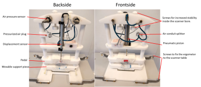 |
Increasing-load leg extensions in supine position using a novel pneumatic ergometer for dynamic 1H and 31P NMR muscle evaluations at 3T
Alfredo Liubomir Lopez Kolkovsky1,2, Beatrice Matot1,2, Yves Fromes1,2, Eric Giacomini1, Pierre G Carlier3, Harmen Reyngoudt1,2, and Benjamin Marty1,2
1NMR Laboratory, Neuromuscular Investigation Center, Institute of Myology, Paris, France, 2NMR Laboratory, CEA/DRF/IBFJ/MIRCen, Paris, France, 3University Paris-Saclay, CEA/DRF/SHFJ, Orsay, France
Evaluating quadriceps function is key in the context of sarcopenia. A pneumatic ergometer was built to allow performing knee extensions in supine position. An increasing-load, 13-min-long, isotonic exercise was successfully performed by volunteers during interleaved 1H MRI/1H MRS/31P MRS acquisitions in the quadriceps. Work, pedal displacement and velocity values per stroke and the maximum voluntary torque were measured with the ergometer. End-of-exercise [PCr] decreased (69±10%) and T2* increased (up to 10.5±7.2% in Rectus Femoris) relative to baseline. Simultaneous vascular, metabolic and physical effort evaluations during an incremental physical test could be a powerful method to investigate muscle quality in aging.
|
|
| 17:05 | 0540.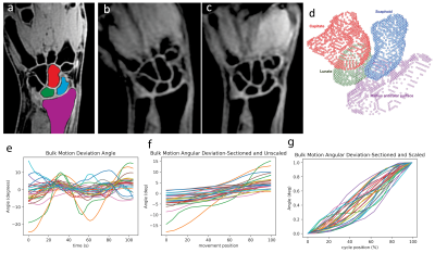 |
Normative Baseline Analysis of Carpal Bone Kinematic Profiles Using 4D MRI
Kevin Koch1, Mohammad Zarenia1, V. Emre Arpinar1, and Rajeev Mannem1
1Radiology, Medical College of Wisconsin, Milwaukee, WI, United States
4D-dynamic MRI were collected and utilized to track unconstrained movement of individual wrist carpal bones in 31 asymptomatic volunteer subjects. Rotational and translational trajectories of the scaphoid, lunate, and capitate bones were collected and processed using a novel multi-subject profile registration and analysis engine. Morphological images were analyzed by an expert radiologist to identify asymptomatic abnormalities within the cohort. Statistical parametric mapping of each profile was performed to identify normative variances of each profile metric and correlation with identified abnormalities. The study presents methodological approaches and normative baseline profile metrics of value to future studies of symptomatic wrists.
|
|
17:07 |
0541.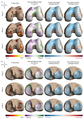 |
Large-scale Analysis of Meniscus Morphology as Risk Factor to Incidence of Osteoarthritis
Kenneth T Gao1,2, Emily Xie1, Vincent Chen1, Francesco Caliva1, Claudia Iriondo1, Richard B Souza1, Sharmila Majumdar1, and Valentina Pedoia1
1Department of Radiology and Biomedical Imaging, University of California, San Francisco, San Francisco, CA, United States, 2University of California, Berkeley-University of California San Francisco Graduate Program in Bioengineering, Berkeley, CA, United States
While meniscal morphology is central to its capacity for weight distribution and stabilization in the knee joint, the relationship between the geometric shape of the meniscus and osteoarthritis is relatively unexplored. With 4,791 subjects from the Osteoarthritis Initiative, this study utilizes an automatic statistical shape modeling technique to identify variations in meniscus morphology as precursors to future development of osteoarthritis. Seven of the 20 shape features extracted were found to be significantly associated with osteoarthritis incidence. These features were further characterized with local changes in cartilage thickness across 8 years.
|
|
| 17:09 | 0542.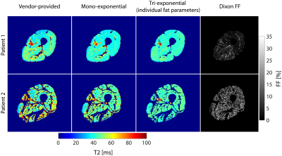 |
Assessing muscle T2 in elderly men with osteosarcopenia
Lena V. Gast1, Oliver Chaudry2, Wolfgang Kemmler1,3, Michael Uder1, Armin M. Nagel1,4, and Klaus Engelke2,3
1Institute of Radiology, University Hospital Erlangen, Friedrich-Alexander-Universität Erlangen-Nürnberg (FAU), Erlangen, Germany, 2Department of Medicine 3, University Hospital Erlangen, Friedrich-Alexander-Universität Erlangen-Nürnberg (FAU), Erlangen, Germany, 3Institute of Medical Physics, Friedrich-Alexander-Universität Erlangen-Nürnberg (FAU), Erlangen, Germany, 4Division of Medical Physics in Radiology, German Cancer Research Center (DKFZ), Heidelberg, Germany In addition to fat fraction (FF), muscular T2 could provide information on disease progression and potential training effects in osteosarcopenia, a combination of osteoporosis and sarcopenia. However, muscular T2 is highly dependent on the amount of muscle fat infiltration, which is usually increased in these patients. Thus, we aimed to assess T2 in thigh muscle tissue of elderly men with osteosarcopenia, comparing vendor-provided T2 maps with custom-fitted approaches also considering fat infiltration. We found highly elevated T2 values in the vendor-provided T2 maps compared to tri-exponentially fitted T2, which might lead to misinterpretation of muscle T2. |
 Back to Meeting Home
 Back to the Program-at-a-Glance
The International Society for Magnetic Resonance in Medicine is accredited by the Accreditation Council for Continuing Medical Education to provide continuing medical education for physicians.
 Back to Meeting Home
 Back to the Program-at-a-Glance