Power Pitch
Pitch: Advanced MRI Techniques in Epilepsy & Psychiatry
Joint Annual Meeting ISMRM-ESMRMB & ISMRT 31st Annual Meeting • 07-12 May 2022 • London, UK

Power Pitch
Pitch: Advanced MRI Techniques in Epilepsy & Psychiatry
Power Pitch Session: How it Works
1st Hour: 2-minute Power Pitches in the Power Pitch Theater.
2nd Hour: 60-minute digital poster presentations at the smaller screens around the perimeter of the Power Pitch Theater.
| 14:30 | 0464.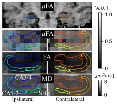 |
Microscopic fractional anisotropy may be sensitive to unilateral hippocampal abnormalities in temporal lobe epilepsy
Nico J. J. Arezza1,2, Jorge G. Burneo3, Ali R. Khan1,2,4, and Corey A. Baron1,2,4
1Medical Biophysics, Western University, London, ON, Canada, 2Centre for Functional and Metabolic Mapping, Robarts Research Institute, London, ON, Canada, 3Epilepsy Program, Clinical Neurological Sciences, Western University, London, ON, Canada, 4School of Biomedical Engineering, Western University, London, ON, Canada
Drug-resistant temporal lobe epilepsy (TLE) can often be treated surgically, but successful intervention depends on the use of imaging to localize the epileptic focus. Here, microscopic fractional anisotropy (μFA), fractional anisotropy (FA), and mean diffusivity (MD) were investigated to assess their sensitivities to abnormalities in TLE. μFA was significantly reduced in the ipsilateral hippocampal subfield containing the cornu ammonis 3/4 and dentate gyrus (p=0.0044), suggesting that it may be sensitive to pathological hippocampal abnormalities. Furthermore, differences between ipsilateral and contralateral μFA and MD, but not FA, correlated with ipsilateral atrophy in that same region.
|
|
| 14:32 | 0465.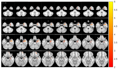 |
MR Fingerprinting Features in Patients with MRI-negative Pharmacoresistant Focal Epilepsy
Ting-Yu Su1, Yingying Tang2, Joon Yul Choi1, Siyuan Hu3, Ken Sakaie4, Hiroatsu Murakami1, Stephen Jones4, Imad Najm1, Dan Ma3, and Zhong Irene Wang1
1Epilepsy Center, Cleveland Clinic, Cleveland, OH, United States, 2Department of Neurology, West China Hospital of Sichuan University, Chengdu, China, 3Biomedical Engineering, Case Western Reserve University, Cleveland, OH, United States, 4Imaging Institute, Cleveland Clinic, Cleveland, OH, United States
MR fingerprinting (MRF) is an advanced quantitative MR technique that allows for efficient acquisition of multiparametric tissue maps. We applied high-resolution 3D MRF to examine T1 and T2 changes in 30 pharmacoresistant focal epilepsy patients with negative MRI, using a voxel-wise group-analysis approach. Forty age-and-gender-matched healthy controls were also included for comparison. Significant T1 increase was detected in the temporal pole, mesial temporal, and superior temporal regions, as well as the orbitofrontal cortex, ipsilateral to the side of the epilepsy. These MRF-detected subtle tissue property changes suggest potential structural damage in the ipsilateral limbic network in MRI-negative pharmacoresistant focal epilepsy.
|
|
| 14:34 | 0466.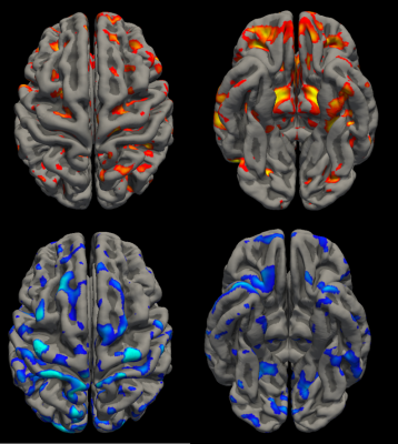 |
Alterations in Temporal Lobe Functional Connectivity Differ Based on Clinical Outcomes in Patients with Mesial Temporal Sclerosis
Anish Vinay Sathe1, Michael Kogan2, KiChang Kang1, Jingya Miao1, Mashaal Syed1, Isaiah Ailes1, Caio Matias1, Feroze Mohamed1, Ashwini Sharan1, and Mahdi Alizadeh1
1Thomas Jefferson University, Philadelphia, PA, United States, 2The University of New Mexico, Albuquerque, NM, United States
Mesial temporal sclerosis (MTS) is a severe form of temporal lobe epilepsy. Resection is the treatment of choice for refractory MTS but can still show seizure recurrence. We used seed-to-voxel analysis via amplitude synchronization to determine alterations in functional connectivity in MTS based on seizure-free status one year post-resection. Positive and negative correlations between temporal lobe structures and other brain regions were calculated and compared. We found changes in connectivity in MTS between ascending reticular activating system structures such as the thalamus and brainstem and temporal lobe structures. We also found differences in connectivity alterations between seizure-free and non-seizure-free patients.
|
|
| 14:36 | 0467.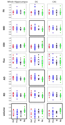 |
Analysis of Hippocampal Subfield Diffusion Imaging for Lateralization in Mesial Temporal Sclerosis
Gustavo Chau Loo Kung1, Andrew Chiu2, Zachary Davey3, Nicole Mouchawar4, Mackenzie Carlson1, Douglas Martin5, Kevin Graber3, Babak Razavi3, Jennifer McNab4, and Michael Zeineh4
1Bioengineering, Stanford University, Stanford, CA, United States, 2Radiology, Stanford Medicine, Stanford, CA, United States, 3Neurology & Neurological Sciences, Stanford Medicine, Stanford, CA, United States, 4Radiology Department, Stanford University, Stanford, CA, United States, 5University Hospitals, 11100 Euclid Ave, OH, United States
We performed subfield characterization of Mesial Temporal Sclerosis (MTS) using diffusion metrics. Analysis of MRI from 57 temporal lobe epilepsy patients was based on categorization from medical records, initial clinical radiological reads, and diagnostic changes after subfield DTI analyses. We observed differences in mean diffusivity, axial diffusivity and orientation dispersion in the dentate gyrus of patients with MTS. Differences between MTS and control patients were more pronounced in left MTS. We demonstrate how these methods may aid in the MRI identification of MTS.
|
|
| 14:38 | 0468.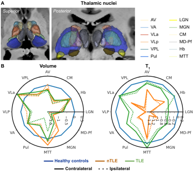 |
Detection of unilateral changes in thalamic volume and microstructure in focal epilepsy using 7T MRI
Benoit Testud1,2, Roy A.M. Haast1,2, Julia Scholly1,2,3,4, Hugo Dary1,2, Arnaud Le Troter1,2, Jean-Philippe Ranjeva1,2, Fabrice Bartolomei3,4, and Maxime Guye1,2
1Aix-Marseille Université, CNRS, CRMBM, Marseille, France, 2APHM La Timone, CEMEREM, Marseille, France, 3Aix-Marseille Université, INSERM, INS, Marseille, France, 4Department of Epileptology, APHM La Timone, Marseille, France
This work evaluated differences in thalamic morphometry and T1 across drug-resistant temporal (TLE) and non-temporal lobe epilepsy (nTLE) patients using 7T MRI. Results show bilateral thalamic atrophy across all patients, with strongest effects observed in TLE patients and ipsilateral pulvinar and mediodorsal nuclei. Moreover, T1 appeared lower, especially in medial, lateral, and posterior nuclei. While T1 was lowest across nTLE patients, an ipsi- vs contralateral effect was uniquely present in TLE patients. Future, joint analyses with other available myelin- and iron sensitive metrics (e.g., tissue susceptibility and diffusivity), connectivity features and clinical features will increase sensitivity to differentiate among patients.
|
|
| 14:40 | 0469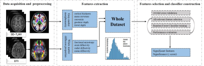 |
Gray and white matter structural examination for diagnosis of MDD and StD in adolescents and young adults: a preliminary radiomics analysis Video Permission Withheld
Huan Ma1, Jianzhong Yang2, Dewei Sun1, Dafu Zhang1, Yan Zhang2, Jing Yuan2, and Xiaoyong Zhang3
1Department of Radiology, The Third Affiliated Hospital of Kunming Medical University, Kunming, Yunnan Province, China, 2Department of Psychiatry, The Second Affiliated Hospital of Kunming Medical University, Kunming, Yunnan Province, China, 3Clinical Science, Philips Healthcare, Chengdu, China, Chengdu, China
We obtained sMRI data from unmedicated adolescents and young adults with MDD, StD and well-matched healthy control subjects, and identified radiomics features from gray and white matter, and establish classification models. The accuracies and AUC were 86.75%, 0.93 for distinguishing MDD from HC, 70.51%, 0.69 for discriminating StD from HC, and 59.15%, 0.66 for differentiating MDD from StD, respectively. These findings provide preliminary evidence that radiomics features of brain structure are valid for discriminating MDD and StD from HCs. The MRI-based radiomics approach, with further improvement and validation, is a potential facilitating method to clinical diagnosis of mental illness.
|
|
| 14:42 | 0470.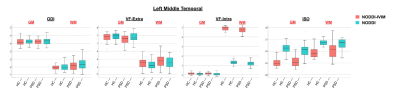 |
Effect of Intravoxel Incoherent Motion on NODDI Diffusion Parameters in both Healthy and Psychotic Spectrum Disorder Populations
Faye McKenna1, Veronica Yu Sui1, and Mariana Lazar1
1Radiology, New York University School of Medicine, New York, NY, United States
We investigated the intravoxel incoherent imaging (IVIM) effect on neurite orientation dispersion and density imaging (NODDI) metric calculations in both healthy controls (N=36) and a clinical population of individuals with a psychotic spectrum disorder (PSD) (N=55) by extending the original model to include a microvascular blood compartment. We found that without controlling for the IVIM effect, several NODDI metrics were possibly misestimated, which subsequently affected group comparisons and results.
|
|
| 14:44 | 0471.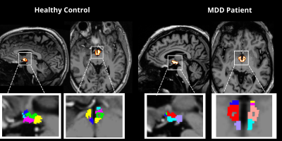 |
Hypothalamic subnuclei segmentation and diffusion tractography in Major Depressive Disorder at 7T
Mackenzie Langan1,2, Ameen Al Qadi1,2, Gaurav Verma1, Bradley Delman3, James Murrough4, Priti Balchandani*1, and Laurel Morris*4
1Biomedical Engineering and Imaging Institute, Icahn School of Medicine at Mount Sinai, New York, NY, United States, 2Icahn School of Medicine, Graduate School of Biomedical Sciences, New York, NY, United States, 3Diagnostic, Molecular and Interventional Radiology, Icahn School of Medicine at Mount Sinai, New York, NY, United States, 4Psychiatry, Icahn School of Medicine at Mount Sinai, New York, NY, United States
We performed a novel 7T analysis of volumetric, microstructural and tractographic measures in the hypothalamus in MDD and matched controls. We uncovered significant differences in FA from left anterior superior and left tubular inferior, and discovered correlations between these regions and clinical measures of depression and anxiety. These findings warrant further investigations of microstructural integrity within the hypothalamus in patients with MDD and provide a basis for exploration of microstructural integrity within other neuropsychiatric disorders characterized by dysregulated mood and anxiety.
|
|
| 14:46 | 0472.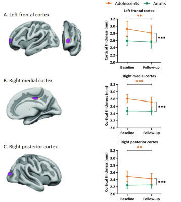 |
Long-term effects of stimulant treatment on regional cortical thickness development in ADHD
Zarah van der Pal1, Kristine Walhovd2,3, Inge Amlien2, Antonia Kaiser1, Hilde Geurts4, Liesbeth Reneman1, and Anouk Schrantee1
1Department of Radiology & Nuclear Medicine, Amsterdam UMC, University of Amsterdam, Amsterdam, Netherlands, 2Department of Psychology, Center for Lifespan Changes in Brain and Cognition, University of Oslo, Oslo, Norway, 3Departments of Radiology and Nuclear Medicine, Oslo University, Oslo, Norway, 4Dutch Autism and ADHD Research Center, Amsterdam Brain and Cognition, University of Amsterdam, Amsterdam, Netherlands
Stimulant medication is commonly used in treatment for attention-deficit/hyperactivity disorder, yet its effects on brain development remain unclear. This study investigated the long-term age-dependent effects of stimulants on cortical development after a 4-year naturalistic follow-up of adolescents and adults with ADHD using T1-weighted scans. Medication use was higher in adolescents than adults, and ADHD symptoms improved in both age groups. In line with literature on cortical development, analyses revealed reductions in apparent cortical thickness in adolescents only. However, we observed no effect of medication use on change in cortical thickness, suggesting previously identified psychostimulant effects may be transient.
|
|
| 14:48 | 0473.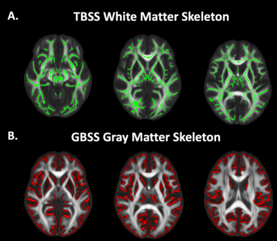 |
Tract- and Gray Matter- Based Spatial Statistics Show Microstructural Alterations in Autism Spectrum Disorder
Marissa DiPiero1,2, Hassan Cordash2,3, Molly B. Prigge4, Jace B. King4, Carolyn K. King4, Nicholas Lange5, Erin D. Bigler6,7,8,9, Brandon A. Zielinski4,6,10, Jeffrey Anderson4, Janet E. Lainhart2,11, Andrew Alexander2,11,12, and Douglas C. Dean III2,12,13
1Neuroscience Training Program, University of Wisconsin-Madison, Madison, WI, United States, 2Waisman Center, Madison, WI, United States, 3School of Computer, Data & Information Sciences, University of Wisconsin-Madison, Madison, WI, United States, 4Radiology, University of Utah, Salt Lake City, UT, United States, 5Psychiatry, Harvard School of Medicine, Boston, MA, United States, 6Neurology, University of Utah, Salt Lake City, UT, United States, 7Psychiatry, University of Utah, Salt Lake City, UT, United States, 8Psychology and Neuroscience Center, Brigham Young University, Provo, UT, United States, 9Neurology, University of California-Davis, Davis, CA, United States, 10Pediatrics, University of Utah, Salt Lake City, UT, United States, 11Psychiatry, University of Wisconsin-Madison, Madison, WI, United States, 12Medical Physics, University of Wisconsin-Madison, Madison, WI, United States, 13Pediatrics, University of Wisconsin-Madison, Madison, WI, United States
Advanced diffusion MRI techniques, such as Neurite Orientation Dispersion and Density Imaging (NODDI), may be used to improve characterization of gray matter (GM) and white matter (WM) microstructure of the brain. In this work, we used Gray Matter Based Spatial Statistics and Tract Based Spatial Statistics to investigate cortical GM and WM microstructural differences in individuals with autism spectrum disorder (ASD). Group differences and age by group interaction models were assessed. We demonstrate wide-spread alterations in GM and WM microstructure in ASD. Findings provide unique evidence of altered neurodevelopmental processes affecting microstructural development in ASD that persist into adulthood.
|
|
| 14:50 | 0474.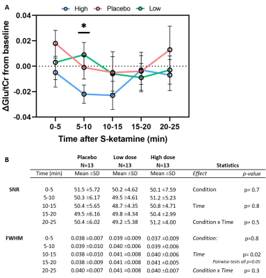 |
Dose-dependent effects of S-ketamine on frontal neurometabolism: a 7T functional MRS study
Daphne Boucherie1, Sean van Mil1, Liesbeth Reneman1, Markus Hollmann2, and Anouk Schrantee1
1Department of Radiology and Nuclear Medicine, Amsterdam University Medical Centres, University of Amsterdam, Amsterdam, Netherlands, 2Department of Anaesthesiology, Amsterdam University Medical Centers, University of Amsterdam, Amsterdam, Netherlands
The therapeutic antidepressant effect of S-ketamine has been linked to changes in glutamatergic neurotransmission. Here, we investigated the value of dynamic functional MRS compared to static MRS in detecting dose-dependent effects of subanesthetic S-ketamine on glutamate in the anterior cingulate cortex. Although significant dose-dependent subjective dissociative effects of S-ketamine were observed, we did not find significantly different glutamate changes between placebo, low-dose S-ketamine and high-dose S-ketamine, using either a “static” or “dynamic” analysis approach. Nevertheless, exploratory analyses suggest that glutamate might be modulated at specific time-points after S-ketamine administration, illustrating the potential additional information obtained from a dynamic approach.
|
 Back to Meeting Home
 Back to the Program-at-a-Glance
The International Society for Magnetic Resonance in Medicine is accredited by the Accreditation Council for Continuing Medical Education to provide continuing medical education for physicians.
 Back to Meeting Home
 Back to the Program-at-a-Glance