Power Pitch
Pitch: Imaging Nerves, Head & Neck
Joint Annual Meeting ISMRM-ESMRMB & ISMRT 31st Annual Meeting • 07-12 May 2022 • London, UK

Power Pitch Session: How it Works
1st Hour: 2-minute Power Pitches in the Power Pitch Theater.
2nd Hour: 60-minute digital poster presentations at the smaller screens around the perimeter of the Power Pitch Theater.
| 14:45 | 0648.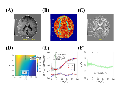 |
Is diffusion tensor principal diffusivity aligned with axon fiber in the human brain white matter?
YUXI PANG1
1Department of Radiology, University of Michigan, Ann Arbor, MI, United States
An angle offset has been identified in proton magnetic resonance transverse relaxation orientation dependencies in the human brain WM in vivo when DTI primary diffusivity direction was used as an internal reference. This angle offset has not yet been accounted for in previous studies. The present work demonstrates that the observed angle offset can be removed using an angle derived from the perpendicular and parallel diffusivities of an axially symmetric diffusion tensor regardless of axon fiber orientations. The finding from this study clearly suggests that the diffusion tensor principal diffusivity direction deviates from an axon fiber orientation in WM.
|
|
| 14:47 | 0649. |
In-Vivo Probabilistic Tractography of the Extracranial Branches of the Trigeminal Nerve
Kellen Mulford1, Sean Moen2, Andrew W. Grande2, David Darrow2, Donald R. Nixdorf3, Pierre-Francois Van de Moortele1, and Can Özütemiz4
1Center for Magnetic Resonance Research, University of Minnesota, Minneapolis, MN, United States, 2Department of Neurosurgery, University of Minnesota, Minneapolis, MN, United States, 3Division of TMD & Orofacial Pain, University of Minnesota, Minneapolis, MN, United States, 4Department of Radiology, University of Minnesota, Minneapolis, MN, United States
Diffusion tensor imaging-based tractography of the peripheral nerves of the face is difficult owing to the complex tissue interfaces which degrade signal and introduce artifact. In this study, we used readout segmented EPI to attempt nerve fiber tracking of the inferior-alveolar, lingual, and infra-orbital branches of the trigeminal nerve. In 6 healthy participants we were able to visualize all three nerves on both sides of the face. This technique could be applies to nerve-injury and neuropathic pain populations to assess for alterations in nerve structure.
|
|
| 14:49 | 0650.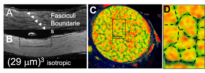 |
Fasciculus Axonal Connective Tissue (FACT) Mapping of Porcine Optic Nerve for Accurate Connectome Mapping at Viable Cost
Sudhir Kumar Pathak1, Yijen L Wu2,3, Vijay Saradhi Gorantla4, Fatih Zor4, Yalcin Kulahci4, Alan Watson5, Yongxin Zhao6, and Walter Schneider1,7
1Psychology, University of Pittsburgh, Pittsburgh, PA, United States, 2Rangos Research Center Animal Imaging Core, Children's Hospital of Pittsburgh of UPMC, Pittsburgh, PA, United States, 3Developmental Biology, School of Medicine, University of Pittsburgh, Pittsburgh, PA, United States, 4Regenerative Medicine, Wake Forest Institute for Regenerative Medicine, Winston-Salem, NC, United States, 5Department of Cell Biology, University of Pittsburgh, Pittsburgh, PA, United States, 6Department of Biological Sciences, Carnegie Mellon University, Pittsburgh, PA, United States, 7Neurosurgery, University of Pittsburgh, 15260, PA, United States
Accurate brain connectome mapping requires tracking fasciculus bundles of axons within tracts. In a porcine optic nerve harvested tissue, we optimized/tested MRI structural and diffusion imaging to demonstrate following fasciculi within the optic tract. On a 7T Bruker magnet, we can clearly identify fasciculus boarders and diffusion paths providing 29 micron precision MRI with clear tissue differentiation. We found that axons travel with no evidence of crossing fasciculi walls. This suggests Fasciculus Axonal Connective Tissue (FACT) imaging can provide full mapping of connectivity between cortical sources supporting comprehensive accurate mapping of connectivity in large mammal tract systems at viable cost.
|
|
| 14:51 | 0651.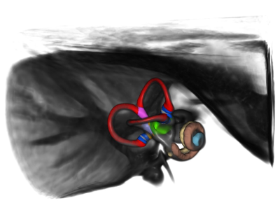 |
In-vivo observations on structural aging of the inner ear – a two-center study
Franziska Thiessen1, Seyed-Ahmad Ahmadi2, Virginia Flanagin2, Joyce Bosmans3, Huseyin Ozenc Taskin4, Vincent Van Rompaey5, Geoffrey Karl Aguirre4, and Peter zu Eulenburg6
1Institut for Neuroradiology, LMU Munich, Munich, Germany, 2Center for Vertigo and Balance Disorders, LMU Munich, Munich, Germany, 3Translational Neurosciences, University of Antwerp, Antwerpen, Belgium, 4Department of Neurology, Perelman School of Medicine, University of Pennsylvania, Philadelphia, PA, United States, 5Department of Otorhinolaryngology-Head & Neck Surgery, University of Antwerp, Antwerpen, Belgium, 6Institute for Neuroradiology, LMU Munich, Munich, Germany
We are on the road to further the role of high-resolution structural neuroimaging in the diagnostics of the most prevalent vestibular disorders. After creating an in-vivo template and atlas space for the inner ear, we have no investigated structural aging across all cochlear and vestibular anatomical regions in a representative cohort (n=87). We significant reduction along the aging process for cochlear width and length as well as some semicircular canal dimensions. Total intracranial volume was a highly relevant covariate in our analysis. Aging also seemed to affect most neuroimaging quality parameters as well and had to be controlled for.
|
|
14:53 |
0652.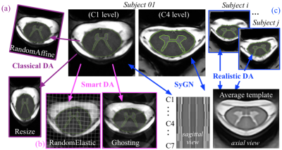 |
A multiclass deep-learning model with realistic data augmentation for improved 7T gray matter/spinal cord segmentation on patients and controls
Nilser J. Laines Medina 1,2, Charley Gros3,4, Julien Cohen-Adad3,4, Arnaud Le Troter1,2, and Virginie Callot1,2,5
1CRMBM, Aix-Marseille Univ, CNRS, Marseille, France, 2CEMEREM, APHM, CHU Timone, Marseille, France, 3NeuroPoly Lab, Institute of Biomedical Engineering, Polytechnique Montréal, Montreal, QC, Canada, 4MILA, Québec AI Institute, Montreal, QC, Canada, 5iLab-Spine, International Associated Laboratory, Marseille-Montreal, France Automated methods for WM/GM segmentation in the spinal cord are now largely available. However, these techniques were mostly developed for conventional systems (≤3T) and do not necessarily perform well on 7T MRI data that feature finer details, contrasts, but also different artifacts or signal dropout. The primary goal of this study was thus to propose a new deep-learning model allowing robust SC/GM multi-class segmentation based on high-resolution 7T T2*-w MR images. The second objective was to highlight the relevance of implementing a realistic hybrid data augmentation strategy to provide better model generalization. |
|
| 14:55 | 0653.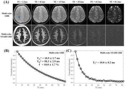 |
Myelin Water Imaging Using a New 3D Short TR Adiabatic Inversion Recovery Prepared Short Echo Time (STAIR-STE) Sequence
Yajun Ma1, Hyungseok Jang1, Alecio F Lombardi 1, Annie Hiniker 2, Roland R Lee 1, Eric Y Chang1,3, Amy J Jak3,4, Dawn Schiehser 3,4, Alan N Simmons 3,4, Brian P Head3,5, Graeme M Bydder1, and Jiang Du1
1Radiology, University of California, San Diego, San Diego, CA, United States, 2Pathology, University of California, San Diego, San Diego, CA, United States, 3Veterans Affairs San Diego Healthcare System, San Diego, CA, United States, 4Psychiatry, University of California, San Diego, San Diego, CA, United States, 5Anesthesiology, University of California, San Diego, San Diego, CA, United States
Myelin water imaging (MWI) has been proposed as a myelin-specific technique to quantify water trapped within or tightly bound to myelin bilayers and thus provides an indirect assessment of myelin content and integrity. In this study, we developed and evaluated a novel 3D short TR adiabatic inversion recovery prepared short echo time (STAIR-STE) Cones sequence for robust MWI on a clinical 3T scanner. Our results show that the 3D STAIR-STE Cones sequence robustly suppresses long-T2 intra/extracellular water signals and provides selective volumetric imaging and quantification of myelin water fraction in the whole brain.
|
|
| 14:57 | 0654.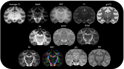 |
Beyond Fractional Anisotropy: A Comparative Study of Myelin Water Fraction and Propagator Anisotropy in the Bonnet Macaque Brain
Laurel A Dieckhaus1, Courtney J Comrie1, Kelsey E McDermott2, Daniel T Gray2, Carol A Barnes2,3,4, and Elizabeth B Hutchinson1
1Biomedical Engineering, University of Arizona, Tucson, AZ, United States, 2Evelyn F. McKnight Brain Institute, University of Arizona, Tucson, AZ, United States, 3Psychology, Neurology and Neuroscience, University of Arizona, Tucson, AZ, United States, 4Psychology and Neurology, University of Arizona, Tucson, AZ, United States
White matter (WM) is commonly studied using the Diffusion Tensor Imaging (DTI) metric Fractional Anisotropy (FA), which has limitations especially in regions where more than one fiber population exists. We compared FA to diffusion techniques (Mean Apparent Propagator (MAP-MRI)) and relaxometry techniques (Myelin Water Fraction (MWF) and Bound Pool Fraction (BPF)) mapping in the normal bonnet macaque brain. The combination of histograms and correlation analysis revealed that MAP-MRI metric, Propagator Anisotropy (PA) and MWF may offer more information about WM than FA alone, while remaining relatively correlated with FA; while BPF did not have strong association with FA or MWF.
|
|
| 14:59 | 0655.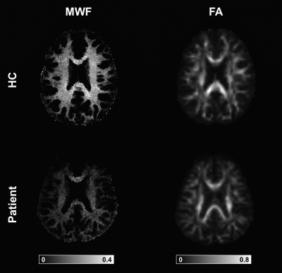 |
Assessing white matter maturation and integrity in children using multi-component 3D-MR Fingerprinting and diffusion imaging
Marta Lancione1,2, Matteo Cencini1,2, Elena Scaffei1,3, Emilio Cipriano1,4, Guido Buonincontri1, Chiara Ticci1, Rosa Pasquariello1, Roberta Battini1,5, Raffaello Canapicchi1, Laura Biagi1,2, Michela Tosetti1,2, and Italian DEvelopmental Age Health Network (IDEA)6
1IRCCS Stella Maris, Pisa, Italy, 2Imago7 Research Foundation, Pisa, Italy, 3Department of Neuroscience, Psychology, Drug Research and Child Health, Neurofarba, University of Florence, Florence, Italy, 4Department of Physics, University of Pisa, Pisa, Italy, 5Department of Clinical and Experimental Medicine, University of Pisa, Pisa, Italy, 6Italian DEvelopmental Age Health Network (RETE IDEA Ministry of Health), Rome, Italy
New biomarkers for myelination could improve the understanding of neurodevelopmental diseases and their diagnosis and treatment. We quantified Myelin Water Fraction (MWF) using MRF in a cohort of children with leukoencephalopathies and age-matched controls and we compared it to DTI-based FA. We obtained normative curves of white matter development with both techniques. MWF discriminated between controls and patients with higher sensitivity than FA, it was more myelin-specific and independent of the degree of axonal packing. Thanks to short scan time and simultaneous acquisition of other quantitative maps, MRF-based MWF may represent a valuable tool to study developmental disorders.
|
|
| 15:01 | 0656.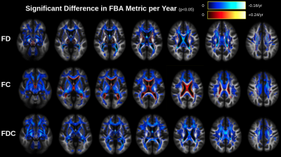 |
Fixel-based analysis of aging white matter reveals selective fiber-specific degeneration
Ana Han1, Jordan A. Chad1,2, Thijs Dhollander3, and J. Jean Chen1,2
1Rotman Research Institute, Baycrest Health Sciences, Toronto, ON, Canada, 2Department of Medical Biophysics, University of Toronto, Toronto, ON, Canada, 3Developmental Imaging, Murdoch Children's Research Institute, Melbourne, Australia
White matter (WM) microstructural aging is often studied with diffusion MRI via voxelwise metrics such as fractional anisotropy (FA). Decreasing FA with advancing age is frequently interpreted as WM microstructural degeneration, but relying on FA for this purpose only makes sense if a voxel contains a single fiber tract. In this work we use fixel-based analysis (FBA) to investigate the association of fiber-specific measures with age among each individual "fixel" within a voxel. We find intra-voxel differences in age associations of crossing fixels, demonstrating that traditional voxel-based analyses are inappropriate for probing age-related fiber degeneration in scenarios of multi-fiber crossings.
|
|
| 15:03 | 0657.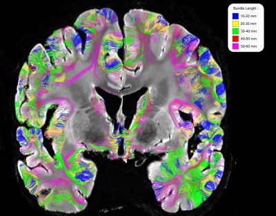 |
Length-dependent spatial distribution of short fiber bundles revealed with the mesoscopic Chenonceau dataset
Alexandros Popov1, Ivy Uszynski1, Bastien Herlin1, Maelig Chauvel1, Igor Maldonado2, Christophe Destrieux2, and Cyril Poupon1
1BAOBAB, NeuroSpin, CEA, Université Paris-Saclay, CNRS, Gif-sur-Yvette, France, France, 2iBrain U1253, Université de Tours, CHU Bretonneau, INSERM, Tours, France Mesoscopic diffusion datasets allow to study the superficial tracts of the human brain. We exploit the new Chenonceau Dataset, an ultra-high resolution diffusion-weighted dataset, to investigate the organization of short fibers ( between 10 and 60 mm ) in the whole brain. To achieve this goal, we process the dense Chenonceau connectogram with a hierarchical clustering algorithm to obtain coherent fiber bundles, sorted over a centimetric range. It leads to the first density mapping of short fibers across the whole human brain. This mapping displays important inter-hemispheric variations, supporting the functional lateralization of various functional networks. |
 Back to Meeting Home
 Back to the Program-at-a-Glance
The International Society for Magnetic Resonance in Medicine is accredited by the Accreditation Council for Continuing Medical Education to provide continuing medical education for physicians.
 Back to Meeting Home
 Back to the Program-at-a-Glance