Power Pitch
Pitch: High-Resolution fMRI & Functional Connectivity
Joint Annual Meeting ISMRM-ESMRMB & ISMRT 31st Annual Meeting • 07-12 May 2022 • London, UK

Power Pitch
Pitch: High-Resolution fMRI & Functional Connectivity
Power Pitch Session: How it Works
1st Hour: 2-minute Power Pitches in the Power Pitch Theater.
2nd Hour: 60-minute digital poster presentations at the smaller screens around the perimeter of the Power Pitch Theater.
| 14:30 | 0258.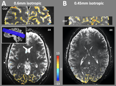 |
Evaluation of Single-shot EPI with Sub-millimeter Resolution fMRI on the Next-Generation 7T brain scanner.
Alexander JS Beckett1,2, An T Vu3,4, Sinyeob Ahn5, Salvatore Torrisi1,2, Jonathan R Polimeni6,7, Essa Yacoub8, Kawin Setsampop9,10, Berkin Bilgic6,7, Shajan Gunamony11,12, Andreas Potthast13, Peter Dietz13, Yulin Chang5, and David A Feinberg1,2
1University of California, Berkeley, CA, United States, 2Advanced MRI Technologies, Sebastopol, CA, United States, 3Radiology, University of California, San Francisco, CA, United States, 4San Francisco Veteran Affairs Health Care System, San Francisco, CA, United States, 5Siemens Medical Solutions, Malvern, PA, United States, 6Athinoula A. Martinos Center for Biomedical Imaging, Massachusetts General Hospital, Charlestown, MA, United States, 7Harvard-MIT Health Sciences and Technology, MIT, Cambridge, MA, United States, 8Center for Magnetic Resonance Research, University of Minnesota, Minneapolis, MN, United States, 9Department of Radiology, Stanford University, Stanford, CA, United States, 10Department of Electrical Engineering, Stanford University, Stanford, CA, United States, 11Imaging Centre of Excellence, University of Glasgow, Glasgow, United Kingdom, 12MR CoilTech Limited, Glasgow, United Kingdom, 13Siemens Healthcare GmbH, Erlangen, Germany
The newly developed “Impulse” head gradient (200 mT/m Gmax, 900 T/m/s) on the NexGen 7T scanner has been specifically designed to allow for high-resolution single-shot EPI with minimal distortions and blurring by minimizing echo spacing. In conjunction with the increased SNR and reduced g-factor noise afforded by the 96 channel receive array coil, mesoscale fMRI at 0.45mm and 0.6mm isotropic resolutions can be robustly achieved in human subjects.
|
|
| 14:32 | 0259.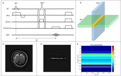 |
Spin-echo line-scanning at 7T
Luisa Raimondo1, Tomas Knapen1,2, Serge O Dumoulin1,2, Wietske van der Zwaag1, and Jeroen C.W Siero1,3
1Spinoza Centre for Neuroimaging, Amsterdam, Netherlands, 2VU University, Amsterdam, Netherlands, 3Radiology, University Medical Centre, Utrecht, Netherlands
We implemented spin-echo line-scanning (SELINE) fMRI through a simple rotation of the spin-echo refocusing gradient to a plane perpendicular to the excited slice and removing the phase encode gradient. This technique promises a combination of high spatio-temporal resolution and specificity of functional responses to the microvasculature. We compared SELINE data to the corresponding gradient-echo version (GELINE). We demonstrate that SELINE showed much improved line selection compared to GELINE, albeit at the cost of a significant drop in functional sensitivity. The low functional sensitivity needs to be addressed before SELINE can be applied.
|
|
| 14:34 | 0260.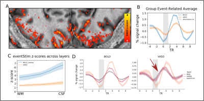 |
High temporo-spatial resolution VASO reveals differential laminar reactivity to event-related stimuli at 7T
Sebastian Dresbach1, Renzo Huber1, and Rainer Goebel1
1Faculty of Psychology and Neuroscience, Maastricht University, Maastricht, NL, Maastricht, Netherlands
Due to its high specificity, VASO plays a major role in laminar fMRI. To mitigate its lower sensitivity compared to GE-BOLD researchers mostly employ block-designs to increase SNR. Here, we developed a VASO sequence with a short TR (895ms volume acquisition) and showed that it provides the means to capture layer-specific haemodynamic responses with high spatio-temporal resolution. During event-related stimulation, we show reliable responses in visual and somatosensory cortices. Furthermore, the short TR and high specificity of VASO enabled us to show differences in laminar reactivity and onset times, thus demonstrating the high value of event-related designs using CBV-based fMRI.
|
|
| 14:36 | 0261.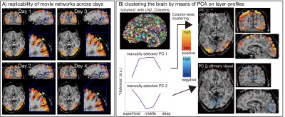 |
Towards whole brain layer-fMRI connectivity: methodological advancements for functional layer connectomics
Kenshu Koiso1,2, Sebastian Dresbash1, Christopher J Wiggins3, Omer Faruk Gulban1,4, Yoichi Miyawaki2,5, Benedikt A Poser1, and Renzo Huber1
1The Faculty of Psychology and Neuroscience, Maastricht University, Maastricht, Netherlands, 2Graduate School of Informatics and Engineering, The University of Electro-Communications, Tokyo, Japan, 3Scannexus, Maastricht, Netherlands, 4Brain Innovation, Maastricht, Netherlands, 5Center for Neuroscience and Biomedical Engineering, The University of Electro-Communications, Tokyo, Japan Laminar-specific fMRI allows neuroscientists to address research questions of directional functional connectivity within and across brain areas. While recent sequence developments allow improvements in coverage and mitigations of venous biases, previous attempts of whole-brain connectome datasets turned out to be too artifact-dominated (Mueller 2021) to be neuroscientifically applicable. Here we present a new and improved sequence used for acquiring a relatively large open dataset of whole-brain laminar connectivity. Its purpose is to:
|
|
| 14:38 | 0262.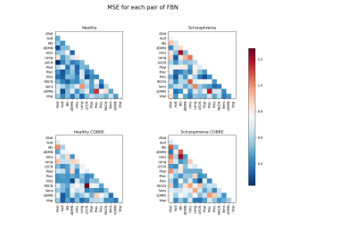 |
Normative modelling detects abnormal functional connectivity in schizophrenia
Duarte Saraiva1 and Hugo Ferreira1
1Faculty of Sciences of the University of Lisbon, Lisboa, Portugal
Machine learning (ML) applications on the diagnosis of neuropsychiatric disorders (NPD) have not reached clinical practice yet, as the continuous spectrum of NPD demands more complex, non-binary classification approaches. Herein, a ML-based normative model was created from healthy subjects, which “fails” when tested on schizophrenia patients. In particular, abnormal functional connectivity patterns were found in such patients, in agreement to what has been described in the literature. Moreover, a clustering method and analysis at the individual level indicate that subgroups may exist within the schizophrenia spectrum, suggesting that a personalized and precision-based diagnosis is within reach for such NPD.
|
|
| 14:40 | 0263.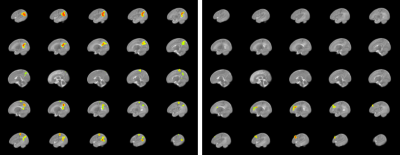 |
The relation between brain anatomy, functional connectivity, and emotional behavior in 3- and 9-month old infants
Layla Banihashemi1, Vanessa Jean Schmithorst1, Michele A Bertocci1, Alyssa Samolyk1, Joao Santos1, Amelia Versace1, Megan Taylor1, Gabrielle English1, Jessie Northrup1, Vincent Lee1, Richelle Stiffler1, Harris Aslam1, Ashok Panigrahy1, Alison Hipwell1, and Mary Phillips1
1University of Pittsburgh, Pittsburgh, PA, United States
Uncinate fasciculus, forceps minor, and cingulum bundle volumes measured in infants at 3 months of age via DWI tractography were found to predict infant emotionality at 3 months and 9 months of age. These relations were found to be mediated and/or suppressed by functional connectivity from the orbitofrontal cortex, dorsolateral prefrontal cortex, and posterior default mode. Therefore, modulating connectivity among regions in the default mode, central executive, and salience networks might be promising future approaches for interventions to maintain infant emotionality and reduce risk of future psychopathology later in childhood and adolescence.
|
|
| 14:42 | 0264.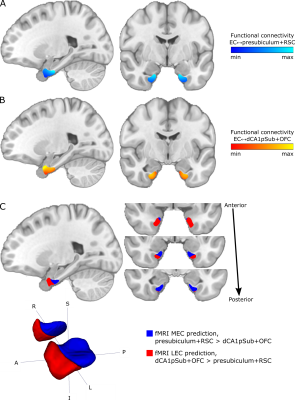 |
Investigating structural and functional connectivity of human entorhinal subregions using DTI and fMRI
Ingrid Framås Syversen1,2, Daniel Reznik3, Tobias Navarro Schröder1, and Christian F. Doeller1,3,4
1Kavli Institute for Systems Neuroscience, NTNU - Norwegian University of Science and Technology, Trondheim, Norway, 2Department of Diagnostic Imaging, Akershus University Hospital, Lørenskog, Norway, 3Max Planck Institute for Human Cognitive and Brain Sciences, Leipzig, Germany, 4Institute of Psychology, Leipzig University, Leipzig, Germany
Despite previous attempts to localize the human homologues of the medial (MEC) and lateral entorhinal cortex (LEC) using fMRI and DTI separately, there are still uncertainties related to the choice of imaging modality and seed regions used. In this study, we investigated both structural connectivity from DTI and functional connectivity from fMRI between the EC and associated brain regions. Differential EC connectivity to these regions was then used to predict the locations of the human homologues of MEC and LEC. Our results from both DTI and fMRI showed a qualitatively similar subdivision into posteromedial and anterolateral EC, supporting previous studies.
|
|
| 14:44 | 0265.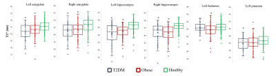 |
Type 2 diabetes mellitus and obesity are associated with altered T2* and resting-state brain activity: a UK Biobank study
Guocheng Jiang1,2, Walter Swardfager2,3, Abdullah Al-Ozairi4, Ebaa Al-Ozairi 5, Sandra E Black 2,6, and Bradley J MacIntosh1,2
1Department of Medical Biophysics, University of Toronto, Toronto, ON, Canada, 2Hurvitz Brain Sciences, Sunnybrook Research Institute, Toronto, ON, Canada, 3Department of Pharmacology and Toxicology, University of Toronto, Toronto, ON, Canada, 4Department of Psychiatry, Kuwait University, Kuwait City, Kuwait, 5Dasman Diabetes Institute, Kuwait City, Kuwait, 6Department of Medicine, University of Toronto, Toronto, ON, Canada
T2*-weighted image is a versatile and common MR imaging readout. We studied anatomical and functional brain features in three groups: Type 2 diabetes mellitus (T2DM), obese, and healthy adults. We observed that T2DM and obese adults are associated with lower regional T2* relative to healthy adults. We further found that an adverse lipid profile in obese adults was associated with decreased regional brain intrinsic activity. This work helps to characterize how T2DM and obesity affects brain using susceptibility-weighted and resting-state functional MRI approaches.
|
|
| 14:46 | 0266.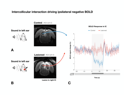 |
Intercollicular interactions drive ipsilateral negative BOLD responses upon monaural auditory stimulation
Frederico Severo1, Mafalda Valente1, and Noam Shemesh1
1Champalimaud Research, Champalimaud Centre for the Unknown, Lisboa, Portugal
Negative BOLD responses (NBRs) in rat Inferior Colliculus (IC) were recently observed upon monaural auditory stimulation, but their origins and importance remain poorly understood. Intercollicular communication is proposed as a prominent mechanism for auditory processing, including sound localization/lateralization & in gain control regulation. Here, we investigated intercollicular interaction via monoaural stimulation at 9.4T. Rats exhibited NBRs in the ipsilateral IC and positive BOLD responses (PBRs) in the contralteral IC. When the contralateral hemisphere was lesioned, the NBRs vanished in the ipsilateral IC. Our findings suggest that intercollicular interaction is essential for ipsilateral negative BOLD responses and for auditory processing.
|
|
| 14:48 | 0267.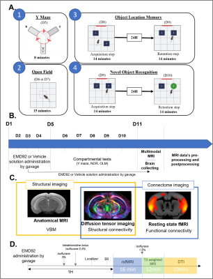 |
Multimodal brain MRI characterization of a mouse model of Down syndrome and evaluation of a pharmacological treatment.
Julien CAILLETTE1, Laëtitia Degiorgis1, Marion Sourty1, Marion Rame1, Helin Atas-Ozcan2, Laurent Meijer3, Yann Herault2, and Laura-Adela Harsan1,4
1ICube, University of Strasbourg-CNRS, Strasbourg, France, 2IGBMC, University of Strasbourg-CNRS-INSERM, Illkirch-Graffenstaden, France, 3Perha Pharmaceuticals, Perharidy Peninsula, Roscoff, France, 4Department of Biophysics and Nuclear Medicine, University Hospital of Strasbourg, Strasbourg, France DYRK1A protein-kinase overexpression was identified as key player in cognitive impairments observed in Down syndrome (DS) patients and in animal models. Inhibition of this kinase rescued cognitive deficits in the Dp1Yey mouse model of DS, but the associated mechanisms remain unknown. Here we used multimodal brain MRI and characterized the morphology, microstructure and functional connectivity (FC) of Dp1Yey mice. Voxel-based morphometry revealed brain-wide morphological alterations in Dp1Yey mice, while DTI analysis revealed white matter changes. Further, rsfMRI showed that pharmacologic DYRK1A inhibition modifies the functional connectivity for memory related nodes, synergistic with cognitive improvements observed in behavioral tests. |
|
| 14:50 | 0268.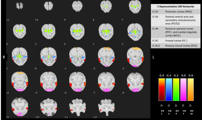 |
Alterations in intrinsic functional networks in squirrel monkey brain produced by dorsal column lesion of spinal cord using resting state fMRI
Anirban Sengupta1, Arabinda Mishra1, Feng Wang1, Li Min Chen1, and John C Gore1
1Vanderbilt University Medical Center, Nashville, TN, United States
The goal was to study how intrinsic functional networks within squirrel monkey brain undergo changes in connectivity after a dorsal column lesion (DCL) of the spinal cord. We used independent component analysis (ICA) to decompose whole brain fMRI data into spatially independent functional networks. Thirteen networks were identified, many of which resemble networks from human and macaque brain studies. Inter-network connectivity was computed before and after DCL of the cervical spinal cord. Changes in inter-network connectivity were not restricted to sensorimotor areas but spread over other cortical areas. Also, one of the connectivity increased significantly, possibly indicating compensation post lesion
|
|
| 14:52 | 0269.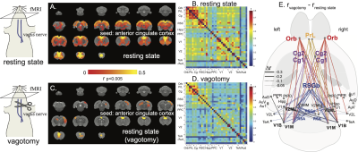 |
Autonomic Function Regulates the Default-Mode Network in Rats
Jiayue Cao1, Xiaokai Wang1, and Zhongming Liu1,2
1Biomedical Engineering, University of Michigan, ANN ARBOR, MI, United States, 2Electrical engineering and computer science, University of Michigan, ANN ARBOR, MI, United States
The default mode network (DMN) is central to cognition. Consistent across species, functional connectivity (FC) of DMN exhibits partly similar patterns across brain states, including wakefulness, sleep, sedated or anesthetized states. Regions in DMN are also involved in regulating visceral organs. It is likely that DMN facilitates visceral physiology to maintain homeostasis in states of unconsciousness. However, the evidence for the association between autonomic function and DMN is sparse and piecemeal. Here, we use fMRI in rat models to investigate how vagal nerve de-innervation and stimulation affect the DMN and its interaction with other regions related to autonomic function.
|
 Back to Meeting Home
 Back to the Program-at-a-Glance
The International Society for Magnetic Resonance in Medicine is accredited by the Accreditation Council for Continuing Medical Education to provide continuing medical education for physicians.
 Back to Meeting Home
 Back to the Program-at-a-Glance