Power Pitch
Pitch: Preclinical Neuro
Joint Annual Meeting ISMRM-ESMRMB & ISMRT 31st Annual Meeting • 07-12 May 2022 • London, UK

Power Pitch Session: How it Works
1st Hour: 2-minute Power Pitches in the Power Pitch Theater.
2nd Hour: 60-minute digital poster presentations at the smaller screens around the perimeter of the Power Pitch Theater.
09:15 |
0413.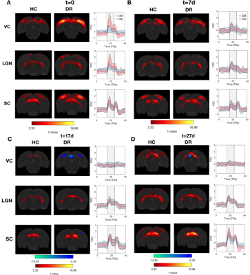 |
Mapping the time course of cortical reorganization and the influence of visual experience during plasticity.
Joana Carvalho1, Francisca Fernandes2, and Noam Shemesh3
1Preclinical MRI Laboratory, Champalimaud Foundation, Lisbon, Portugal, 2Preclinical MRI Laroratory, Champalimaud Centre for the Unknown, Lisbon, Portugal, 3Champalimaud Centre for the Unknown, Lisbon, Portugal
During development, the organization of the brain’s underlying circuitry is shaped by sensory experience. However, how the adult brain reorganizes remains much less understood. Here, we performed a longitudinal BOLD-fMRI study assessing plasticity in the adult rat brain using a chronic visual deprivation model and a novel set-up for mapping detailed visual responses. We show that dark rearing (DR) boosts neural responses to visual stimuli but delays retinotopic and spatial frequency (SF) specialization in the visual pathway. Our findings suggest that DR shifts the stability/plasticity balance and that fMRI can map dynamic processes in the visual system.
|
|
| 09:17 | 0414.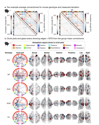 |
Mouse rsfMRI connectome fingerprinting recovers subject as well as genetically encoded Ca2+ indicator loci
Francesca Mandino1, Corey Horien2, Xilin Shen1, David O'Connor3, Xinxin Ge4, Peter Herman1, An Qu1, John Onofrey1,3,5, Michael C Crair6,7, Xenophon Papademetris1,3, Todd R Constable1,8, and Evelyn Lake1
1Radiology and Biomedical Imaging, Yale University, New Haven, CT, United States, 2Interdepartmental Neuroscience Program, Yale University, New Haven, CT, United States, 3Biomedical Engineering, Yale University, New Haven, CT, United States, 4Department of Physiology, UCSF, San Francisco, CA, United States, 5Urology, Yale University, New Haven, CT, United States, 6Kavli Institute for Neuroscience, Yale University, New Haven, CT, United States, 7Neuroscience, Yale University, New Haven, CT, United States, 8Neurosurgery, Yale University, New Haven, CT, United States
We find that the connectome, from resting-state functional magnetic resonance imaging data, can be used to identify (ID) individuals across species (humans and mice). This finding hints at the potential to use these data for individualized medicine and for translational research. To this end, we interrogate how ID rates differ across species, and how the manipulations we introduce into animal models – the use of clones and genetically encoded calcium indicators (GECI) – impact the connectome. We find species-specific ID rates differ, but only require small portions of the connectome, and that GECI loci can be recovered using this framework.
|
|
| 09:19 | 0415.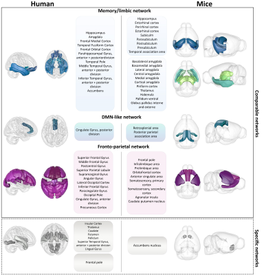 |
Translational exploration of dynamic functional connectivity changes in Alzheimer’s disease patients and Thy-Tau22 mice at the prodromal stage
Laetitia Degiorgis1, Elena Chabran1, Marion Sourty1, Frédéric Blanc1,2, and Laura-Adela Harsan1,3
1ICube Laboratory UMR 7357, Strasbourg University and CNRS, Strasbourg, France, 2Centre Mémoire de Ressources et de Recherche (CM2R), Strasbourg University Hospitals, Neurology and Geriatrics units, Strasbourg, France, 3Department of Biophysics and Nuclear Medicine, Strasbourg University Hospitals, Strasbourg, France
Between-species comparisons become essential to validate findings from animal models. In case of Alzheimer’s disease (AD) especially important is to evaluate their predictive potential for the sporadic form and to assess their relevance for the early stages of pathology. Therefore, we used dynamic resting-state functional connectivity to investigate and comparatively assess changes in early AD patients and prodromal Thy-Tau22 mice, a mouse model of tauopathy. We highlighted between-species similarities showing less dynamic communication of brain areas in diseased groups than in controls, and comparable modifications of memory and default mode-like networks in early AD and prodromal Thy-Tau22 mice.
|
|
| 09:23 | 0416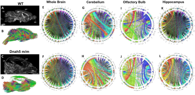 |
Insights into Communicating Congenital Hydrocephalus on Neurodevelopment in a Mouse Model of Ciliopathy Video Permission Withheld
Yijen L Wu1, Margaret Caroline Stapleton1, Ashok Panigrahy2, and Cecilia Lo1
1Developmental Biology, University of Pittsburgh, Pittsburgh, PA, United States, 2Radiology, Children's Hospital of Pittsburgh, Pittsburgh, PA, United States
Mice homozygous to the partial loss-of-function Dnah5 allele, a component in the ependymal motile cilia, developed congenital hydrocephalus. Volumetric analysis showed enlarged aqueduct, indicating that the hydrocephalus was not caused by collapsed aqueduct, but rather by altered cerebrospinal fluid homeostasis. Furthermore, specific brain regions displayed dysplasia, including hippocampus, olfactory bulb and cerebellum. Diffusion tractography followed by topology analysis showed altered neuronal network organization in these brain areas. Our study suggests that ependymal cilia may play a crucial role in grey matter and white matter development and neuronal network organization.
|
|
| 09:25 | 0417.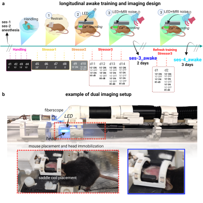 |
Longitudinal simultaneous cortex wide Ca2+ imaging and whole brain functional magnetic resonance imaging in awake mice across the lifespan
Francesca Mandino1, Xilin Shen1, David O'Connor2, Bandhan Mukherjee3, Kristin DeLuca3, Ashley Owens3, An Qu1, John Onofrey1,2,4, Xenophon Papademetris1,2, Stephen M. Strittmatter5, and Evelyn Lake1
1Radiology and Biomedical Imaging, Yale University, New Haven, CT, United States, 2Biomedical Engineering, Yale University, New Haven, CT, United States, 3Neurology and Neuroscience,, Yale University, New Haven, CT, United States, 4Urology, Yale University, New Haven, CT, United States, 5Neurology and Neuroscience, Yale University, New Haven, CT, United States
There is a clear need for imaging protocols in awake animals. Yet, there are only a few approaches in mice, despite their usefulness as translational research subjects. We introduce the first protocol for longitudinal simultaneous fMRI and Ca2+ imaging in awake mice. The same animals are imaged four times (from 4 to 12 months of age) – twice under anesthesia, and twice awake. Our framework includes two acclimation protocols: an initial intensive training followed by a ‘refresher' course. We find a marked improvement in motion between the two awake sessions. Implications for future dual-imaging experiments in awake mice are considered.
|
|
| 09:27 | 0418.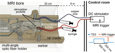 |
Whole brain mapping of transcranial electrical stimulation-induced effects by BOLD-fMRI in rats
Mihaly Voroslakos1, Tanzil Mahmud Arefin2, Jiangyang Zhang2, Leeor Alon2, and Gyorgy Buzsaki1
1Neuroscience Institute, NYU Langone Health, New York, NY, United States, 2Department of Radiology, NYU School of Medicine, New York, NY, United States
Transcranial Electrical Stimulation (TES) is a noninvasive method that can modulate neuronal activity. Despite 20 years of intensive research, the basic mechanisms, and the extent of TES influence on brain functions is still not well understood. To gain a better understanding of the TES effects, we combined neurostimulation with BOLD-fMRI in rats. We have designed an MR-compatible TES setup, including a deuterium-based stimulation electrode system. Our results revealed BOLD responses to TES and altered cortical and cortico-subcortical network responses induced by a variety of stimulation patterns and intensities.
|
|
| 09:29 | 0419.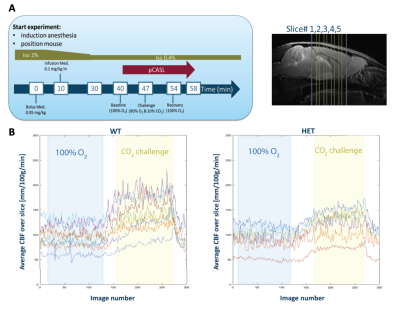 |
Impaired brain perfusion and cerebrovascular reactivity in the zQ175 mouse model of Huntington’s Disease, a longitudinal pCASL-MRI study
Tamara Vasilkovska 1,2, Verdi Vanreusel1, Somaie Salajeghe1, Johan van Audekerke1,2, Lydiane Hirschler3, Dorian Pustina4, Roger Cachope4, Haiying Tang4, Longbin Liu4, Celia Dominguez4, Ignacio Munoz-Sanjuan4, Annemie Van der Linden1,2, and Marleen Verhoye1,2
1Bio-Imaging Lab, University of Antwerp, Antwerp, Belgium, 2µNEURO Research Centre of Excellence, University of Antwerp, Antwerp, Belgium, 3C.J. Gorter Center for High Field MRI, Leiden University Medical Center, Leiden, Netherlands, 4CHDI Management/CHDI Foundation, Princeton, NJ, United States Abnormal cerebral vasculature and consequent diminished cerebrovascular function have been shown to play a role in Huntington’s Disease (HD). However, how these impairments reflect on cerebral blood flow (CBF) dynamics and vascular reactivity (CVR) have been poorly understood. Towards this, we performed a longitudinal study measuring CBF and CVR, using pCASL, in the zQ175 KI Huntington’s disease (HD) mouse model at pre-manifest (3/4.5 months), disease onset (6 months) and manifest (9 months) stage. Our results demonstrate decreased CBF under CO2 challenge at disease onset and decreased CVR in manifest HD stage in the zQ175 KI mice compared to age-matched controls. |
|
| 09:31 | 0420.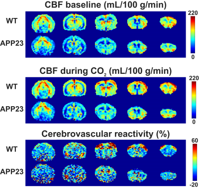 |
Impairments in cerebral blood flow and cerebrovascular reactivity in the APP23 mouse model of cerebral amyloidosis
Leon P Munting1, Mariel G Kozberg1, Lydiane Hirschler2, Jan M Warnking3, Emmanuel L Barbier3, Andre van der Kouwe4, Joseph B Mandeville4, Christian T Farrar4, Steven M Greenberg1, Brian J Bacskai1, and Susanne J van Veluw1
1Massachusetts General Hospital, BOSTON, MA, United States, 2Leiden University Medical Center, Leiden, Netherlands, 3Grenoble Institut des Neurosciences, Grenoble, France, 4Athinoula A. Martinos Center for Biomedical Imaging, Boston, MA, United States
Patients with cerebral amyloid angiopathy (CAA) have accumulations of amyloid-beta in the cerebral vasculature, which ultimately lead to stroke and dementia. MRI markers of CAA include microbleeds and reduced cerebrovascular reactivity (CVR). The APP23 mouse model with vascular amyloid-beta accumulation is known to have microbleeds and reduced cerebral blood flow (CBF). Here, using pseudo-Continuous Arterial Spin Labeling (pCASL)-MRI with a 10 % CO2 stimulation, we show that besides lower CBF, APP23 mice also have reduced CVR. Furthermore, in our data, a higher microbleed count was associated with lower CBF, but not with CVR.
|
|
| 09:33 | 0421.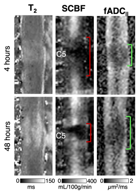 |
Differential information of perfusion and diffusion MRI following traumatic spinal cord injury in the rat.
Briana P Meyer1, Seung-Yi Lee1, and Matthew D Budde2
1Biophysics, Medical College of Wisconsin, Milwaukee, WI, United States, 2Neurosurgery, Medical College of Wisconsin, Milwaukee, WI, United States
Spinal cord tissue perfusion plays a central role in the acute care of spinal cord injury (SCI). We previously optimized pseudo-continuous arterial spin labeling (pCASL) MRI for the rat spinal cord. This work represents the first longitudinal study combining pCASL and filtered diffusion weighted MRI to examine the relationship between spinal cord blood flow and filtered axial diffusivity following a rat model of SCI. This work reveals unique and differing spatiotemporal dynamics between the two contrasts.
|
|
| 09:35 | 0422.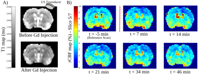 |
Evidence of a cortical hypoperfusion spreading following local ultrasound Blood Brain Barrier opening in the rat brain.
Wafae Labriji1, Julien Clauzel1, Carla Cirillo1, Maxime Lafond2, Cyril Lafon2, Lydiane Hirschler3, Jan Warnking3, Emmanuel L. Barbier3, Isabelle Loubinoux1, and Franck Desmoulin1,4
1ToNIC, Toulouse NeuroImaging Center (UMR 1214 - Inserm / UPS), Toulouse, France, 2LabTAU, Inserm, Centre Léon Bérard, Université Lyon 1, Lyon, France, 3GIN, Grenoble Institut des Neurosciences, Inserm, Université Grenoble Alpes, Grenoble, France, 4CREFRE, Inserm, Université Toulouse III Paul Sabatier, ENVT, Toulouse, France
The use of ultrasound combined with microbubbles to open the blood-brain barrier is an increasingly common method in clinical and preclinical research. This work describes the effect of this BBB opening method on brain perfusion in rats, using pCASL sequence. Surprisingly, through a dynamic analysis, we observed a long time hypoperfusion spreading in the brain cortex following sonication. A similar phenomenon has been described in studies where cortical spreading depression (CSD) has been induced.
|
|
| 09:37 | 0423.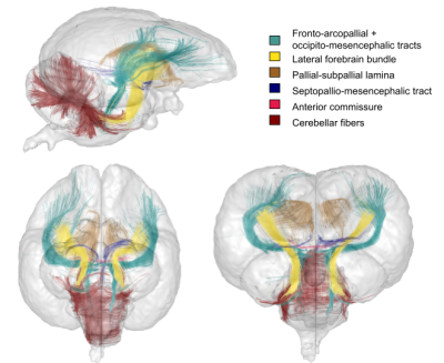 |
Towards an ultra-high resolution structural connectivity atlas of the parrot brain using extreme high-field 17.2T diffusion MRI
Ivy Uszynski1, Luisa Ciobanu1, Solène Bardin1, Pierre Estienne2, Kei Yamamoto2, and Cyril Poupon1
1BAOBAB, NeuroSpin, Université Paris-Saclay, CNRS, CEA, Gif-sur-Yvette, France, 2Neuro-PSI, Université Paris-Saclay, CNRS UMR 9197, Saclay, France
The cognitive abilities of parrots are known to be highly developed to the extent of matching those exhibited by crows and primates. However, little is known about the anatomy and structural connectivity of the parrot’s brain. In this work, we propose to use the power of diffusion MRI along with the strong gradients of a unique 17.2T preclinical MRI system to deliver a first white matter atlas of the parrot brain.
|
|
| 09:39 | 0424.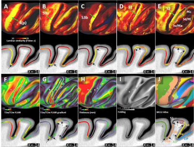 |
Laminar Myeloarchitectonic Mapping using T1- and T2-weighted MRI in Macaque Monkeys
Joonas A. Autio1, Takayuki Ose1, Akiko Uematsu1, Takuro Ikeda1, Masahiro Ohno1, Yuki Masamoto1, Henry Kennedy2,3, David C. Van Essen4, Matthew F. Glasser4,5, and Takuya Hayashi1
1Center for Biosystems Dynamics Research, RIKEN, Kobe, Japan, 2Inserm, Stem Cell and Brain Research Institute, Lyon, France, 3State Key Laboratory of Neuroscience, Institute of Neuroscience, Shanghai, China, 4Department of Neuroscience, Washington University School of Medicine, St Louis, MO, United States, 5Department of Neuroscience, Washington University School of Medicine, St Louis, MT, United States
Classically, many cortical areas have been defined by their distinctive myeloarchitectonic laminar profiles. Here, we explore laminar transitions by adjusting the structural MRI resolution according to the average width of standard six cortical layers and by attenuating Gibbs’ ringing artefact using T1w/T2w-FLAIR ratio in anesthetized macaque monkeys. We demonstrate that laminar similarity measures, together with the ‘Human Connectome Project’-style midthickness weighted gradients, reveal sharp transitions between several cortical areas in heavily and lightly myelinated brain areas providing mutually supportive information of cortical organization.
|
 Back to Meeting Home
 Back to the Program-at-a-Glance
The International Society for Magnetic Resonance in Medicine is accredited by the Accreditation Council for Continuing Medical Education to provide continuing medical education for physicians.
 Back to Meeting Home
 Back to the Program-at-a-Glance