Oral Session
Outstanding Abstracts from the Annual Meeting
Joint Annual Meeting ISMRM-ESMRMB & ISMRT 31st Annual Meeting • 07-12 May 2022 • London, UK

Oral Session
Outstanding Abstracts from the Annual Meeting
08:00 |
0591.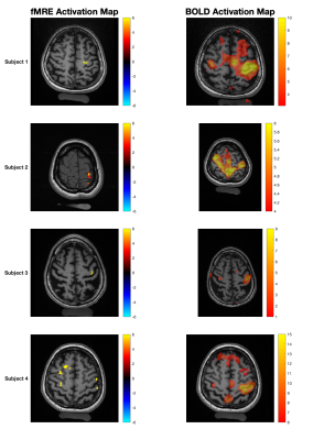 |
Imaging Neuronal Activity at Fast Timescales in Humans using MR Elastography Video Unavailable 1Radiology, Brigham & Women's Hospital, Boston, MA, United States, 2Harvard Medical School, Boston, MA, United States, 3Athinoula A. Martinos Center for Biomedical Imaging, Charlestown, MA, United States, 4Radiology, Massachusetts General Hospital, Boston, MA, United States, 5Neurosurgery, Brigham & Women's Hospital, Boston, MA, United States, 6Laboratory for Vascular Translational Science (LVTS), Institut National de la Santé et de la Recherche Médicale (INSERM), Paris, France A stimulus-interleaved magnetic resonance elastography (MRE) sequence was utilized to demonstrate fast functionally mediated localized changes in shear wavelength during a motor task. Four healthy adult subjects underwent a visual-cue mediated right-hand finger-tapping task, switching between tapping and no-tapping blocks every 2 seconds. Areas of greatest significance between the stimulus states were localized to the left primary motor cortex. A decreased stiffness of ~30% was observed during task performance compared to rest. Compared to traditional BOLD fMRI with a 20-second block, functional MRE areas of greatest statistical difference demonstrated greater percentage change and greater spatial localization. |
|
| 08:03 | 0134.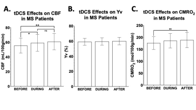 |
Neuronal Response to Transcranial Direct Current Stimulation (tDCS) in Multiple Sclerosis Video Unavailable 1Radiology, NYU Grossman School of Medicine, New York City, NY, United States, 2Neurology, NYU Grossman School of Medicine, New York City, NY, United States, 3Soterix Medical Inc., New York City, NY, United States, 4Department of Biomedical Engineering, City College of New York, New York City, NY, United States
Transcranial direct current stimulation (tDCS) represents an innovative therapeutic tool for neurological diseases such as multiple sclerosis (MS). In this study, MRI measurements of cerebral blood flow (CBF), venous oxygenation (Yv) and cerebral metabolic rate of oxygen (CMRO2) of MS patients and healthy controls (HCs) were acquired pre-, during- and post-tDCS to investigate its simultaneous effects. Whilst HCs showed an immediate increase (~5%) in CMRO2 (during- versus pre-tDCS, p<0.001), MS patients showed a delayed response to tDCS (7.7% CMRO2 increase pre- versus post-tDCS; p=0.007). We suggest that neuronal response to tDCS represents a potential biomarker of neuronal plasticity.
|
|
| 08:06 | 4370.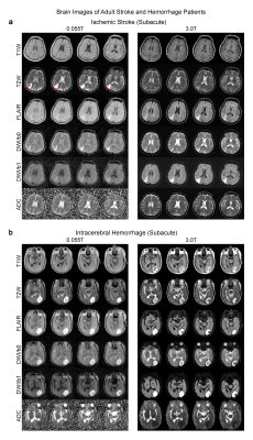 |
A Feasibility Study of 0.055T MRI for Neuroimaging and Comparison with Clinical 3T MRI Video Unavailable 1Laboratory of Biomedical Imaging and Signal Processing, The University of Hong Kong, Hong Kong SAR, China, 2Department of Electrical and Electronic Engineering, The University of Hong Kong, Hong Kong SAR, China, 3Department of Diagnostic Radiology, Li Ka Shing Faculty of Medicine, The University of Hong Kong, Hong Kong SAR, China, 4Department of Surgery, Li Ka Shing Faculty of Medicine, The University of Hong Kong, Hong Kong SAR, China, 5Department of Medicine, Li Ka Shing Faculty of Medicine, The University of Hong Kong, Hong Kong SAR, China, 6School of Biomedical Sciences, Li Ka Shing Faculty of Medicine, The University of Hong Kong, Hong Kong SAR, China
Magnetic resonance imaging is a key diagnostic tool in modern healthcare, yet its accessibility is low with the vast majority of clinical MRI scanners being placed in highly specialized radiology departments, large centralized imaging centers, and housed on ground floors of hospitals and clinics. In this study, we deployed our recently developed 0.055T brain ultra-low-field MRI scanner to demonstrate preliminary feasibility in diagnosing tumor and stroke cases with direct comparisons to 3T clinical MRI results. The development of such ULF MRI technologies will enable patient-centric and site-agnostic MRI scanners to fulfill the unmet clinical needs across various global healthcare sites.
|
|
| 08:09 | 0255.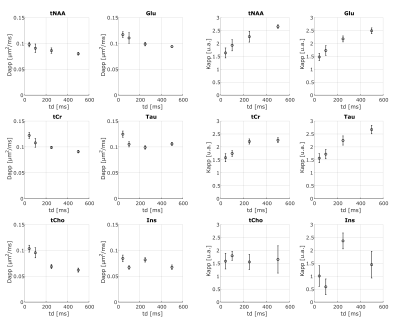 |
Investigating exchange, structural disorder and restriction in Gray Matter via water and metabolites diffusivity and kurtosis time-dependence Video Unavailable 1Université Paris-Saclay, Commissariat à l’Energie Atomique et aux Energies Alternatives (CEA), Centre National de la Recherche Scientifique(CNRS), Molecular Imaging Research Center (MIRCen), Laboratoire des Maladies Neurodégénératives, Fontenay aux Roses, France, 2Centre for Medical Image Computing (CMIC), Department of Computer Science, University College London, London, United Kingdom, 3Cardiff University Brain Research Imaging Centre (CUBRIC), School of Psychology, Cardiff University, Cardiff, United Kingdom, 4School of Computer Science and Informatics, Cardiff University, Cardiff, United Kingdom
This work reports first-time measurements of the time-dependent diffusivity and kurtosis of both water and metabolites in vivo in the mouse gray matter (GM). Our aim is to exploit the complementary information provided by the diffusion of water and intracellular metabolites to investigate and potentially disentangle the role of exchange, structural disorder and restriction in GM. Our results show evidence that water diffusion-time dependence in GM is mostly driven by 1D short-range disorder, potentially alongside exchange. Conversely, metabolites’ diffusion-time dependence is exclusively driven by cellular restrictions, paving a new way to quantify noninvasively microstructural restrictions in GM.
|
|
| 08:12 |
0162.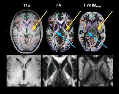 |
In vivo demonstration of generalized anisotropy profiles for resolving boundaries between subcortical gray and white matter Video Unavailable 1Athinoula A. Martinos Center for Biomedical Imaging, Massachusetts General Hospital & Harvard Medical School, Charlestown, MA, United States
We investigate the use of generalized anisotropy profiles (GAPs) for delineating boundaries between subcortical gray and white matter in vivo. Replicating results from a previous ex vivo study, we show that GAPs, which are computed from the diffusion propagator, provide more informative contrasts than T1- and T2-weighted images or conventional diffusion metrics. An undersampled Cartesian-grid sampling of q-space can be used to obtain these profiles with a reasonable scan time. Thus, GAPs show promise as a multi-channel contrast that could be used in the future to improve structural segmentation.
|
|
| 08:15 | 0019.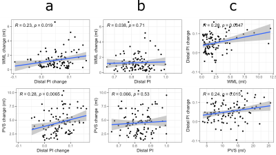 |
Longitudinal associations between 4D flow MRI intracranial pulsatility, white matter lesions, and perivascular space dilation across 5 years Video Unavailable 1Department of Radiation Sciences, Umeå University, Umeå, Sweden, 2Umeå Center for Functional Brain Imaging (UFBI), Umeå University, Umeå, Sweden, 3Department of Clinical Science, Neurosciences, Umeå University, Umeå, Sweden, 4Department of Integrative Medical Biology (IMB), Umeå University, Umeå, Sweden, 5Department of Applied Physics and Electronics, Umeå University, Umeå, Sweden
Small vessel disease (SVD) is associated with elevated vascular stiffness and pulsatility, but longitudinal studies are lacking. Using a 4D flow MRI approach sensitive to age-differences in pulsatility in large and small cerebral arteries, we evaluated 5-year changes in pulsatility in relation to changes in white matter lesions (WML) and perivascular spaces (PVS), radiological markers of SVD. Pulsatility change correlated with WML and PVS volume change, but pulsatility at baseline did not predict WML and PVS progression. However, WML and PVS volumes at baseline predicted 5-year pulsatility change, suggesting that microvascular dysfunction associated with SVD accelerates pulsatility increases.
|
|
| 08:18 |
3729.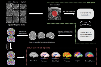 |
Hemodynamics, brain volume and cortical development in fetuses with complex congenital heart disease Video Unavailable 1Radiology, Shandong Provincial Hospital Affiliated to Shandong University, Jinan, China, 2Key Laboratory for Biomedical Engineering of Ministry of Education, Department of Biomedical Engineering, College of Biomedical Engineering & Instrument Science, Zhejiang University, Hangzhou, China, 3Shandong Provincial Hospital Affiliated to Shandong First Medical University, Jinan, China, 4Siemens Healthineers Ltd, Beijing, China
Fetuses with complex congenital heart disease (CHD, n=23) and healthy gestational age-matched controls (n=25) were investigated. Hemodynamics were assessed by Doppler examinations. Fetal brain MR images were segmented to get global and regional morphological measurements. Results showed that fetuses with CHD have abnormally higher umbilical artery pulsation index and smaller global and regional brain volumes as early as 20-30 weeks; however, thickness, mean curvature, and sulcal depth of different brain lobes showed no statistical difference from the healthy controls. These results add to growing evidence of antenatal brain abnormalities of the CHD fetuses during the second and early third trimesters.
|
|
| 08:21 | 0036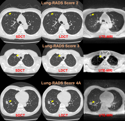 |
MRI with UTE: Capability for Nodule Detection and Lung RADS Classification as Compared with Standard- and Reduced-Dose CTs in Screening Cohort Video Permission Withheld
Yoshiharu Ohno1,2, Masao Yui3, Kaori Yamamoto3, Daisuke Takenaka4, Takeshi Yoshikawa4, Masato Ikedo3, Saki Takeda5, Akiyoshi Iwase5, Yuka Oshima1, Nayu Hamabuchi1, Satomu Hanamatsu1, Yuki Obama1, Hiroyuki Nagata1, Takahiro
Ueda1, Hirotaka Ikeda1, Kazuhiro Murayama2, and Hiroshi Toyama1
1Radiology, Fujita Health University School of Medicine, Toyoake, Japan, 2Joint Research Laboratory of Advanced Medical Imaging, Fujita Health University School of Medicine, Toyoake, Japan, 3Canon Medical Systems Corporation, Otawara, Japan, 4Diagnostic Radiology, Hyogo Cancer Center, Akashi, Japan, 5Radiology, Fujita Health University Hospital, Toyoake, Japan
No report has been found to compare nodule detection and Lung RADS classification capabilities in lung cancer screening cohort among pulmonary MR imaging with UTE, low-dose CT (LDCT) and standard-dose CT (SDCT). We hypothesized that pulmonary MR imaging with UTE has a similar potential to detect pulmonary nodules and evaluate Lung-RADS classification and can apply lung cancer screening as well as CT. The purpose of this study was to compare the capability for nodule detection and Lung RADS classification among pulmonary MR imaging with UTE, LDCT and SDCT in lung cancer screening population.
|
|
| 08:24 | 0145.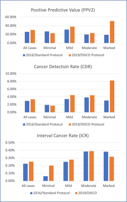 |
High Spatiotemporal Resolution Breast MR Increases Cancer Detection and Reduces Unnecessary Biopsies in Patients with Marked BPE Video Unavailable 1Memorial Sloan Kettering Cancer Center, New York, NY, United States
Contrast-enhanced breast MRI is a powerful tool for breast cancer detection, but can be limited in the setting of marked background parenchymal enhancement (BPE), which can mask underlying lesions. We found that among patients with marked BPE, positive predictive value (PPV2) and cancer detection rate (CDR) were statistically significantly increased when the high spatial/high temporal resolution protocol, rather than the standard protocol, was used. The interval cancer rate was not significantly different between these groups.
|
|
| 08:27 |
0723.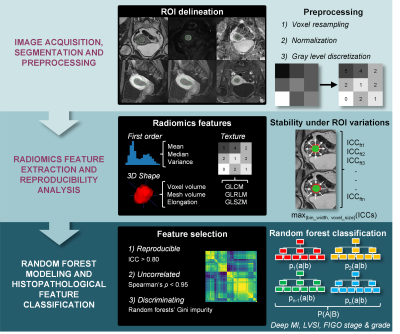 |
Development of MRI-based 3D Radiomics Signatures for Preoperative Risk Stratification of Patients with Histology-proven Endometrial Cancer Video Unavailable 1Medical Physics Unit, McGill University, Montreal, QC, Canada, 2Cancer Research UK Cambridge Institute, University of Cambridge, Cambridge, United Kingdom, 3Department of Diagnostic Radiology, McGill University, Montreal, QC, Canada, 4Department of Radiology, Kobe University Graduate School of Medicine, Kobe, Japan, 5Department of Radiology, Cochin Hospital, AP-HP.Centre, Paris, France, 6Faculté de Médecine, Université de Paris, Paris, France, 7Department of Precision Medicine, GROW - School for Oncology and Developmental Biology, Maastricht University, Maastricht, Netherlands, 8Department of Computer Science, Université de Sherbrooke, Sherbrooke, QC, Canada, 9Augmented Intelligence & Precision Health Laboratory, Research Institute of McGill University Health Centre, Montreal, QC, Canada, 10Research Institute of McGill University Health Centre, Montreal, QC, Canada, 11Department of Obstetrics and Gynecology, McGill University Health Centre, Montreal, QC, Canada, 12School of Computer Science, McGill University, Montreal, QC, Canada
Radiomics analysis on standard MRI prior to surgery holds potential to help identifying high-risk histopathological features of endometrial carcinoma, including FIGO stage, deep myometrial invasion, lymphovascular space invasion and tumor grade, thus supporting preoperative risk stratification for optimal patient management. This dual-center retrospective study evaluated the role of radiomics to assess high-risk phenotypes of endometrial cancer in women who underwent 1.5-T MRI before hysterectomy. Radiomics-based machine learning models provided consistent clinically acceptable performance for differentiating early from advanced FIGO stage endometrial carcinoma and for differentiating low- from high-risk histopathological markers in two independent datasets from different institutions on preoperative MRI.
|
|
| 08:30 |
0666.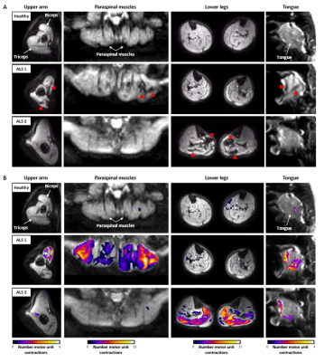 |
Whole-body fasciculation detection in Amyotrophic Lateral Sclerosis (ALS) using motor unit MRI (MUMRI) Video Unavailable 1Newcastle University Translational and Clinical Research Institute, Newcastle University, Newcastle upon Tyne, United Kingdom, 2Newcastle Biomedical Research Centre, Newcastle University, Newcastle upon Tyne, United Kingdom, 3Northern Medical Physics and Clinical Engineering, Freeman Hospital, Freeman Hospital, Newcastle upon Tyne NHS Foundation Trust, Newcastle upon Tune, United Kingdom, 4Department of Neuroradiology, Royal Victoria Infirmary, Newcastle upon Tyne, United Kingdom, 5Directorate of Clinical Neurosciences, Royal Victoria Infirmary, Newcastle upon Tyne, United Kingdom
The spontaneous contraction of motor units in muscle, i.e. fasciculation, has been recognised as an important diagnostic marker in amyotrophic lateral sclerosis (ALS). Fasciculation can be imaged with a novel MRI technique called motor unit MRI. This technique uses a diffusion weighted sequence on which fasciculation presents as short-living signal voids. We demonstrated an increased fasciculation rate in ALS patients compared to healthy controls by assessing the four body regions relevant in the diagnosis of ALS. The affected body regions differed between patients. This is in line with the heterogeneous disease onset and supports our proposed whole-body approach.
|
|
| 08:33 | 0510.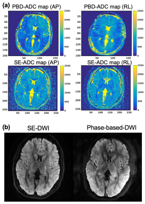 |
Phase-based 3D diffusion mapping using RF phase-modulated gradient echo imaging Video Unavailable 1Radiology, University of Wisconsin-Madison, Madison, WI, United States, 2Biomedical Engineering, University of Wisconsin-Madison, Madison, WI, United States, 3Medical Physics, University of Wisconsin-Madison, Madison, WI, United States, 4Emergency Medicine, University of Wisconsin-Madison, Madison, WI, United States, 5Medicine, University of Wisconsin-Madison, Madison, WI, United States A novel 3D diffusion mapping method using RF phase-modulated gradient echo imaging was developed by encoding diffusion weighting into signal phase. We developed closed form signal equations based on configuration theory. A four-pass acquisition and lookup table-based reconstruction is performed to estimate diffusion and T2 simultaneously. Simulation results revealed excellent agreement of signal magnitude and phase between Bloch equation and the developed equations. Phantom and in vivo studies demonstrated the feasibility of the proposed approach. These results demonstrate that the proposed method can enable qualitative and quantitative diffusion imaging with smaller gradient amplitude compared to the conventional diffusion-weighted imaging. |
|
| 08:36 | 0271.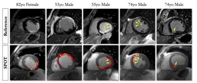 |
Improved Myocardial Scar Visualization with Two-minute Free-breathing Joint Bright- and Black-blood Late Gadolinium Enhancement Imaging Video Unavailable 1IHU LIRYC, Electrophysiology and Heart Modeling Institute, Université de Bordeaux – INSERM U1045, Bordeaux, France, 2Department of Cardiovascular Imaging, Hôpital Cardiologique du Haut-Lévêque, CHU de Bordeaux, Bordeaux, France, 3Department of Diagnostic and Interventional Radiology, Lausanne University Hospital and University of Lausanne, Lausanne, Switzerland, 4INRIA, Université Côte d’Azur, Sophia Antipolis, France, 5Department of Cardiac Electrophysiology, Hôpital Cardiologique du Haut-Lévêque, CHU de Bordeaux, Bordeaux, France, 6CIBM Center for Biomedical Imaging, Lausanne, Switzerland
Black-blood late gadolinium enhancement (LGE) techniques are increasingly being used to uncover myocardial scar patterns that may be otherwise confused with blood signal on conventional bright-blood LGE, especially when involving the subendocardium. With signal-attenuated blood and dark muscle representation, these techniques may however be hampered by a lack of anatomical information, making spatial localization of myocardial injuries a challenging task. Here we aim to assess the clinical performance of a novel LGE technique that combines both bright- and black-blood imaging in a single time-efficient free-breathing exam to obtain LGE images with unprecedented scar contrast and localization.
|
|
| 08:39 | 3101.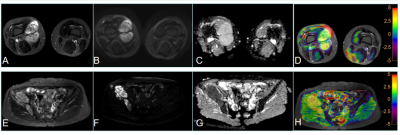 |
Preliminary assessment of 3D APT-weighted combined with diffusion weighted imaging in characterization of bone and soft tissue tumors Video Unavailable 1The First Affiliated Hospital of Zhengzhou University, Zhengzhou, China, 2Philips Healthcare, Beijing, 100102, China, Beijing, China
Amide proton transfer weighted imaging (APTWI) is a noninvasive emerging molecular MRI technique based on chemical exchange saturation transfer (CEST) between amide protons of proteins and polypeptides and free water protons. This work investigated and evaluated the ability of APT parameter in distinguishing benign from malignant bone and soft tissue tumors. Results indicated that APTWI combined with diffusion weighted imaging showed a significantly improved differentiation between benign and malignant tumors.
|
|
| 08:42 | 5036.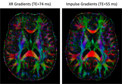 |
Evaluation of High Resolution Diffusion MRI on the Next-Generation 7T scanner Video Unavailable 1Radiology and Biomedical Imaging, University of California, San Francisco, San Francisco, CA, United States, 2Radiology, San Francisco Veteran Affairs Health Care System, San Francisco, CA, United States, 3Helen Wills Neuroscience Institute, University of California, Berkeley, Berkeley, CA, United States, 4Advanced MRI Technologies, Sebastopol, CA, United States, 5Siemens Medical Solutions USA, Inc., Melvern, PA, United States, 6Siemens Healthcare, Erlangen, Germany
The newly developed head gradient (200 mT/m Gmax, 900 T/m/s SR) on the new NexGen 7T system provides many potential benefits for high-resolution diffusion imaging. Faster, stronger gradients can significantly reduce diffusion encoding times and TE for up to a 3-fold improvement SNR while also reducing TR and total scan time. We demonstrate these benefits in high resolution diffusion imaging applications with b-values of up to 10,000 s/mm2 at 7T.
|
|
| 08:45 | 0223.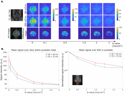 |
300 mT/m diffusion MRI 'below the neck': First results in healthy prostate Video Unavailable 1Cardiff University Brain Research Imaging Centre (CUBRIC), Cardiff University, Cardiff, United Kingdom, 2Siemens Healthcare Ltd, Camberly, United Kingdom, Camberly, United Kingdom, 3Siemens Healthcare GmbH, Erlangen, Germany, Erlangen, Germany, 4Centre for Medical Image Computing, University College London, London, United Kingdom, 5Image Sciences Institute, University Medical Center Utrecht, Utrecht, Netherlands
This work showcases the first ever use of ultra-strong gradient in ’below the neck’ human MRI. It assesses the benefits, compared to more commonly-available gradient strengths, of 300mT/m gradients for comprehensive characterization of the prostate gland. Leveraging such gradient amplitudes enables unique tissue contrasts, with a potential to improve specificity and/or sensitivity of current MRI methods for prostate cancer characterisation and detection.
|
 Back to Meeting Home
 Back to the Program-at-a-Glance
The International Society for Magnetic Resonance in Medicine is accredited by the Accreditation Council for Continuing Medical Education to provide continuing medical education for physicians.
 Back to Meeting Home
 Back to the Program-at-a-Glance