ISMRT Poster Presentations
ISMRT Poster Presentations - Clinical
Joint Annual Meeting ISMRM-ESMRMB & ISMRT 31st Annual Meeting • 07-12 May 2022 • London, UK

ISMRT Poster Presentations
ISMRT Poster Presentations - Clinical
| 17:00 | 5051.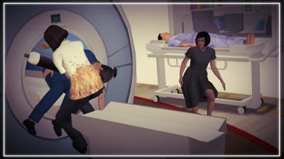 |
Gamified Learning in MRI Safety Video Unavailable 1Neuroradiology, National Neuroscience Institute, Singapore, Singapore
Magnetic Resonance (MR) safety plays an important role in ensuring patients and staff safety and is important for all staff to be competent prior to working in the MRI environment. To make MR safety training more engaging and motivated, game-based learning was adopted to simulate situations that are dangerous and impossible to re-enact in the real world. Learner acts as avatar and explore the MR safety concepts through a 3D version of the National Neuroscience Institute (NNI), Neuroradiology department. Learner will identify hazards through mini-games and role-play as Chief Risk Officer to identify gaps from a MR related accident
|
|
| 17:00 | 5052. | I’m Late, I’m Late, For a Very Important Date: Pointers for Effective Patient Communication in MRI Video Unavailable 1Radiology, Uniformed Services University, Bethesda, MD, United States, 2Radiology, Walter Reed National Military Medical Center, Bethesda, MD, United States, 3Geneva Foundation, Tacoma, WA, United States
Many of us have or are now working in a high volume fast paced MR environment. But what happens when your patient is anxious, in pain, angry or any other of the many obstacles we as MR Technologist/Radiographers may encounter? First impressions will set the mood for the entire procedure and possibly the whole time your patient is at your institution. Being kind, patient and willing to listen to your patients will go a long way when dealing with anxious patients. You can make a difference.
|
|
| 17:00 | 5053.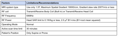 |
SAR Considerations of MRI Patients with Implanted Deep Brain Stimulator Video Unavailable 1Neuroradiology, National Neuroscience Institute Singapore, Singapore, Singapore
Magnetic Resonance Imaging (MRI) has been a widely used modality in patients with Deep Brain Stimulator (DBS) for a variety of neurological diseases. Radiofrequency heating still presents as a major concern in patient’s safety and exists as the most common limiting factor. This poster would help individuals to gain a deeper understanding about the importance of SAR considerations in patients with DBS whilst undergoing MRI without compromising patients safety and scan quality.
|
|
| 17:00 | 5054.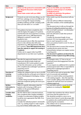 |
Improving MRI in patients with Cochlear Implants (CIs) Video Unavailable 1Radiology Department, Glan Clwyd Hospital, Bodelwyddan, Wales, United Kingdom, 2Betsi Cadwaladr University Health Board, Bangor, Wales, United Kingdom, 3British Cochlear Implant Group MRI Working Group, Bradford, United Kingdom, 4Sir Peter Mansfield Imaging Centre, University of Nottingham, Nottingham, United Kingdom, 5Hearing Theme, NIHR Nottingham Biomedical Research Centre, Nottingham, United Kingdom, 6Hearing Sciences, School of Medicine, University of Nottingham, Nottingham, United Kingdom
A cochlear implant (CI) contains an implanted magnet placed under the scalp. Deaf children often receive a CI in their first year and as such are likely to need MRI in their lifetime. The presence of an implanted magnet raises safety concerns around MRI, with MRI often being avoided in favour of other imaging techniques that are less diagnostically powerful and use ionising radiation. The British Cochlear Implant Group (BCIG) MRI Working Group was assembled with the aim of collecting existing best practice and produce a set of guidance for the safe and effective clinical MR in individuals with CIs.
|
|
| 17:00 | 5055.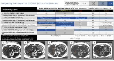 |
Chemical Shift Proton Density Fat Fraction Imaging: End-user perspectives on liver fat assessment Video Unavailable 1Canon Medical Research USA, Mayfield Village, OH, United States, 2Cardiology, Johns Hopkins University, Baltimore, MD, United States, 3Canon Medical Systems Corporation, Tochigi, Japan
CS-PDFF is an indispensable method of assessing steatosis. However, each site must make informed decisions when selecting assessment methodology, as both ROI style and location can impact measurements. This abstract discusses how well ROIs of various styles characterize average PDFF and, additionally, how each ROI style tolerates the influence of various confounding factors (e.g.: artifacts and elevated noise). We also explore potential contributing factors to localized variations in hepatic PDFF, as well as their relative importance. Finally, select normative values from our cohort are reported, with a focus on observed differences as related to ROI placement and size.
|
|
| 17:00 | 5056. | Techniques for Preparing and Scanning Neonatal Patients for MRI Video Unavailable 1MRI, Robert wood Johnson University Hospital, New Brunswick, NJ, United States
Neonatal Imaging can be challenging in MRI. New texts are often unsure of how to ensure the babies safety and "compliance". We present here are some basic techniques on how to prepare and scan neonatal patients in MRI
|
|
| 17:00 | 5057.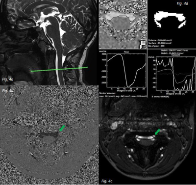 |
Proposal for an MRI protocol for cerebrospinal flows Video Unavailable 1Radiology, CHU amiens picardie, AMIENS, France, 2CHU amiens picardie, AMIENS, France
Flow MRI is useful for imaging flow from the LCS and blood in the craniospinal system. Currently, at the Amiens University Hospital, we are performing these sequences in clinical routine in patients with pathologies involving potential alterations in cerebral hemohydynamics such as hydrocephalus, intracranial hypertension, Arnold Chiari's malformation. This poster presents the specifics of the MRI flow sequence applied to brain flows.
|
|
| 17:00 | 5058.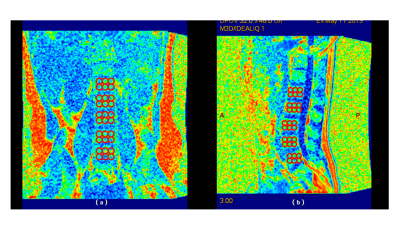 |
Iterative decomposition of water and fat with echo asymmetry and least-squares estimation (IDEAL-IQ) for evaluation of early bone mass changes in senile osteoporosis patients Video Unavailable 1Department of Radiology Imaging, The Second Affiliated Hospital of Xiamen Medical College, Xiamen, China, 2GE Healthcare, Beijing, China
BMD is the basis of the diagnostic criteria of OP recommended byWHO. However, limitations in the prediction and evaluation of fracture in postmenopausal_women, because BMD can not fully reflect the bone quality that affects bone strength and fracture risk. We found significant differences in the values among three groups. FF, fat-phase and water-phase value had good performance, while R2*value had poor performance in discriminating osteopenia and OPgroups. Overall, IDEAL-IQtechniqueis expected to provide certain reference indexes, which can noninvasively and quantitatively evaluate the bone metabolism of lumbar vertebrae, such as fat content, and provide accurate information for differential diagnosis of OP.
|
|
| 17:00 | 5059.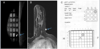 |
A conclusion on how computer-aided localization of a breast lesion impact Breast MRI-guided biopsies when compared with manual localization techniques Video Unavailable 1Radiology, Memorial Sloan Kettering Cancer Center, Forest Hills, NY, United States, 2Memorial Sloan Kettering Cancer Center, New York, NY, United States
Patients with suspicious lesions on MRI that cannot be detected with another modality may undergo MRI guided biopsy. The MRI biopsy may induce anxiety and may be uncomfortable with the breast in compression which may be exacerbated by the length of the procedure. Software has been developed by certain companies to streamline and increase ease of the MRI-guided biopsy. We performed a study to see if this software decreases the length of the procedure. We also will see if software leads to differences recommendation, concordance, and subsequent cancer if the biopsy was benign.
|
|
| 17:00 | 5060.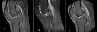 |
Radial-Cartesian K-Space Acquisition for Consistent High-Resolution and High-Quality Knee MRI Video Unavailable 1GE Healthcare, Bangalore, India, 2GE Healthcare, Waukesha, WI, United States
Knee MRI is the most common Musculo-skeletal MRI [1]. It is a non-invasive tool for the evaluation of disorders such as meniscal, ligamental, soft tissue, bone and bone marrow injuries and abnormalities. Acquisition of a high-resolution and high-quality MRI is key to boost the confidence of the radiologist in diagnosis. In this manuscript we have presented how radial MRI, commercially referred to as PROPELLER (GE)/ BLADE (Siemens)/ MULTIVANE (Philips) etc. parameters, patient positioning and immobilization can be adapted for the available hardware such as receiver coil to yield high-resolution and high-quality knee MRI.
|
|
| 17:00 | 5061.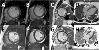 |
Role of cardiovascular magnetic resonance in differentiating peripartum cardiomyopathy from pregnancy unmasking of underlying cardiomyopathy Video Unavailable 1Cape Universities Body Imaging Centre, University of Cape Town, Cape Town, South Africa, 2Division of Human Biology, Department of Biomedical Engineering, University Of Cape Town, Cape Town, South Africa, 3Cape Heart Institute, Faculty of Health Sciences,, University of Cape Town, Cape Town, South Africa, 4Division of Cardiology, Department of Medicine, University of Cape Town, Cape Town, South Africa, 5Department of Radiation Medicine, Groote Schuur Hospital, University of Cape Town, Cape Town, South Africa Peripartum cardiomyopathy (PPCM) is a form of heart failure associated with pregnancy.1,2 We evaluated cardiovascular magnetic resonance (CMR) scans on 6 patients with PPCM and incomplete recovery. CMR showed an increase in left ventricular volumes and impairment of left ventricular ejection fraction (6-39%) in all patients. Right ventricular dysfunction was present in 5 patients. Linear mid-wall late gadolinium enhancement was present in all cases. Two patients had PPCM, one had dilated cardiomyopathy (DCM) with hypertrophy, 2 had DCM with left ventricular non-compaction [>6 months postpartum], and one had familial DCM. CMR is useful in distinguishing PPCM from other DCM phenotypes.
|
|
| 17:00 | 5062.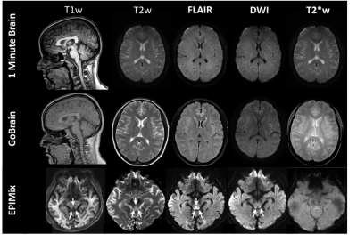 |
EPIc-reMIX, an Accelerated Routine Brain Imaging Protocol Video Unavailable 1Philips Healthcare, Gainesville, FL, United States
EPIMix is a multi-contrast EPI sequence developed which generates the standard brain imaging contrasts in 78 seconds. Studies using accelerated brain protocols like EPIMix and GoBrain have shown similar diagnostic performance to the routine clinical protocols. However, EPIMix suffers from EPI geometric distortions and like GoBrain is limited to 2D acquisitions. EPIc-reMIX, an accelerated brain imaging protocol, was created to leverage the speed of EPIMix and GoBrain while improving image quality and adding 3D-imaging capabilities.
|
|
| 17:00 | 5063.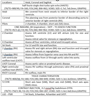 |
Cardiovascular Magnetic Resonance (CMR) in the diagnosis of Sarcoidosis: A Case report Video Unavailable 1Cape Universities Body Imaging Centre, University of Cape Town, Cape Town, South Africa, 2Division of Human Biology, Department of Biomedical Engineering, University of Cape Town, Cape Town, South Africa, 3Department of Radiation Medicine, Groote Schuur Hospital, Cape Town, South Africa, 4Department of Medicine, faculty of Health Sciences, University of Cape Town, Cape Town, South Africa, 5The Hatter Institute, Faculty of Health Sciences, University of Cape Town, Cape Town, South Africa
Sarcoidosis is defined as a multisystem granulomatous disease of unknown origin, characterized by noncaseating granulomas. In cardiac sarcoidosis (CS), granulomas have been linked to ventricular tachyarrhythmias and electrical instability. Cardiovascular magnetic resonance (CMR) can provide diagnostic information and aid the clinical management. Here, we report a case of a middle-aged male patient with intermittent complete heart and with a history of hypertension and diabetes mellitus. He was referred for evaluation of CS. Subsequently, the patient underwent successful cardiac pacemaker implant and remains clinically well.
|
 Back to Meeting Home
 Back to the Program-at-a-Glance
The International Society for Magnetic Resonance in Medicine is accredited by the Accreditation Council for Continuing Medical Education to provide continuing medical education for physicians.
 Back to Meeting Home
 Back to the Program-at-a-Glance