ISMRT Poster Presentations
ISMRT Poster Presentations - Research
Joint Annual Meeting ISMRM-ESMRMB & ISMRT 31st Annual Meeting • 07-12 May 2022 • London, UK

ISMRT Poster Presentations
ISMRT Poster Presentations - Research
| 17:00 | 5064.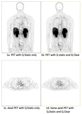 |
Reducing motion artifacts on abdominal PET/MR images Video Unavailable 1Radiology, University of California, San Francisco, San Francisco, CA, United States, 2Research, Stanford University, Palo Alto, CA, United States, 3PET/MR Clinical Development, GE Healthcare, Waukesha, WI, United States
Motion can pose a significant challenge for positron emission tomography (PET) reconstructions. A block sequential regularized expectation maximization reconstruction algorithm (Q.Clear, GE Healthcare) is used to increase the signal-to-noise ratio (SNR) of PET images. Respiratory motion compensation Quiescent Phase Gating (QPG), also called Q.Static, is used to reduce motion-induced artifacts by extracting data from the quiescent period. Q.Static performed without Q.Clear increases image noise as only ~50% of the counts are utilized for image reconstruction. However, Q.Static performed with Q.Clear improves image quality for whole-body PET without increasing the scan time.
|
|
| 17:00 | 5065.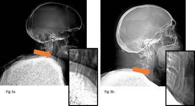 |
Are low dose CT scouts suitable for MRI safety screening of subjects with unknown medical history? Video Unavailable 1King's College Hospital, london, United Kingdom, 2Royal Marsden Hospital, London, United Kingdom
Increasing numbers of patients are presenting for MRI who cannot complete the safety questionnaires. CT scout images have low radiation dose, and are faster and easier to acquire compared to plain film x-rays, the recommended modality for MR safety screening for patients with unknown history. This study assesses the performance of CT scout images in detecting and identifying a range of head/neck and body implants. Using 40 head/neck and 40 body CT scouts, two Readers reviewed the images for possible internal implants. 88% of implants were found and correctly identified, concluding that the majority of visible implants are detectable.
|
|
| 17:00 | 5066.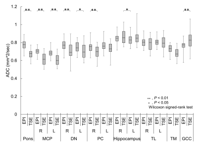 |
New reference ADC data for the normal brain with distortion correction in TSE- and EPI-DWI Video Permission Withheld
Yasuo Takatsu1,2,3, Masafumi Nakamura4, Hajime Sagawa5, Yuichi Suzuki6, Nobuyuki Mori7, Shunichi Motegi8, and Tosiaki Miyati9
1Department of Radiological Technology, Faculty of Health and Welfare, Tokushima Bunri University, Sanuki-city, Japan, 2Department of System Control Engineering, Graduate School of Engineering, Tokushima Bunri University, Sanuki-city, Japan, 3Division of Health Sciences, Graduate School of Medical Sciences, Kanazawa University, Kanazawa, Japan, 4Otsu City Hospital, Otsu, Japan, 5Division of Clinical Radiology Service, Kyoto University Hospital, Kyoto, Japan, 6Department of Radiology, The University of Tokyo Hospital, Tokyo, Japan, 7Department of Radiology, Osaka Red Cross Hospital, Osaka, Japan, 8Department of Radiological Sciences, International University of Health and Welfare, Ohtawara, Japan, 9Division of Health Sciences, Graduate School of Medical Sciences, Kanazawa University., Kanazawa, Japan
We performed echo planar imaging (EPI) and turbo spin echo (TSE) diffusion-weighted imaging (DWI) using magnetic resonance imaging to obtain basic clinical data of the apparent diffusion coefficient (ADC) in 26 parts of normal brains and compared the datasets using our retrospective distortion correction technique. The ADC was significantly higher measured by EPI-DWI than by TSE-DWI. The signal-to-noise ratio of EPI-DWI was significantly higher than that of the TSE-DWI. Care must be taken when measuring ADCs near the base of the skull, such as the brain stem, where the SNR of the imaging technique is likely to decrease or distort.
|
|
| 17:00 | 5067.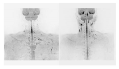 |
Reductin of cervical motion and ADC inaccuracy by use of neck fixation device for DWIBS Imaging Video Unavailable 1National Hospital Organization Ibarakihigashi Hospital, Toukai, Japan, 2Graduate School of Ibaraki Prefectural University of Health Sciences, Ami, Japan, 3National Hospital Organization Mito Medical Center, Ibaraki-machi, Japan
The effectiveness of a neck fixation device to improve the image quality of DWIBS was investigated. Healthy volunteers were examined while chewing with and without a neck fixation device using 3-T MRI system. The amount of mandibular bone movement and the ADC values of some body parts were decreased by the fixation device. These are thought to improve the image quality and the ADC measurement accuracy. The technique using a neck fixation device helps improve image quality of DWIBS in the head and neck region.
|
|
| 17:00 | 5068.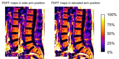 |
Influence of arm position on proton density fat fraction in vertebral bone marrow using chemical shift-encoded magnetic resonance imaging Video Unavailable 1Graduate School of Medical Technology, Teikyo University, Tokyo, Japan, 2Central of Radiology, Teikyo University Hospital, Tokyo, Japan, 3Department of Radiological Technology, Juntendo University, Tokyo, Japan
Reports have shown radio frequency magnetic field (B1) inhomogeneity during spine imaging. Proton density fat fraction (PDFF) measurement using chemical shift-encoded magnetic resonance imaging has been performed using a low flip angle to avoid T1 bias. However, differences in arm positions during MRI can cause changes in B1 heterogeneity. This study evaluated the influence of arm position (side and elevated arm positions) on the PDFF obtained from the lumbar spine, where B1 heterogeneity tends to occur. The PDFF was slightly but significantly higher in the elevated arm position than in the side arm position.
|
|
| 17:00 | 5069. | Health complaints subjectively associated with static magnetic field and acoustic noise exposure among MR personnel in Sweden Video Unavailable 1Department of Nursing, Umeå University, Umeå, Sweden, 2Department of Radiation Sciences, Radiation Physics, Umeå University, Umeå, Sweden, 3Department of Radiology, Institute of Clinical Sciences, Sahlgrenska Academy, University of Gothenburg, Gothenburg, Sweden, 4Department of Radiology, Sahlgrenska University Hospital, Gothenburg, Sweden, 5Department of Medical Imaging and Physiology, Skåne University Hospital, Lund, Sweden, 6Department of Diagnostic Radiology, Clinical Sciences, Lund University, Lund, Sweden
Our aim was to explore the prevalence of health complaints subjectively associated with static magnetic field and acoustic noise exposure among MR personnel in Sweden, using CT personnel as a control group. Utilizing a cross-sectional survey, 529 respondents answered items regarding symptom prevalence and its attribution, acoustic noise at work, health factors and work-environmental details including stress. Respondents were categorized into three groups (MR personnel, CT personnel, and mixed personnel (both MR and CT)) and data were tested with logistic regression to evaluate risk associations with symptoms.
|
|
| 17:00 | 5070.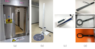 |
Evaluation of the characteristics of magnetic detectors and metal detectors for MRI examinations Video Unavailable 1Radiological Technology, Gunma Prefectural College of Health Sciences, Maebashi, Japan, 2Radiological Technology, Nagoya University Hospital, Nagoya, Japan, 3Diagnostic imaging, Institute of Brain and Blood Vessels Mihara Memorial Hospital, Isesaki, Japan, 4Imaging Center, St. Marianna University School of Medicine Hospital, Kawasaki, Japan, 5Radiological Technology, Tokyo Women's Medical University Hospital, Shinjuku, Japan
The purpose of this study is to evaluate magnetic detectors and metal detectors to understand their performance and characteristics. The measurement objects were smartphones, disposable pocket warmers, scissors, ballpoint pens, safety pins, and hair bands with rings. The detection performance of each object was examined at several facilities using a gate-type, a pole-type and, a handheld-type magnetic detectors and a handheld-type metal detector. There were differences in detectability depending on the size and type of magnetic material. The sensitivity range and objects that are easy to detect may also differ depending on the type of detector.
|
|
| 17:00 | 5071. |
A comparison of child and adult perceptions and experiences of having an MRI at 7T Video Unavailable 1London Collaborative Ultra high field System (LoCUS), London, UK, Kings College London, London, United Kingdom, 2Guys and St Thomas’ NHS Foundation Trust, Kings College London, London, United Kingdom, 3Perinatal Imaging and Health, Kings College London, London, United Kingdom, 4Centre for the Developing Brain, School of Biomedical Engineering and Imaging Sciences, Kings College London, London, United Kingdom, 5Biomedical Engineering Department, School of Biomedical Engineering and Imaging Sciences, Kings College London, London, United Kingdom, 6Radiology Department, Great Ormond Street Hospital for Children, London, United Kingdom, 7Department of Forensic and Neurodevelopmental Sciences, Institute of Psychiatry, Psychology and Neuroscience, Kings College London, London, United Kingdom, 8MR Research Collaborations, Siemens Healthcare Limited, London, United Kingdom, 9MRC Centre for Neurodevelopmental Disorders, Kings College London, London, United Kingdom
The aim of this study was to gather data from children on their subjective experiences when undergoing high field MRI and compare this to adult data collected with similar questionnaires. Seventeen children and twenty-six healthy adults had brain imaging at 7T. Their experiences which included: (a) acoustic noise, (b) anxiety, (c) metallic taste (d) vertigo (dizziness) and e) involuntary eye movement (nystagmus) or flashing lights were evaluated. We found that children scanned at 7T reported similar experiences to adults.
|
|
| 17:00 | 5072.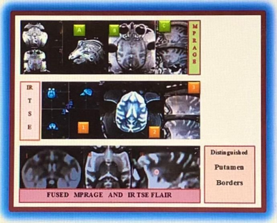 |
Non-Invasive MRI of Putamen Using Fused T1 MPRAGE and IR TSE FLAIR Video Unavailable 1MRI Research, University of Pennsylvania, Philadelphia, PA, United States, 2MRI Interventions, ClearPoint, Irvine, CA, United States, 3Comparative Medicine Services Core, CHOP Research Institute, Philadelphia, PA, United States, 4Neurosurgery, University of Pennsylvania, Philadelphia, PA, United States
The innovative method of fusing T1-weighted magnetization prepared rapid acquisition gradient echo (MPRAGE) and inversion recovery turbo spin echo fluid-attenuated inversion recovery (IR TSE FLAIR) provides the whole brain image with improved tissue contrast and distinguishes the boundaries within the subcortical structures of the basal ganglia. The presented experiment uses non-invasive MRI-guided imaging to obtain an adequate therapeutic coverage infusing the AAV test article to the right and left Putamen. Results have shown that fused MPRAGE and IR TSE FLAIR acquisitions obtained non-invasively provides more reliable and precise imaging of the right and left Putamen targeted for gene therapy.
|
|
| 17:00 | 5073.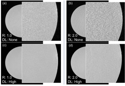 |
Noise Power Spectrum and Modulation Transfer Function in Deep Learning Based Reconstruction Method Video Unavailable 1Radiological Technology, Kobe City Medical Center General Hospital, Kobe, Japan, 2Diagnostic Radiology, Kobe City Medical Center General Hospital, Kobe, Japan
The purpose of this study was to evaluate the effects of Deep Learning (DL) method in MRI using Noise Power Spectrum and Modulation Transfer Function. To evaluate the effect of DL method, we compared the results without DL method and the results with DL method in the NPS and the MTF using phantom MR images. Deep learning Reconstruction method in MRI has the ability to reduce the average NPS value by more than 48.7% in denoising and improve the spatial resolution by 50% at all DL intensities.
|
|
| 17:00 | 5074.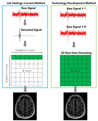 |
Reducing MRI scan time to increase patient throughput Video Unavailable 1Radiology, UCSF, San Francisco, CA, United States, 2Chemistry nd Chemical Biology, Cornell University, Ithaca, NY, United States We have developed a novel wavelet-based signal processing algorithm capable of denoising signals with extremely low signal-to-noise ratios. The idea is that if raw MRI signals can be denoised in real-time prior to image generation, high quality images can potentially be obtained at drastically shorter scan-times, thereby improving patient comfort and increasing imaging throughput. While conventional signal processing methods are often limited by inadequate noise removal or signal distortion, our method does not suffer from such drawbacks, as it performs localized noise removal based on capturing randomness. This represents an opportunity to increase MRI throughput within existing infrastructure. |
|
| 17:00 | 5075.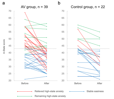 |
Anxiety relaxation during MRI with a patient-friendly audiovisual system Video Unavailable 1Department of Radiology, Chiba University Hospital, Chiba, Japan, 2Diagnostic Radiology and Radiation Oncology, Graduate School of Medicine, Chiba University, Chiba, Japan, 3Department of Neurosurgery, National Hospital Organization Chiba Medical Center, Chiba, Japan, 4Research Center for Child Mental Development, Chiba University, Chiba, Japan
Many patients experience anxiety, not limited to claustrophobia, before magnetic resonance imaging (MRI) examination. Thus, to examine improvements to the environment during MRI examinations, this study employed a non-randomized controlled trial and evaluated whether a patient-friendly audiovisual system in the MR scanner room reduces patient anxiety. As a result, the patient-friendly audiovisual system compared to the traditional environment reduced anxiety in patients undergoing MRI examinations. Thus, this system can be used to provide a more patient-centered environment and may relieve the negative perceptions associated with MRI examinations.
|
|
| 17:00 | 5076.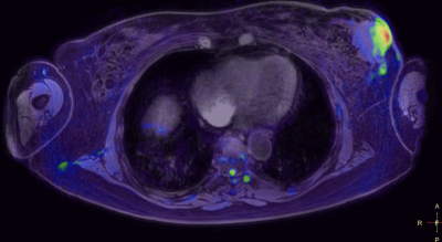 |
Whole Body DWI at 3T with Inline Geometric Distortion Correction and Broadband RF Inversion Video Unavailable 1Mermaid Beach Radiology, Gold Coast, Australia, 2Philips Healthcare ANZ Clinical Science, Sydney, Australia
The purpose of this work is to reverse geometric distortion uniformly across all stations of whole body Diffusion Weighted Imaging (DWI) at 3T for near perfect co-registration with cartesian imaging and correct alignment between stations for improved diagnostic confidence. This translation of a well established and published research technique translated in to clinical in-line application for geometric distortion correction in Whole Body DWI applications at 3T has revolutionised our confidence further in challenging regions of the body such as the thorax as well as vastly improved geometric stitching of multiple stations.
|
|
| 17:00 | 5077.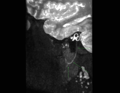 |
3D Cranial Nerve Imaging with Background Suppression at 3T Video Unavailable 1Mermaid Beach Radiology, Gold Coast, Australia, 2Philips Healthcare ANZ Clinical Science, Sydney, Australia
Clinical presentations of many cranial nerve symptoms are scanned without an obvious gross pathology. The gold standard in MRI of high resolution multiplanar imaging with and without contrast often result in no obvious anatomical or post contrast injection enhancement changes. It involves a 3D fast spin echo Short Tau Inversion Recovery (STIR) with addition Motion Sensitive Driven Equilibrium (MSDE) preparation pulses for blood vessel/flow suppression. This sequence is improving our diagnostic confidence of cranial nerve neuropathies/neuritis with validation of perceived clinical indications.
|
|
| 17:00 | 5078.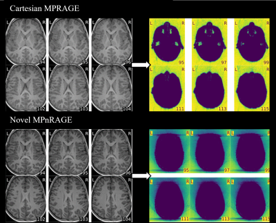 |
Constructing an Image Quality Comparison Video Unavailable 1Radiology, Boston Children's Hospital, Boston, MA, United States, 2Computational Radiology Laboratory, Boston Children's Hospital, Boston, MA, United States
Magnetic resonance imaging (MRI) has become an increasingly popular imaging exam for pediatric patients due to its high resolution, multiplanar capabilities, and lack of ionizing radiation [1]. MRI is highly beneficial by providing detailed imaging to assist in the diagnosis and treatment plan for patients, but before a sequence becomes part of an established clinical protocol, there are many trials performed and data collected. Novel sequences, such as the motion robust imaging technique MPnRAGE [2], are emerging to help alleviate clinical imaging dilemmas, like motion artifacts, but this data needs to be examined and understood to make advancements.
|
|
| 17:00 | 5079. | Evaluating Preparation Methods for Clinical MRI Scans in Young Children without Anesthesia Video Unavailable 1Alberta Children's Hospital, Calgary, AB, Canada
We compared the efficacy of three different preparation methods for clinical MRI scans without sedation in young children (3-7 years): at-home resources, a session with a child-life specialist, or a mock MRI session with a child life specialist. Success rates were excellent at 91%, with no differences between training groups. Subtle group differences in fear/anxiety ratings suggest a slight benefit of the mock MRI session in reducing anxiety before scans.
|
 Back to Meeting Home
 Back to the Program-at-a-Glance
The International Society for Magnetic Resonance in Medicine is accredited by the Accreditation Council for Continuing Medical Education to provide continuing medical education for physicians.
 Back to Meeting Home
 Back to the Program-at-a-Glance