Weekday Course
Physics for Clinicians II
Joint Annual Meeting ISMRM-ESMRMB & ISMRT 31st Annual Meeting • 07-12 May 2022 • London, UK

| Physics for Clinicians II.A | |||
| 09:15 | Image Reconstruction for Clinicians
Katherine Wright
This presentation will review basic and advanced MR image reconstruction methods. Raw data in MRI are not collected directly in the image domain. They are collected in k-space, where each data point contains information about the entire object being imaged. k-space data must be transformed into an image, a process called image reconstruction, which is most often performed by applying the Fourier Transform. We will review properties of k-space and the relationship between k-space and image domains, as these are key to understanding most reconstruction methods. Next, we will review several reconstruction methods that are used on clinical MRI scanners.
|
||
| 09:45 | 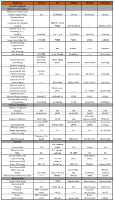 |
Techniques & Terminology Across Vendors
Matthias Gunther
MRI is the medical imaging modality with by far the largest diversity of “imaging modes” (aka MR sequences). There seems to be the habit in the MR community to compete in terms of fantasy when it comes to sequence naming. Over the past, this has led to a huge variety of different acronym denoting MR sequence, which are closely related, but still carry different names. Very minor changes seem to justify the creation of new acronyms. This makes the field of MR sequences challenging. Fortunately, dictionaries of acronyms exist to guide one through this jungle (for links see reference section).
|
|
| Physics for Clinicians II.B | |||
| 10:15 | Addressing Patient-Related Artifacts: Implants & Motion
Ives Levesque
This lecture will review the manifestations, fundamental physics, and mitigation strategies of patient-related artifacts, namely those that arise from motion and from implants. In the first portion, artifacts arising from implants will be addressed, focusing on susceptibility induced signal dropout, image distortion, and signal displacement. In the second portion, we will turn to the broad category of motion-related artifacts to explore how various sources of motion corrupt MR images. Possible “confound” artifacts will be included. Approaches for mitigation of these artifacts will be discussed, emphasizing the practical tradeoffs and the latest solutions proposed in research.
|
||
| 10:45 | Translating New Methods to the Clinic: A Practical Guide
Jamie MacKay
This session will outline the key practical steps in translating a novel MRI method into the clinic. The challenges at each step will be discussed, with an emphasis on the important difference between clinical validation and demonstration of clinical utility.
My talk will be illustrated by examples of MRI methods which have been successful & unsuccessful at each stage of the translation process. In particular, we will consider whole-body MRI and quantitative cartilage MRI as exemplars of MRI methods which have been more or less successful in translation to the clinic. |
||
 Back to Meeting Home
 Back to the Program-at-a-Glance
The International Society for Magnetic Resonance in Medicine is accredited by the Accreditation Council for Continuing Medical Education to provide continuing medical education for physicians.
 Back to Meeting Home
 Back to the Program-at-a-Glance
 View the Presentation