Weekday Course
2D-4D Flow & Perfusion in the Body: Methods & Clinical Applications
Joint Annual Meeting ISMRM-ESMRMB & ISMRT 31st Annual Meeting • 07-12 May 2022 • London, UK

Weekday Course
2D-4D Flow & Perfusion in the Body: Methods & Clinical Applications
| Blood Flow & Perfusion in the Body: Methods | |||
| 16:45 | Current Methods for 2D, 3D & 4D Flow in Body Organs
Hideki Ota
Clinical application of flow imaging has been accepted, and various MR acquisition techniques are applied for imaging vessels of body organs. Depending on clinical purposes, blood flow should be visualized as static images for vascular morphology and/or measured for hemodynamic evaluation. In this talk, the following will be introduced: 1) basic knowledge of associations between flow and signals, 2) non-contrast-enhanced inflow-dependent MR angiography and contrast-enhanced MR angiography for qualitative flow visualization, and 3) principles of 2D PC and 4D flow MRI for flow visualization and measurement. Future directions of 4D flow MRI to overcome time-consuming process will also be discussed. |
||
| 17:15 | Current Methods for Perfusion & Permeability MRI in Body Organs
Octavia Bane
Dynamic contrast-enhanced (DCE-)MRI uses the unique physiology of liver perfusion (dual input from the hepatic artery and the portal vein) to characterize diffuse liver disease, and conditions arising from end-stage liver disease such as portal hypertension and hepatocellular carcinoma. By attending this lecture, course participants will be introduced to: 1) the conceptual difference between perfusion and vascular permeability, and why it is impossible to measure both at the same time; 2) acquisition protocol requirements for DCE-MRI; 3) the main pharmacokinetic models used with DCE-MRI in diffuse and focal liver disease; 4) examples of clinical applications of DCE-MRI in the liver.
|
||
| Blood Flow & Perfusion in the Body: Applications | |||
| 17:45 | 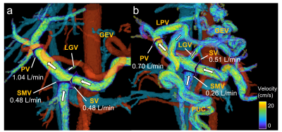 |
Clinical Applications of 4D Flow MRI in the Portal Venous System
Thekla Oechtering
Assessment of hemodynamics in the portal venous system is essential for the diagnosis of many liver pathologies. 4D flow MRI offers a comprehensive approach for understanding pathophysiological mechanisms. It simultaneously and noninvasively acquires time-resolved flow and anatomic information in a 3D imaging volume. Although promising, it is particularly challenging in the portal venous system because of small vessel calibers, slow flow velocities, and respiratory motion. This presentation will describe how to perform and evaluate 4D flow MRI exams of the portal venous system. Moreover, it will discuss potential clinical applications and promising quantitative parameters that could help diagnose various pathologies.
|
|
| 18:15 |  |
Clinical Applications of cardiac & Lung MRI - a focus on pulmonary hypertension Video Permission Withheld
Andy Swift
This talk covers a number of the clinical applications of blood flow and perfusion imaging in the lungs. There will be a focus on pulmonary hypertension and parenchymal lung diseases of emphysema and interstitial lung disease. The use of several MRI sequences will be discussed, non contrast approached phase contrast MRI, arterial spin labelling and PREFUL, in addition contrast enhanced approaches such and dynamic contrast enhanced angiography and magnetic resonance angiography. The feasibility of these approaches and their diagnostic and prognostic value will be discussed.
|
|
 Back to Meeting Home
 Back to the Program-at-a-Glance
The International Society for Magnetic Resonance in Medicine is accredited by the Accreditation Council for Continuing Medical Education to provide continuing medical education for physicians.
 Back to Meeting Home
 Back to the Program-at-a-Glance
 View the Presentation